Introduction
Cherubism is a human genetic disorder that causes bilateral symmetrical enlargement of the maxilla and the mandible in children. It is caused by mutations in SH3BP2 . The exact pathogenesis of the disorder is an area of active research. Sh3bp2 knock-in mice were developed by introducing a Pro416Arg mutation (Pro418Arg in humans) in the mouse genome. The osteoclast phenotype of this mouse model was recently described.
Methods
We examined the bone phenotype of the cherubism mouse model, the role of Sh3bp2 during bone formation, osteoblast differentiation, and osteoblast function.
Results
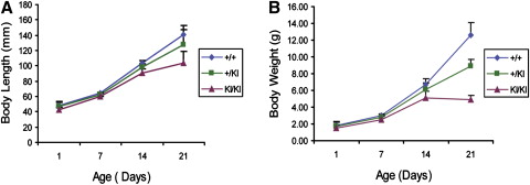
We observed delays in early postnatal development of homozygous Sh3bp2 KI/KI mice, which exhibited increased growth plate thickness and significantly decreased trabecular bone thickness and bone mineral density. Histomorphometric and microcomputed tomography analyses showed bone loss in the cranial and appendicular skeletons. Sh3bp2 KI/KI mice also exhibited a significant decrease in osteoid formation that indicated a defect in osteoblast function. Calvarial osteoblast cell cultures had decreased alkaline phosphatase expression and mineralization, suggesting reduced differentiation potential. Gene expression of osteoblast differentiation markers such as collagen type I, alkaline phosphatase, and osteocalcin were decreased in osteoblast cultures from Sh3bp2 KI/KI mice.
Conclusions
These data suggest that Sh3bp2 function regulates bone homeostasis through not only osteoclast-specific effects, but also effects on osteoblast differentiation and function.
Editor’s comment
Cherubism is a genetic disorder that shows up in children about 3 to 4 years of age. Approximately 200 cases have been reported in medical journals. The name originates from the chubby-faced angels or cherubs of Renaissance paintings. Symmetrical cyst-like cavities in the maxilla and the mandible are the hallmark of cherubism. The cavities fill with soft fibrous tissue, which is histologically indistinguishable from giant cell granulomas. Expansion of the fibrous tissue can cause characteristic facial deformities in cherubism patients. The fibrous lesions usually regress spontaneously after puberty, but, in severe cases, after the active phase of the disorder, some facial deformity remains, and the patient might need surgical correction.
This study won the 2009 Thomas M. Graber Award of Special Merit (basic science category) for Padma M. Mukherjee, a graduate of the University of Connecticut. Her goals were to further characterize the bone phenotype of cherubism knock-in mice and to examine the role of Sh3bp2 in osteoblast differentiation and function. The data demonstrate for the first time that Sh3bp2 is important for bone formation and osteoblast differentiation and function. Cherubic lesions develop during the mixed dentition, when there is an inflammatory response and tooth movement (exfoliation of deciduous teeth and eruption of permanent teeth). It will therefore be interesting to study the effects of the cherubism mutation in tooth movement experiments. In future experiments, the authors hope to correlate these results to the human phenotype.
During the peer review process, one reviewer noted that “Findings like these hasten the day when orthodontists will be able to assay patients for high- or low-functioning bone cells through identification of marker genes and proteins, and truly assess biological compatibility for orthodontic treatment and employ individualized therapeutics.” In simple terms, this study provides the basis for determining individual qualitative and quantitative differences in bone, which currently are almost wholly unappreciated clinically.