(1)
Canberra, ACT, Australia
Summary
The most common peri-radicular lesions of the jaws are inflammatory lesions caused by infection of the dental pulp. Peri-apical granuloma, chronic peri-apical abscess, peri-apical pocket cysts, radicular cysts, extra-radicular infections, foreign body reactions and fibrous scar tissue typically present as unilocular peri-apical radiolucencies of varying size and degrees of circumscription. The need to differentiate can only be based on a careful history taking, clinical signs and symptoms and equivocal pulpal sensibility testing followed by commencement of nonsurgical root canal treatment to tip the balance in favour of healing.
Clinical Relevance
Clinically peri-apical lesions cannot be differentially diagnosed as either cystic or non-cystic lesions based on conventional radiographs. An accurate diagnosis can only be made following histopathological serial sectioning of the lesion in its entirety. Unlike true radicular cysts, peri-apical pocket cysts (Bay cysts) may heal after nonsurgical root canal therapy. The clinician must also be aware of other radiopaque jaw lesions that mimic lesions of endodontic origin that may not respond to conventional treatments requiring careful follow-up and in some cases surgical exploration and biopsy to confirm the true nature of the problem.
4.1 Overview of Peri-apical Pathologies in Endodontics
A cyst, derived from the Greek Krystis meaning sac or bladder, is defined as a closed pathological cavity, lined by an epithelium that contains a liquid or semisolid material [1]. Radicular cysts are inflammatory jaw cysts that commonly occur at the peri-apex of a necrotic, infected pulp as a direct sequel to apical granuloma, although a granuloma need not always develop into a cyst. Occasionally they may also be found on the lateral aspects of the root in relation to a lateral accessory canal [2].
Persistent peri-radicular radiolucencies of endodontic origin are commonly due to intra-radicular infection [3], extra-radicular infection [4–6], foreign body reactions [7], true cysts [8, 9] and fibrous scar tissue [10]. Orthograde root canal treatment or re-treatment with the intention to remove/eliminate microorganisms below a critical threshold conducive to healing is the first line of treatment in all cases. Lesions associated with extra-radicular infections, true cysts and foreign body reactions can only be managed by peri-radicular surgery. Peri-apical lesions that heal by fibrous scar tissue require no treatment (Fig. 4.1).
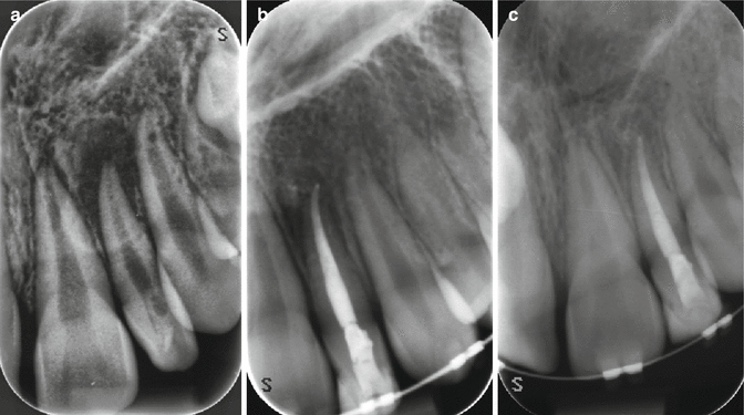
Fig. 4.1
Clinical radiographs showing resolution of peri-apical endodontic lesion following nonsurgical root canal therapy. Note (a) preoperative view of tooth 22 showing distinct peri-radicular lesion, (b) 6-month review radiograph following completion showing significant reduction in lesion size and (c) 12-month follow-up demonstrating intact periodontal ligament space associated with the peri-apex
Many lesions that occur in the jaw including lesions of endodontic origin (inflammatory cysts, granulomas, abscesses or fibrous scars) have similar radiological appearances often making it difficult to differentiate among them (Table 4.1 and Fig. 4.2). Although rare, other clinically confusing peri-apical lesions have been documented including lesions of malignancy. It is therefore imperative to have an understanding of the pathogenesis of common endodontic lesions and their management to ensure that misdiagnosis is avoided. Careful history taking and clinical findings should be evaluated taking into consideration radiographic findings to help in narrowing down the differential diagnosis. Follow-up is important not only to ensure that any therapeutic treatment approach has been successful but also to confirm the correct diagnosis has been made [11, 12].
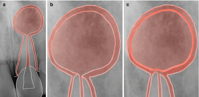
Table 4.1
Surgical sieve for radiolucent and mixed lesions of the jaw
|
Well-circumscribed radiolucent lesions
|
|
Developmental
|
|
Nasopalatine duct cyst
|
|
Dentigerous cyst
|
|
Odontogenic keratocyst
|
|
Inflammatory
|
|
Peri-apical granuloma
|
|
Peri-apical pocket cyst
|
|
Radicular cyst
|
|
Neoplastic
|
|
Multiple myeloma
|
|
Ameloblastoma
|
|
Traumatic
|
|
Traumatic bone cyst
|
|
Metabolic
|
|
Giant cell lesions of hyperparathyroidism
|
|
Idiopathic
|
|
Aneurysmal bone cyst
|
|
Central giant cell granuloma
|
|
Ill-defined radiolucent lesions
|
|
Inflammatory
|
|
Acute osteomyelitis
|
|
Neoplastic
|
|
Osteogenic sarcoma
|
|
Chondrosarcoma
|
|
Metastatic lesions
|
|
Lesions with mixed and/or variable radiological appearances
|
|
Developmental
|
|
Fibrous dysplasia
|
|
Inflammatory
|
|
Condensing osteitis
|
|
Neoplastic
|
|
Osteochondroma (benign)
|
|
Ossifying (cement-ossifying) fibroma
|
|
Osteosarcoma (malignant)
|
|
Normal anatomical structures
|
|
Mental foramen
|
|
Maxillary sinus
|
|
Nutrient canals
|
Fig. 4.2
Diagrams showing (a) non-vital tooth with a peri-radicular lesion that cannot be distinguished radiographically from a peri-apical granuloma, abscess or cyst. Note (b) a peri-apical pocket cyst that by definition has an epithelium-lined cavity that communicates with the root canal system and (c) a radicular cyst where the epithelium-lined cavity is completely enclosed from the root canal system. The former will often resolve following nonsurgical root canal therapy, whereas the latter may require surgical enucleation
Two distinct categories of radicular cysts have been reported in the literature making a distinction between those cavities containing completely enclosed epithelium (true radicular cyst) and those containing epithelium-lined cavities that are open to the root canals (bay cyst or peri-apical pocket cyst) (Fig. 4.2). The former is an independent entity resulting in a lesion that is self-sustaining and no longer dependent on the presence or absence of root canal infection. As a result surgical excision would have to be performed following conventional root canal treatment to ensure healing. In the latter nonsurgical root canal treatment carried out effectively would eliminate the infection within the root canal space, ensuring peri-apical pocket cysts would heal [8, 9].
Currently the gold standard for diagnosis of a peri-apical lesion is based on clinical and histological findings using serial sectioning of the lesion in its entirety [11]. In the past, assumptions were made based on radiological findings to differentiate between a true radicular cyst and peri-apical granuloma, which were scientifically unsound.
Often lesion size was measured and larger radiographic diameters were more likely thought of as cystic as opposed to granulomatous in nature, which was not always the case [13–15]. Radiological appearances of cysts are often described as round or ovoid radiolucencies surrounded by a narrow radiopaque demarcating margin, which extends from the lamina dura of the involved tooth. In infected or rapidly growing cysts this margin may not be present and these features are therefore not correlative with the definitive diagnosis of a true radicular cyst [16].
Several surgical approaches exist for the management of cystic lesions of the jaws including enucleation, marsupialisation and decompression. Enucleation comprises of complete removal of the cyst lining which is a definitive treatment modality not usually requiring further intervention. The risk of morbidity is higher as nearby structures and teeth may be damaged. Marsupialisation is the conversion of a cyst into a pouch. The cystic roof is removed in its entirety, and the cut edges of the remaining cyst are sutured to the adjacent soft tissue lining the oral cavity, maxillary sinus or nasal cavity into a continuous pouch. Surgical decompression is a minimally invasive technique whereby a large cystic lesion is converted into a small one with the aim of any future surgical intervention having reduced morbidity when considering enucleation. Decompression involves the insertion of a decompression stent/drainage tube into the peri-apical lesion, regular irrigation, periodic length adjustment and maintenance of the drain for varying lengths of time. It is contraindicated in cases of large dental granulomas or any solid cellular lesion for the reason that there is an absence of a fluid-filled cavity to decompress [17–21].
There have been numerous reports of non-odontogenic benign and malignant lesions presenting in the peri-apical area mimicking lesions of endodontic origin. The frequency and distribution of radiolucent jaw lesions in a retrospective study revealed that most non-healing lesions submitted for biopsy were classified as either apical granulomas (40.4 %) or apical cysts (33.1 %), and they were often from the maxillary anterior jaw. Over 20 % of the reported non-healing radiolucent lesions submitted had a more severe pathologic implication, such as odontogenic keratocysts (8.8 %), central giant cell lesions (1.3 %), ameloblastomas (1.2 %) and even the small but important number of metastatic lesions (0.26 %). Most of these lesions were located in the posterior mandible. Granulomas or cysts (73 %) were often from the anterior maxillary jaw. Thus non-healing radiolucent jaw lesions other than granulomas or cysts were reported over 20 % of the time and may have more severe pathological implications, suggesting the value of differential diagnoses [22].
Common cysts that mimic lesions of endodontic origin include odontogenic keratocysts [23], residual cysts [24], lateral periodontal cysts [25] and nasopalatine duct cysts [26].
Benign aggressive lesions that cause locally destructive lesions mimicking peri-apical pathosis include central giant cell granulomas [27, 28], central ossifying fibroma [29], calcifying epithelial odontogenic tumour (Pindborg tumour) [30, 31], osteoblastoma [32] and central odontogenic fibroma [33].
Benign cemento-osseous dysplasia including peri-apical cemental dysplasia can develop around the apices of the teeth, representing a well-recognised diagnostic challenge that is difficult to distinguish from peri-apical granulomas in the early stages [34, 35].
Granulomatous inflammation distinguished from granulation tissue associated with peri-apical granulomas is a distinct entity that has been reported in the literature elicited by foreign material. It has been proposed that foreign body-induced granulomatous inflammation at the peri-apex of the teeth may result in endodontic treatment that fails to heal [7, 36, 37].
A variety of misdiagnosed malignant neoplastic lesions have been reported in the endodontic literature, masquerading at the peri-apex of the teeth similar to lesions of endodontic origin. As a result of clinical and radiographic similarities, these lesions may be mistaken with inflammatory or infectious diseases of the jaws, creating a dilemma for the clinician. Histopathological evaluation is of paramount importance when the lesion fails to respond to routine treatments [12, 31, 38–45].
4.2 Differential Diagnosis of Radiolucent Lesions of the Jaw
The most common peri-radicular lesions of the jaws are peri-apical granuloma, peri-apical cyst (radicular and peri-apical pocket cyst) and the chronic dental abscess, which are inflammatory in origin due to the demise of the dental pulp. All three lesions typically present as unilocular, peri-radicular radiolucencies of varying sizes. Unequivocal results of clinical signs and symptoms including pulp vitality testing and radiographic diagnosis often lead to commencement of root canal treatment which tips the balance for healing to occur. The need for follow-up is a prerequisite not only to determine the outcome of treatment but also to ensure that a correct diagnosis was reached. In the cases of lesions that fail to respond, a surgical approach may be necessary to confirm histopathologically the underlying reason for failure and also rule out the possibility of other lesions that mimic endodontic pathology.
4.3 Lesions of Endodontic Origin
Peri-apical granuloma
A chronic inflammatory reaction that consists of granulomatous tissue predominantly infiltrated with lymphocytes, plasma cells and macrophages. The lesion is maintained by persistent necrotic pulpal contents resulting in a well-circumscribed radiolucent lesion around the peri-apex of a tooth. Occasionally the lesion may present laterally in response to a lateral canal or perforation. Nonsurgical endodontic therapy will eradicate the lesion (Fig. 4.3).
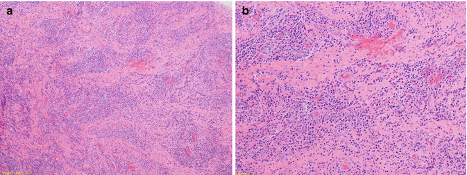
Fig. 4.3
Clinical photomicrographs showing distinct histopathological features of a peri-apical granuloma. Typical features include chronically inflamed granulation tissue at the apex of a non-vital tooth surrounded by fibrous connective tissue and lymphatic (plasma cells, neutrophils, mast cells, etc.). Note light microscopy (a) ×100, (b) ×200 (Courtesy of Drlan Clarke Capital Pathology Canberra ACT)
Peri-apical condensing osteitis
Reactive hyperplasia at the peri-apex of a tooth in response to a low-grade pulpal infection can lead to condensing osteitis. Typically seen in young adults and teenagers, affecting mandibular molars and premolars most commonly. The bone is compact and dense with minimal chronic inflammation appearing more radiopaque at the peri-apex of the tooth. Endodontic treatment is required to resolve the problem.
Acute apical abscess
An abscess is defined as a ‘localised collection of pus’ that can occur around the peri-apex of an endodontically infected tooth. An abscess is a focus of acute inflammation characterised by a distinct collection of polymorphonuclear leukocytes within a pre-existing chronic inflammatory lesion (e.g. granuloma). An acute peri-apical abscess is a very painful condition that is characterised by intense throbbing; extreme pain to touch, biting and percussion; palpation tenderness; and increased mobility of the tooth. An intra-oral or extra-oral swelling may be evident. Radiographically there may not be any signs.
Chronic apical abscess (phoenix abscess)
Chronic apical periodontitis with suppuration
These terms denote a peri-apical abscess that arises from an acute exacerbation of a chronic inflammatory peri-apical lesion (Fig. 4.4).
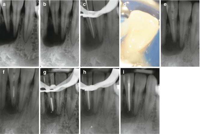
Fig. 4.4
Clinical radiographs and photograph demonstrating case diagnosed as chronic apical abscess. Note (a) and (b) parallel peri-apical and mesial 20° shift showing extensive peri-radicular radiolucent lesion extending from the peri-apex of teeth 32, 31 and 41. Following pulp sensibility testing, a diagnosis of chronic apical periodontitis 31 was made. Teeth 32 and 41 were deemed vital.(c) MAF preparation was completed using hand files, (d) patency filing was used and pus exudate was noted, (e) intra-canal medication was placed for 3 months, and (f) review appointment showed dressing resorbed and peri-apical lesion reduced considerably in size. (g–i) Nonsurgical endodontic treatment completed. Patient has been placed on further review
The lesion appears as a well-demarcated radiolucency at the peri-apex of a tooth. Histopathologically a variable mixture of fibrovascular connective tissue, scar and chronic inflammatory cells is present with foci of neutrophils, oedema and liquefaction necrosis. Often drainage may be evident through a sinus intra-orally with or without symptoms. Nonsurgical endodontic treatment will eradicate the lesion.
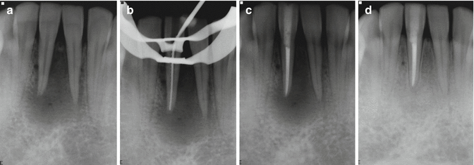
Peri-apical pocket cyst
An inflammatory lesion that contains a sac-like epithelium-lined cavity that is open to and continuous with the root canal. Pocket cysts are more likely to heal after nonsurgical endodontic therapy of the tooth since the treatment removes the source of irritation. A well-defined radiolucency will be associated either apically or laterally and indistinguishable from peri-apical granuloma or radicular cyst. Nonsurgical treatment will often eradicate the lesion (Fig. 4.5).
Stay updated, free dental videos. Join our Telegram channel
VIDEdental - Online dental courses


