and Jasdeep Kaur1
(1)
Earth and Life Sciences Vrije Universiteit Amsterdam and ILEWG, Amsterdam, The Netherlands
Abstract
Many years ago, anatomists, anthropologists, forensic pathologists, and forensic odontologists started to find the correlations between the surface soft tissues of the face and the underlying bone structure of the skull. Different methods of facial reconstruction are based on these hypothesized relationships. The main objective of all facial reconstructions for forensic investigations is to reconstruct an individual’s face, usually when no other identifying or antemortem evidence is available, in a sufficient manner to illustrate a similarity to the missing or deceased person, to help in his or her identification. This chapter refers to the different methods of facial reconstruction and craniofacial identification.
4.1 Introduction
Craniofacial identification is a forensic process where a photograph of a missing person is compared with a skull to determine its identity. There are different methods to sculpture a face onto the skull, all of which are based on the reproduction of a possibly recognizable face using the photos (Phillips and Smuts 1996). Facial reconstruction is a method used in forensic investigations to assist in identification. This chapter is focused on the role of craniofacial identification in forensic investigations.
4.2 Anthropometry (Bromby 2003)
The use of facial parameters as a form of evidence is new, and its reliability in medicolegal cases is debatable. Identification from photographs using facial anthropometry has been used in present-day medicolegal court cases (Bromby 2003). An investigative medicolegal report will usually contain multiple comparisons (Bromby 2003). According to Bromby, “Facial mapping is defined as the process of identifying which individual through the facial features and can include video/photo superimposition, anthropometric measurements and morphological comparisons” (2003, p. 302).
4.3 Photo-anthropometry
Photo-anthropometry is defined as the analysis of anthropometric landmarks, dimensions, and angles to quantify facial characteristics and proportions from a photograph (Ïscan 1993). The main objective of this concept is to get information concerning the real objects illustrated in a photograph from this two-dimensional image. It is frequently used in the examination of traffic accidents, major disasters, and crime scenes.
4.4 Morphology
When the morphological characteristics of an individual are documented, the height, weight, and body type are the noted first. Hair, racial, and ethnic facial appearances, the color of the individual’s skin, and any distinctive individuality such as tattoos, surgical scars, and other scars, congenital deformities, and so forth are documented if feasible. It has been reported that camera angles and camera distance can all affect the appearance of the individual and make identification more difficult. It has been reported that emotions such as anger, fear, happiness, and surprise can significantly change facial features (Ïscan 1993). Physiological and pathological changes such as disease, aging, and exposure to the elements also affect morphology (Donofrio 2000).
4.5 Superimposition
Superimposition is a method of identification that involves photographic and video superimposition. If no medical or dental records are available, but one has an idea about the identity of the deceased person, then a skull and a photograph can be superimposed as well as two photographs. Before the person’s identity has been formally determined, dental and other records, if available, should be examined for further verification. Video superimposition on dead persons is better used to exclude someone rather than to provide positive identification because it can certainly be stated that a skull and photograph may not be of the identical individual (Vanezis and Brierley 1996). In order to ascertain a positive identification, the two images should be taken with the same viewpoint or at least from a very similar position (Vanezis and Brierley 1996). When both images are from living individuals, as opposed to one being an image of a skull, landmarks vary according to the position from which the image was obtained (Shahrom et al. 1996). Landmarks include the nasal cavity, bite line, and eye sockets on a skull, which are usually aligned (Yoshino et al. 2000). It has been reported that special attention is paid to the curves of the forehead, the depth of the nasal bridge, the shape and projection of the nasal bones, the lower face and chin prominence, and the height of the vault, when comparing a skull to a lateral photograph, while attention is focused on the orbital size and shape, the breadth of the nasal bridge, the width of the nasal aperture, the total facial length and width, the ratio of mid-face to upper or lower face length, and the mandibular shape with a frontal photograph. As the images are in position, one image is superimposed on top of the other, keeping the horizontal, vertical, or diagonal alignment (Austin-Smith and Maples 1994).
The skull landmarks (George 1993) that are often used follow:
Craniometric landmarks (Fig. 4.1):
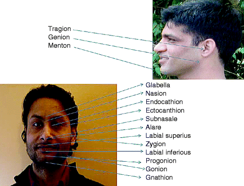
Fig. 4.1
Craniometric landmarks
-
Dacryon (Da): the point of junction of the frontal, maxillary, and lacrimal bones on the lateral wall of the orbit
-
Frontomalar temporal (Fmt): the most lateral point of junction of the frontal and zygomatic bones
-
Glabella (G): the most prominent point between the supraorbital ridges in the midsagittal plane
-
Gnathion (Gn): a constructed point midway between the most anterior (Pog) and most inferior (Me) points on the chin
-
Gonion (Go): a constructed point; the intersection of the lines tangent to the posterior margin of the ascending ramus and the mandibular base, or the most lateral point at the mandibular angle
-
Nasion (N): the midpoint of the suture between the frontal and the two nasal bones
-
Nasospinale (Ns): the point where a line drawn between the lower margins of the right and left nasal apertures is intersected by the midsagittal plane (MSP)
Stay updated, free dental videos. Join our Telegram channel
VIDEdental - Online dental courses


