Introduction
The purpose of this study was to assess differences in dentoalvolar cortical bone thickness between hyperdivergent and hypodivergent young adults.
Methods
Pretreatment cone-beam computed tomography images of 57 patients, including 30 hypodivergent subjects (22 women, 8 men) and 27 hyperdivergent subjects (20 women, 7 men), were analyzed. The data were imported into imaging software (version 10.5; Dolphin Imaging Systems, Chatsworth, Calif); standardized orientations were used to measure buccal and lingual cortical bone thicknesses at 16 interradicular sites of the maxilla and the mandible. Total alveolar ridge thickness and medullary space thickness were measured at the same sites.
Results
T tests showed significant ( P <0.05) group differences, with hypodivergent subjects having significantly thicker buccal cortices. The lingual cortex of the maxilla was also significantly thicker in the hypodivergent than in the hyperdivergent subjects. Alveolar ridge thickness was significantly greater at all sites of the hypodivergent mandible and at the anterior 2 sites of the hypodivergent maxilla. Medullary thickness was significantly greater only in the hypodivergent mandibles between the first molars and the second premolars, and between the first and second premolars. Buccal cortical bone was significantly thicker than lingual cortical bone in the mandible; lingual bone was significantly thicker in the maxilla.
Conclusions
Cortical bone tends to be thicker in hypodivergent than in hyperdivergent subjects. This explains the concomitant differences in alveolar ridge thickness. Medullary space thickness is largely unaffected by facial divergence.
Although facial morphology is largely genetically determined, individual differences in cortical thickness are partially due to functional demands. Cortical bone adapts to the strains to which it is subjected. As delineated by the mechanostat hypothesis of Frost, there is a range of strain values that maintain the form and mass of the bone; strains above this range induce bone production; strains below the maintenance range lead to bone loss.
The forms of the mandible and the maxilla—specifically, the density and thickness of the cortical plate—adapt to masticatory forces. Smaller functional loads might be expected to produce less strain on the maxilla and the mandible. In turn, less strain might be expected to produce less dramatic bony adaptations. A relationship between muscle forces and bony adaptation could explain the correlations that have been reported between muscle function and cortical bone thickness.
Facial divergence has also been related to the masticatory muscles. A naturally occurring example is found in subjects with muscular dystrophy. Their weakened musculature directly affects the structure and position of the maxilla and the mandible. Numerous animal and human studies have shown associations between increased facial divergence and decreased muscle function.
Whereas muscle forces have been related to cortical thickness and facial divergence, limited research has been conducted relating cortical bone thickness to facial divergence. Short-faced modern Japanese people have thicker cortical bones around their mandibular first and second molars than do their long-faced counterparts. Tsunori et al also reported thicker cortical bone in short-faced than in long-faced Asiatic Indian dry skull mandibles. Whether this is related to population or function, or restricted to the mandible, remains unknown. Based on a sample of 111 subjects 10 to 65 years of age, hyperdivergent subjects were recently shown to have thinner mandibular cortical bone 8 to 10 mm below the alveolar crest than do average or hypodivergent subjects, although the differences were not consistent.
Facial type differences in posterior alveolar ridge thickness also require further study. It has been shown that total ridge thickness of the anterior maxillary and mandibular alveoli are narrower in long-faced than in short-faced subjects. The upper third of the mandible between the first premolar and the first premolar is narrower in hyperdivgergent than in average or hypodivergent subjects.
The purpose of this study was to compare interradicular cortical bone thicknesses and alveolar ridge thicknesses of young adults. The goal is to provide reference data for orthodontists placing miniscrew implants for treatment purposes.
Material and methods
Cephalograms generated from pretreatment cone-beam computed tomography (CBCT) scans (i-CAT Classic; Imaging Sciences International, Hatfield, Pa) were evaluated to identify 57 hyperdivergent and hypodivergent white subjects (42 women and 15 men). The images were taken in a single 360° rotation at a scan time of 20 seconds, 120 kVp, and 0.4 mm voxel size. Exclusion criteria included (1) missing or unerupted permanent teeth in the quadrant measured, (2) periapical or periradicular pathologies or radiolucencies of either periodontal or endodontic origin, (3) severe facial or dental asymmetries, (4) vertical or horizontal periodontal bone loss, and (5) a significant medical or dental history (eg, use of bisphosphonates, bone altering medications, or diseases). The scans were selected based on age (≥15 years), race, and facial divergence. The sample was restricted to adults because they have thicker cortices than do adolescents.
The subjects were classified as hyperdivergent or hypodivergent if they had mandibular plane angles greater than 29° or less than 21°, respectively. These angles represent subjects who fall beyond ±1 SD of the normative values of Tweed. Of the 57 CBCT images, 30 subjects (22 women, 8 men) were hypodivergent, and 27 subjects (20 women, 7 men) were hyperdivergent. The mean ages and mandibular plane angles of the 2 groups were 27.4 and 27.5 years, and 14.6° and 32.7°, respectively.
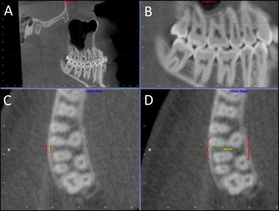
The digital imaging and communications in medicine (DICOM) multi-files of each CBCT scan were imported into 3-dimensional software (version 10.5; Dolphin Imaging Systems, Chatsworth, Calif) for analysis. A blinded investigator (K.A.H.) performed all evaluations. The images were oriented in 3 planes of space so that the buccal and lingual cortical plates could be measured 5 mm from the crest of the alveolar ridge. The vertical height of 5 mm was chosen because adequate amounts of bone for miniscrew implant placement are generally found at the middle of the root. To initially orient the image, the midsagittal plane was centered on the axial slice, and the sagittal slice was rotated so that the Frankfort horizontal plane was parallel to the true horizontal ( Fig 1 , A ). The sagittal view was used to locate the interradicular sites 5 mm from the crest of the alveolar ridge ( Fig 1 , B ). The cortical plate thickness was measured by using the axial slice ( Fig 1 , C ). The slices were oriented so that the shortest distances defining the buccal and lingual cortices were measured.

Buccal and lingual thicknesses were measured in both the maxilla and the mandible midway between the first and second molars, the second premolar and the first molar, the first premolar and the second premolar, and the canine and the first premolar. These interradicular sites have all been used for placement of miniscrew implants.
With the same orientations, the alveolar ridge thickness was measured at the same vertical level as the cortical thickness in both the maxilla and the mandible ( Fig 1 , D ). Ridge thickness was defined as the distance between the buccal and lingual sites on the periosteal surface, between the teeth previously specified to measure cortical thickness. Medullary thickness was defined as ridge thickness minus the sum of buccal and lingual cortical thicknesses. Only the right side of each CBCT scan was evaluated because there are no side differences in cortical thickness for either the maxilla or the mandible.
Bone thickness at each site was measured twice and averaged. To quantify error, replicate analyses were performed on 10 randomly chosen subjects. Paired t tests showed systematic errors from −0.56 to 0.49 mm, none of which was statistically significant. Method errors, which were computed based on the differences between the replicate measures squared and divided by 2 times the sample size (Σ deviations 2 /2n), ranged from 0.12 to 0.45 mm. The poorest reliability was for the maxillary alveolar ridge thickness between the right second premolar and the first molar.
Statistical analysis
Based on the skewness and kurtosis statistics, the distributions were judged to be normal. T tests were used to evaluate differences between hyperdivergent and hypodivergent subjects. Paired t tests were used to compare bone within regions. The cortical thicknesses of the 2 most anterior and 2 most posterior sites in each region were averaged to evaluate regional differences.
Results
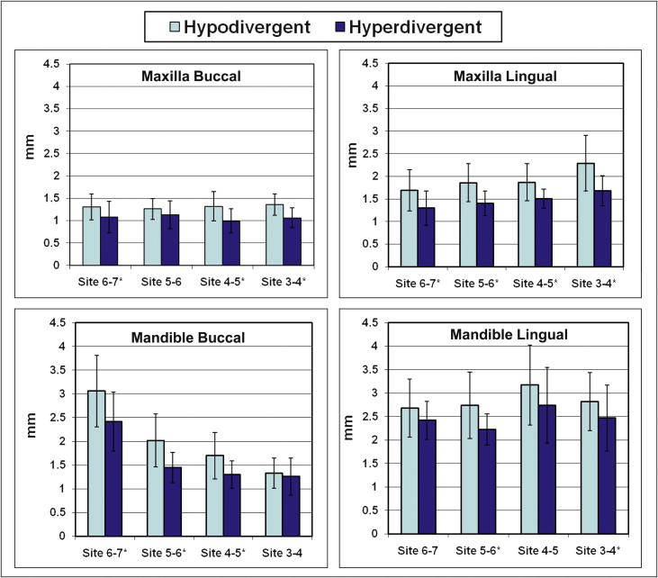
In general, cortical bone was thicker in the hypodivergent than in the hyperdivergent subjects ( Table I ; Fig 2 ). The only maxillary site not significantly ( P <0.05) thicker in the hypodivergent subjects was the buccal site between the first molar and the second premolar. In the mandible, only the buccal sites between the molars and the premolars, and the lingual sites between the second premolar and the first molar showed statistically significant group differences.
| Jaw | Cortex | Site | Hypodivergent | Hyperdivergent | Difference between face types | |||
|---|---|---|---|---|---|---|---|---|
| Mean | SD | Mean | SD | Mean | P value | |||
| Maxilla | Buccal | 6-7 | 1.31 | 0.29 | 1.08 | 0.35 | 0.23 | 0.008 ∗ |
| 5-6 | 1.26 | 0.23 | 1.13 | 0.31 | 0.13 | 0.075 | ||
| 4-5 | 1.32 | 0.33 | 0.99 | 0.27 | 0.33 | <0.001 ∗ | ||
| 3-4 | 1.36 | 0.24 | 1.06 | 0.22 | 0.30 | <0.001 ∗ | ||
| Lingual | 6-7 | 1.69 | 0.46 | 1.30 | 0.38 | 0.39 | 0.001 ∗ | |
| 5-6 | 1.86 | 0.42 | 1.40 | 0.27 | 0.46 | <0.001 ∗ | ||
| 4-5 | 1.87 | 0.41 | 1.51 | 0.21 | 0.36 | <0.001 ∗ | ||
| 3-4 | 2.29 | 0.62 | 1.68 | 0.33 | 0.61 | <0.001 ∗ | ||
| Mandible | Buccal | 6-7 | 3.06 | 0.75 | 2.42 | 0.62 | 0.64 | 0.001 ∗ |
| 5-6 | 2.02 | 0.56 | 1.45 | 0.32 | 0.57 | <0.001 ∗ | ||
| 4-5 | 1.70 | 0.49 | 1.30 | 0.29 | 0.41 | <0.001 ∗ | ||
| 3-4 | 1.33 | 0.32 | 1.26 | 0.39 | 0.08 | 0.412 | ||
| Lingual | 6-7 | 2.68 | 0.62 | 2.42 | 0.41 | 0.26 | 0.065 | |
| 5-6 | 2.74 | 0.71 | 2.23 | 0.33 | 0.52 | 0.001 ∗ | ||
| 4-5 | 3.17 | 0.85 | 2.62 | 0.81 | 0.43 | 0.086 | ||
| 3-4 | 2.82 | 0.62 | 2.47 | 0.70 | 0.35 | 0.050 ∗ | ||

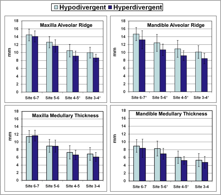
Alveolar ridge thickness was also greater at all sites in the hypodivergent than in the hyperdivergent subjects ( Table II ; Fig 3 ). Differences were statistically significant at all mandibular sites and at the 2 anterior maxillary sites. Medullary thickness of the maxilla showed negligible differences between the hypodivergent and hyperdivergent subjects. The only statistically significant differences in medullary thickness were in the mandible, between the first molar and the second premolar, and between the second premolar and the first premolar.
| Site | Hypodivergent | Hyperdivergent | Difference between face types | ||||
|---|---|---|---|---|---|---|---|
| Mean | SD | Mean | SD | Mean | P value | ||
| Maxilla | |||||||
| Alveolar ridge | 6-7 | 14.45 | 1.48 | 14.07 | 1.42 | 0.38 | 0.333 |
| 5-6 | 12.61 | 1.43 | 11.66 | 1.56 | 0.96 | 0.190 | |
| 4-5 | 10.49 | 1.58 | 9.13 | 1.24 | 1.35 | 0.001 ∗ | |
| 3-4 | 9.98 | 1.38 | 8.64 | 0.93 | 1.34 | <0.001 ∗ | |
| Medullary thickness | 6-7 | 11.44 | 1.54 | 11.66 | 1.40 | −0.22 | 0.577 |
| 5-6 | 8.97 | 1.68 | 8.92 | 1.59 | 0.05 | 0.906 | |
| 4-5 | 7.30 | 1.75 | 6.63 | 1.22 | 0.67 | 0.103 | |
| 3-4 | 6.87 | 1.66 | 6.10 | 1.18 | 0.75 | 0.055 | |
| Mandible | |||||||
| Alveolar ridge | 6-7 | 14.67 | 1.65 | 13.22 | 2.32 | 1.46 | 0.008 ∗ |
| 5-6 | 12.44 | 2.17 | 10.67 | 1.45 | 1.77 | 0.001 ∗ | |
| 4-5 | 10.92 | 2.14 | 9.20 | 1.25 | 1.55 | 0.003 ∗ | |
| 3-4 | 10.10 | 1.61 | 8.46 | 1.34 | 1.64 | <0.001 ∗ | |
| Medullary thickness | 6-7 | 8.93 | 1.63 | 8.37 | 2.34 | 0.56 | 0.297 |
| 5-6 | 8.29 | 1.92 | 6.94 | 1.38 | 1.35 | 0.004 ∗ | |
| 4-5 | 6.05 | 1.58 | 5.26 | 0.98 | 0.79 | 0.032 ∗ | |
| 3-4 | 5.34 | 1.67 | 4.75 | 1.51 | 0.59 | 0.171 | |

The lingual cortical bone of the maxilla was consistently thicker than the buccal cortical bone, with all sites showing statistically significant differences ( Table III ). The cortical bone of the mandible in the area of the first and second molars was the only area with a significantly thicker buccal than lingual cortex; the other sites were significantly thicker on the lingual side.
| Site | Buccal | Lingual | Difference between sites | ||||
|---|---|---|---|---|---|---|---|
| Mean | SD | Mean | SD | Mean | P value | ||
| Maxilla | 6-7 | 1.20 | 0.37 | 1.51 | 0.46 | −0.31 | <0.01 ∗ |
| 5-6 | 1.20 | 0.28 | 1.64 | 0.42 | −0.44 | <0.01 ∗ | |
| 4-5 | 1.16 | 0.34 | 1.70 | 0.37 | −0.54 | <0.01 ∗ | |
| 3-4 | 1.22 | 0.27 | 2.00 | 0.59 | −0.78 | <0.01 ∗ | |
| Mandible | 6-7 | 2.76 | 0.76 | 2.56 | 0.54 | 0.20 | 0.039 ∗ |
| 5-6 | 1.75 | 0.55 | 2.50 | 0.62 | −0.75 | <0.01 ∗ | |
| 4-5 | 1.53 | 0.44 | 2.92 | 0.87 | −1.39 | <0.01 ∗ | |
| 3-4 | 1.30 | 0.35 | 2.66 | 0.67 | −1.36 | <0.01 ∗ | |
Stay updated, free dental videos. Join our Telegram channel

VIDEdental - Online dental courses


