Fig. 9.1
(a) The facial morphology in a 2-month-old unoperated infant with unilateral complete cleft lip and palate (UCCLP). (b) The facial morphology in a 2-month-old unoperated infant with bilateral complete cleft lip and palate (BCCLP)

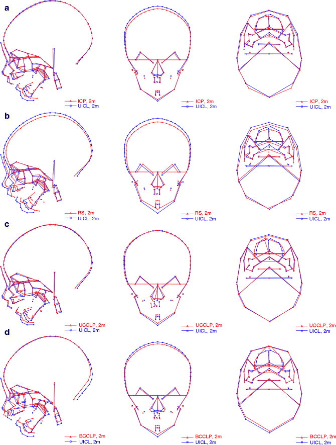
Fig. 9.2
(a–d) Mean plots in three projections (lateral, frontal, and axial) of the four different cleft groups superimposed on the control group with UICL. The lateral mean plots are aligned on the n-s line and registered at s. The frontal mean plots are aligned on the latero-orbital line and registered at the center point of that line. The axial mean plots are aligned on a line between the two tuber points and registered at the center point of that line. Superimposition of the mean plots for the 2-month-old (a) ICP and UICL groups, (b) RS and UICL groups, (c) UCCLP and UICL groups, and (d) BCCLP and UICL groups
Table 9.1
Summary and comparison of the most important findings in the primary anomaly in children with RS, ICP, BCCLP, and UCCLP
|
Anomaly
|
RS
|
ICP
|
BCCLP
|
UCCLP
|
|---|---|---|---|---|
|
Maxilla
|
||||
|
Decreased length measured to premaxilla (sp-pm)a
|
+b
|
+
|
–c
|
–
|
|
Retrognathia measured to premaxilla (s-n-ss)d
|
+
|
+
|
–e
|
–
|
|
Decreased posterior length (ci-pm)f
|
+
|
+
|
+
|
+
|
|
Retrognathia measured to base of jaw (s-n-ci)g
|
+
|
+
|
+
|
+
|
|
Decreased posterior height
|
+
|
+
|
+
|
+
|
|
Increased width
|
+
|
+
|
++
|
++
|
|
Nasal cavity
|
||||
|
Increased width
|
+
|
+
|
++
|
++
|
|
Mandible
|
||||
|
Decreased length
|
+++
|
++
|
++
|
+
|
|
Retrognathia
|
+++
|
++
|
++
|
+
|
|
Pharyngeal airway
|
||||
|
Reduced size
|
+++
|
++
|
+
|
+
|
9.2.1 Cleft Lip (CL)
Isolated CL involves only structures of the embryonic primary palate. The craniofacial morphology in CL subjects has been shown to be fairly normal except for the small region of the cleft including the premaxilla and the incisors. In unoperated bilateral complete CL, the premaxilla may, however, protrude markedly. In unilateral complete CL, the protrusion is less pronounced but asymmetric. In subjects with unoperated unilateral incomplete cleft lip (UICL), the protrusion of the premaxilla is negligible (Hermann et al. 1999a). The interorbital distance in CL subjects seems to be slightly increased compared to the norm (Cohen 1997). The basal part of the maxilla has a normal prognathism in relation to the anterior cranial base, and the mandible is of normal size, shape, and inclination (Dahl 1970; Hermann et al. 1999a). Following lip surgery, the premaxilla is molded into a normal position, and maxillary prognathism measured to point A or ss (subspinale) is normal (Dahl 1970; Han et al. 1995; Hermann et al. 1999a, b, 2000). In conclusion, subjects with UICL have a very close to normal craniofacial morphology from infancy to adult age, and consequently, we have used our group of infants with UICL as a control group in the study of deviations in craniofacial morphology and growth of infants and young children with ICP, RS, UCCLP, and BCCLP since no actual normative cephalometric data for Danish infants and young children are available.
9.2.2 Cleft Palate (CP)
Isolated cleft palate (ICP) involves only structures of the embryonic secondary palate. In Fig. 9.2a, the mean facial diagrams of the ICP group are superimposed on the mean facial diagram of a group of age-matched infants with UICL (control group). The major deviations in the ICP group were: reduced length and posterior height of the maxilla, maxillary retrognathia, increased width of the maxilla and the nasal cavity, and reduced length of the mandible with mandibular retrognathia. Thus, the ICP group revealed bimaxillary retrognathia. The sagittal jaw relationship was, however, normal. In addition, in the ICP group, the upper airway dimensions were reduced. Bimaxillary retrognathia and a short mandible were previously documented in unoperated older children and adults with ICP (Dahl 1970; Bishara 1972).
9.2.3 Robin Sequence (RS)
Robin sequence (RS) is defined as a triad of symptoms: isolated cleft palate, micrognathia, and glossoptosis (Gorlin et al. 2001). RS may be part of several syndromes, e.g., Treacher-Collins syndrome (Kreiborg and Cohen 1996; Cohen 1997). In this chapter, only nonsyndromic cases of RS will be discussed. We consider this group as a subgroup of the ICP group (Hermann et al. 2003a). In Fig. 9.2b, the mean facial diagram of the RS group at 2 months of age is superimposed on the mean facial diagram of the control group. The major deviations in the RS group were decreased length and posterior height of the maxilla, maxillary retrognathia, increased width of the maxilla and nasal cavity, and very short mandible with marked mandibular retrognathia. Thus, the RS group revealed bimaxillary retrognathia; the retrognathia was, however, most marked for the mandible, and the sagittal jaw relation was increased. In addition, the RS group had a significantly smaller cranial base angle (n-s-ba) resulting in a smaller depth of the bony nasopharynx than the controls, and the upper airway dimensions were markedly reduced. The degree of maxillary retrognathia was similar in the RS and the ICP group. However, the mandibular retrognathia in the RS group was even more marked than in the ICP subjects. It would seem that RS subjects probably represent the extreme part of the ICP population in terms of mandibular retrognathia and upper airway constriction. As mentioned above, we consider the RS group as a special subgroup of the ICP group. Accordingly, we believe the bimaxillary retrognathia to be intrinsically associated with the cleft of the secondary palate.
9.2.4 Cleft Lip and Palate (CLP)
Combined clefts of the lip, alveolus, and palate involve structures of both the embryonic primary palate and secondary palate. In Fig. 9.2c, the mean craniofacial morphology in 2-month-old unoperated infants with unilateral complete cleft lip and palate (UCCLP) was compared to the control group (Hermann et al. 1999a). The major deviations in the UCCLP group were decreased posterior length and height of the maxilla; retrognathia of the basal part of the maxilla with relative protrusion of the premaxilla; the width of the maxilla and nasal cavity was markedly increased and the premaxilla deviated to the noncleft side; and the mandible was short and retrognathic. Thus, the UCCLP group revealed bimaxillary retrognathia combined with a relative protrusion of the premaxilla, which deviated to the noncleft side. In addition, in the UCCLP group, the upper airway dimensions were reduced.
Increased width of the midface and nasal cavity was previously reported in unoperated UCCLP infants (Han et al. 1995) and in unoperated adults with UCCLP (Motohashi et al. 1994). Relative protrusion and asymmetry of the premaxilla have also been reported in unoperated UCCLP children, adolescents, and adults (Ortiz-Monasterio et al. 1959, 1966; Bishara et al. 1976, 1985, 1986; Capelozza et al. 1993). The relative protrusion and deviation are probably due to overgrowth in the premaxillary-vomerine complex (Pruzansky 1971; Friede and Morgan 1976; Friede 1978) and due to the lack of structural integrity of the maxilla on one side. This relative protrusion of the premaxilla explains why we found the measurements s-n-ans (S-N-ANS) and s-n-ss (S-N-A) in the infant UCCLP group to be comparable to the values in the control group, despite the fact that the UCCLP group showed significant maxillary retrognathia measured to the basal part of the maxilla.
Dahl et al. (1982) and Hermann et al. (2003a, b) analyzed facial morphology in 2-month-old infants with unoperated bilateral complete cleft lip and palate from our sample. Fig. 9.2d illustrates the mean facial diagram of the BCCLP group superimposed on the mean facial diagram of the control group. The most obvious features in the BCCLP group were protrusion of the premaxilla both in relation to the anterior cranial base and in relation to the basal part of the maxilla; the length of the basal part of the maxilla and posterior maxillary height were decreased; retrognathia of the basal part of the maxilla; markedly increased width of the maxilla and nasal cavity; a short and retrognathic mandible. Thus, the BCCLP group revealed bimaxillary retrognathia with a truly protruding premaxilla. In other words, the protruding premaxilla was situated in a totally retrognathic face with a fairly normal sagittal jaw relationship. In addition, the upper airway dimensions were reduced.
The extreme protrusion of the premaxilla is probably the result of marked overgrowth in the premaxillary-vomerine complex secondary to total lack of structural integrity in the region.
For comparison, Mars and Houston (1990) and da Silva Filho et al. (1998) described groups of adult unoperated patients with BCCLP and found extreme protrusion of the premaxilla and a very convex profile measured as the ANB angle. No measurements were performed to describe the position of the body of the maxilla. Da Silva-Filho et al. (1992a, 1998) also found the mandible to be short and retrognathic and discussed whether this finding was related to the primary anomaly or if it was caused by secondary functional adaptations.
The retrognathia of the basal part of the maxilla and the short and retrognathic mandible found in our sample are, in our opinion, variations intrinsically associated with the cleft of the secondary palate as discussed above.
9.3 Discussion and Conclusions
The Danish study of craniofacial morphology in untreated cleft infants is the hitherto most comprehensive and well-controlled since it covers a whole population, which is homogeneous and in which central registration of clefts has been carried out for more than 65 years, a registration which has been shown to be highly reliable and nearly complete. Furthermore, all cleft infants are surgically treated at one hospital by one surgeon using the same techniques. All infants were examined with state-of-the-art three-projection cephalometry using the hitherto most comprehensive cephalometric analysis covering all craniofacial regions, and the methods were validated. The study included more than 600 children, and even after breakdown into subgroups, the sample sizes were adequate for statistical testing (except maybe for the RS group). Based on these facts, the findings related to the infant craniofacial morphology at 2 months of age, prior to any surgical or orthopedic treatment, must be considered to represent the “true” malformation, primarily caused by intrinsic factors.
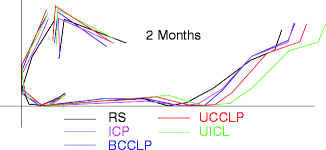
In Table 9.1, the most important findings in the primary anomaly in the Danish infants with RS, ICP, BCCLP, and UCCLP are given, revealing a rather clear pattern. The findings support the suggestion of Dahl (1970) and others that facial clefts should be classified based on the embryonic facial development, i.e., into clefts involving the primary palate only (CL), clefts involving the secondary palate only (CP), and clefts involving structures of both the primary and the secondary palate (CLP). The postnatal facial morphology in these groups differs greatly. Infants with cleft of the secondary palate, with or without cleft of the primary palate, shared a number of characteristic morphological traits when compared to the norm: decreased posterior length of the maxilla, maxillary retrognathia, decreased posterior height of the maxilla, increased width of the maxilla and the nasal cavity, decreased length of the mandible, mandibular retrognathia, and reduced size of the pharyngeal airway. As seen from Table 9.1 and Fig. 9.3, the mandibular involvement was most pronounced in the RS group followed by the ICP and BCCLP groups and, finally, the UCCLP group. A similar pattern was observed for the reduced size of the pharyngeal airway.


Fig. 9.3
Mean plots of the mandible in the RS, ICP, BCCLP, UCCLP, and UICL groups. Superimposition was made on the mandibular line (ML) registered at pogonion (pg)
Stay updated, free dental videos. Join our Telegram channel

VIDEdental - Online dental courses


