Fig. 13.1
Non-eroded healthy cervical dentin which is covered by a thin layer of acellular-afibrillar cement
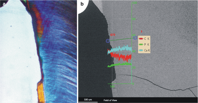
Fig. 13.2
Eroded cervical dentin with complete destruction of the cement layer; (a) polarized light microcopy; (b) EDS line scan of the same lesion. There is a light decrease of the mineral content of the surface
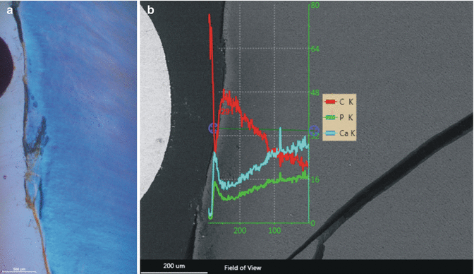
Fig. 13.3
Eroded cervical dentin with beginning demineralization; (a) polarized light microscopy which shows a small hypermineralized superficial layer and a small demineralized zone underneath; (b) EDS line scan through the eroded lesion demonstrating the superficial hypermineralized layer and the demineralized area
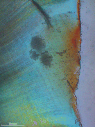
Fig. 13.4
Initial cervical root caries lesion
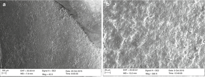
Fig. 13.5
Eroded cervical dentin area; (a) SEM overview of an eroded cervical area beneath the enamel – cementum junction. The surface appears to be rough; (b) higher magnification of the eroded area. The surface has a rugged structure. Dentin tubules are opened
The superficial erosive demineralization results in dissolution of the mineral, but the dentin collagen fibers are remaining (Breschi et al. 2002) and protect the underlying dentin from further demineralization as this collagenous matrix is a diffusion barrier (Shellis et al. 2010). The superficial layer of eroded dentin is a spongelike relative stable structure, and the superficial cervical contour is preserved. However, eroded cervical dentin is vulnerable to mechanical influences like toothbrushing. The surface morphology of eroded dentin varies from irregular ragged surface (Fig. 13.5) to a rather smooth surface. The progression of cervical dentin erosion depended from the oral health of the host, lifestyle, and eating habits (Ganss et al. 2014).
13.2 Restoration of Cervical Dentin Erosions
Generally, like enamel, dentin is capable to remineralize. Prerequisite for remineralization is a sufficient supply of Ca2+ and PO4 3− ions. The natural supply derives from the hyper-saturated saliva. Fluoride promotes remineralizaion of hydroxyapatite and is widely used in the prevention and restoration of dentin erosions. The mechanism of the action of fluoride on remineralization of dentin is still not fully understood. Experimental studies demonstrated a positive effect of fluoride on root dentin remineralization (Addy 2005; Arnold et al. 2014; Nyvad and Fejerskov 1982) (Fig. 13.6). Fluorides are widely used for therapeutic interventions of dentin erosions. The majority are fluoridated self-care oral hygiene products. Fluoridated toothpastes for the use of remineralization of cervical dentin have two functions. Fluoride should promote dentin remineralization, whereas other ingredients like pro-arginin or hydroxyapatite are aimed to occlude open dentin tubules for diminishing dentin hypersensitivity (Arnold et al. 2015). However, a newly published meta-analysis showed that the evidence for an effectiveness of these products is rather low (Twetman 2015).
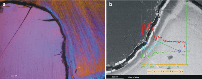

Fig. 13.6
Cervical demineralized dentin after incubation with artificial saliva and 10 ppm NaF. (a) A rather thick remineralized surface layer can be seen on top of the lesion. (b) EDS line scan through the same lesion demonstration a higher Ca, P, and F content within the superficial dentin layer
There are numerous other products available like mouthrinses, pastes, and gels. The effectiveness of fluoridated milk on dentin remineralization has been investigated in an experimental model which has shown that there is a benefit of fluoridated milk in dentin remineralization (Arnold et al. 2014).
Stay updated, free dental videos. Join our Telegram channel

VIDEdental - Online dental courses


