Introduction
The purpose of this study was to evaluate the reliability of measurements made on 3-dimensional digital models obtained with a surface laser scanner (D-250; 3Shape, Copenhagen, Denmark).
Methods
Twenty orthodontic dental casts of permanent dentitions were selected. Three-dimensional images were obtained on this scanner and analyzed by using the Geomagic Studio 5 software (Raindrop Geomagic, Inc, Morrisville, NC). Measurements were made with a digital caliper directly on the dental casts and also digitally on the digital models. Fifteen anatomic dental points were identified, and a total of 11 linear measurements were taken from each cast, including arch length and width. Dependent t tests were used to evaluate intraexaminer reproducibility and measurement accuracy on the digital models.
Results
No statistically significant differences were found between the measurements made directly on the dental casts and on the digital models.
Conclusions
Linear measurements on digital models are accurate and reproducible. Digital models obtained with the surface laser scanner are reliable for measurements of arch width and length.
The fast and continuous advances in computer sciences have resulted in increased usage of new technologies in all levels of modern society. Orthodontics has also been influenced by this phenomenon. Computer-based records are routine in many orthodontic offices. Digital models are increasingly available and provide qualified diagnostic images at a reasonable cost. Digital record storage has several advantages: easy access, need for less physical space, and ability to share information via the Internet with other professionals. With new advances in 3-dimensional dental and orthodontic softwares, orthodontists can examine intra-arch and interarch relationships digitally. Transverse relationships between maxillary and mandibular arches can be better evaluated when 3-dimensional models are viewed in occlusion in different perspectives in the screen. Digital casts also have the advantage of allowing a “virtual treatment” and a “virtual setup.”
Several studies in the literature have verified the accuracy of angular and linear measurements on 3-dimensional digital models with different softwares and found divergent results. Recently, Leifert et al evaluated the accuracy of digital models using OrthoCad software. They measured mesiodistal tooth widths and arch lengths, and found slight but statistically significant differences in some measurements. They concluded that, despite these differences, digital models are clinically acceptable and reproducible when compared with traditional model analyses. Similar results were found by other authors using the same software, Pointstream software, and Emodel software.
Bell et al observed no statistically significant differences between linear measurements made on digital models using the C3D-builder software and conventional models, validating the virtual evaluation. On the other hand, Redlich et al measured 3-dimensional scanned orthodontic models with cross-section planes reconstructed by Teledent software. They observed differences of 1.19 to 3 mm between measurements of space taken from digital and conventional models, showing significantly less crowding when using the linear measurements from the digital models. They concluded that the accuracy of linear measurements on digital models is sometimes questionable, especially in severely crowded dentitions.
No previous study has validated digital models obtained from the 3Shape scanner (D-250; 3Shape, Copenhagen, Denmark) for linear orthodontic measurements. Considering the controversies aforementioned and that digital models are increasingly available for both dental practice and research in orthodontics, the purpose of this study was to determine the accuracy and reproducibility of arch widths and lengths obtained with the surface laser scanner 3Shape.
Material and methods
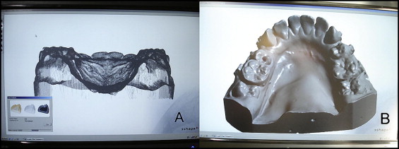
Twenty orthodontic dental models from patients under treatment, in the permanent dentition, were selected. The sample size was calculated before the study; it was estimated that a sample size of 10 to 13 dental models was needed to obtain a statistical power of 95%. These dental casts belonged to a prospective study for which the first premolars had been extracted according to the orthodontic treatment plans. The dental casts were digitized by using the 3Shape D-250 3-dimensional scanner. This scanner operates with a main laser beam, with 2 cameras to capture the image. The images are automatically processed by the 3Shape Sewer Scan software, which generated files with the STL (stereolithography) extension for each dental model ( Fig 1 ).
Two examiners (M.V.S.S. and E.C.V.) were trained sufficiently in using both methods: measuring with a digital caliper, and manipulation and measuring of the virtual images with the software.
The measurements were obtained from conventional dental casts with a digital caliper with 0.01-mm accuracy (Mitutoyo Co, Kanagawa, Japan). Three-dimensional images were analyzed by using Geomagic Studio 5 software (Raindrop Geomagic, Inc, Morrisville, NC). The digital images of the dental models were enlarged on screen to facilitate measurements. All the direct and virtual measurements were made by the 2 examiners.
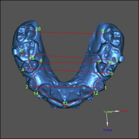
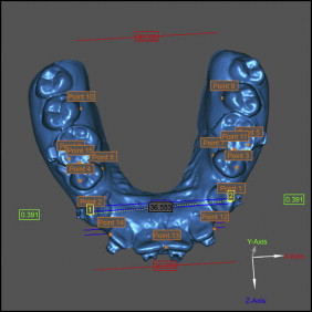
Fifteen anatomic dental points were identified on each pair of casts (plaster and digital models) by using the modified Euclidean distance matrix analysis ( Fig 2 ). Arch widths and lengths were measured as shown in Figure 3 . A total of 11 measurements were made on each dental model.
The dental cast and digital model measurements were repeated for 5 randomly selected patients after 15 days by both examiners. The random errors were evaluated with Dahlberg’s formula, and the systematic errors were investigated with dependent t tests. The results were considered significant at P <0.05. Because there were no significant intraexaminer differences, the average between the 2 operators was used for both methods.
Statistical analysis
Dependent t tests were used for intergroup comparisons. The results were considered significant at P <0.05.
Results
The individual intraexaminer random errors ranged from 0.054 (distance 3-4) to 0.955 (distance 14-15) for the physical measurements, and from 0.064 (distance 1-5) to 0.943 (distance 12-13) for the digital measurements. There were no significant systematic errors for either examiner ( Tables I and II ).
| Measurement 1 (mm) | Measurement 2 (mm) | |||||
|---|---|---|---|---|---|---|
| Measurement | Mean | SD | Mean | SD | Dahlberg | P |
| Caliper | ||||||
| 1-2 | 28.894 | 4.480 | 29.044 | 4.563 | 0.233 | 0.299 |
| 3-4 | 36.88 | 3.701 | 36.918 | 3.939 | 0.054 | 0.895 |
| 5-6 | 43.528 | 4.046 | 43.564 | 3.977 | 0.482 | 0.802 |
| 1-5 | 19.268 | 2.312 | 18.734 | 2.793 | 0.232 | 0.344 |
| 2-6 | 18.454 | 2.293 | 17.654 | 2.744 | 0.694 | 0.119 |
| 7-8 | 27.328 | 2.629 | 27.162 | 2.606 | 0.482 | 0.220 |
| 9-10 | 37.208 | 2.530 | 37.462 | 2.290 | 0.833 | 0.565 |
| 11-12 | 18.722 | 2.348 | 18.618 | 2.422 | 0.694 | 0.621 |
| 12-13 | 12.206 | 1.273 | 12.304 | 1.263 | 0.913 | 0.463 |
| 13-14 | 12.134 | 1.721 | 12.358 | 1.647 | 0.833 | 0.275 |
| 14-15 | 19.18 | 1.834 | 19.342 | 1.985 | 0.955 | 0.557 |
| 3-dimensional image | ||||||
| 1-2 | 28.706 | 4.062 | 28.856 | 4.518 | 0.389 | 0.611 |
| 3-4 | 36.778 | 3.857 | 36.662 | 4.233 | 0.152 | 0.761 |
| 5-6 | 43.288 | 3.858 | 43.446 | 4.502 | 0.624 | 0.768 |
| 1-5 | 19.174 | 2.504 | 19.258 | 2.473 | 0.389 | 0.277 |
| 2-6 | 17.626 | 2.839 | 17.930 | 2.581 | 0.790 | 0.689 |
| 7-8 | 26.976 | 2.421 | 27.370 | 2.339 | 0.624 | 0.144 |
| 9-10 | 37.224 | 2.691 | 37.196 | 2.500 | 0.889 | 0.799 |
| 11-12 | 18.604 | 1.830 | 18.916 | 1.685 | 0.790 | 0.605 |
| 12-13 | 11.640 | 1.404 | 11.822 | 1.382 | 0.943 | 0.356 |
| 13-14 | 12.318 | 1.226 | 12.866 | 1.843 | 0.889 | 0.223 |
| 14-15 | 18.288 | 2.146 | 18.492 | 1.977 | 0.971 | 0.743 |
| Measurement 1 (mm) | Measurement 2 (mm) | |||||
|---|---|---|---|---|---|---|
| Measurement | Mean | SD | Mean | SD | Dahlberg | P |
| Caliper | ||||||
| 1-2 | 28.860 | 4.523 | 28.440 | 4.277 | 0.409 | 0.290 |
| 3-4 | 37.084 | 3.959 | 36.918 | 4.196 | 0.298 | 0.298 |
| 5-6 | 43.774 | 4.313 | 43.228 | 4.295 | 0.544 | 0.253 |
| 1-5 | 19.278 | 2.263 | 18.990 | 2.344 | 0.250 | 0.057 |
| 2-6 | 18.300 | 2.313 | 18.082 | 2.442 | 0.224 | 0.206 |
| 7-8 | 26.994 | 2.773 | 26.996 | 2.705 | 0.061 | 0.973 |
| 9-10 | 37.142 | 2.345 | 37.094 | 2.377 | 0.093 | 0.676 |
| 11-12 | 18.674 | 1.828 | 18.714 | 2.072 | 0.240 | 0.854 |
| 12-13 | 11.996 | 1.404 | 12.142 | 1.297 | 0.181 | 0.270 |
| 13-14 | 12.348 | 1.693 | 12.342 | 1.540 | 0.258 | 0.978 |
| 14-15 | 18.196 | 1.809 | 18.272 | 2.048 | 0.203 | 0.697 |
| 3-dimensional image | ||||||
| 1-2 | 29.102 | 4.516 | 29.044 | 4.563 | 0.106 | 0.562 |
| 3-4 | 36.946 | 3.833 | 37.024 | 4.035 | 0.208 | 0.623 |
| 5-6 | 43.634 | 4.251 | 43.526 | 4.251 | 0.135 | 0.504 |
| 1-5 | 19.012 | 2.627 | 18.950 | 2.636 | 0.064 | 0.659 |
| 2-6 | 17.980 | 2.562 | 18.092 | 2.575 | 0.077 | 0.297 |
| 7-8 | 27.410 | 2.804 | 27.172 | 2.453 | 0.447 | 0.469 |
| 9-10 | 37.128 | 2.527 | 37.228 | 2.542 | 0.128 | 0.359 |
| 11-12 | 19.376 | 1.965 | 19.472 | 1.990 | 0.191 | 0.501 |
| 12-13 | 12.128 | 1.656 | 12.012 | 1.690 | 0.118 | 0.382 |
| 13-14 | 12.610 | 1.730 | 12.582 | 1.616 | 0.068 | 0.726 |
| 14-15 | 19.180 | 1.834 | 19.442 | 1.811 | 0.249 | 0.235 |
Stay updated, free dental videos. Join our Telegram channel
VIDEdental - Online dental courses


