Occlusal radiography
Terminology and classification
Upper standard (or anterior) occlusal
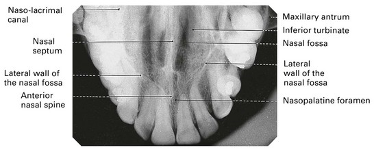
This projection shows the anterior part of the maxilla and the upper anterior teeth.
Main clinical indications
The main clinical indications include:
• Periapical assessment of the upper anterior teeth, especially in children but also in adults unable to tolerate periapical holders
• Detecting the presence of unerupted canines, supernumeraries and odontomes
• As the midline view, when using the parallax method for determining the bucco/palatal position of unerupted canines
• Evaluation of the size and extent of lesions such as cysts or tumours in the anterior maxilla
• Assessment of fractures of the anterior teeth and alveolar bone.
Technique and positioning
The technique can be summarized as follows:
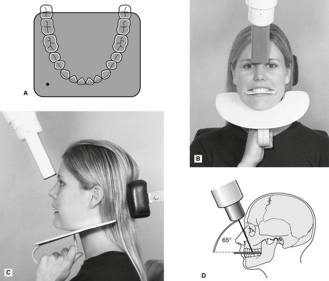
1. The patient is seated with the head supported and with the occlusal plane horizontal and parallel to the floor and is asked to support a protective thyroid shield.
2. The image receptor, suitably barrier wrapped, is placed flat into the mouth on to the occlusal surfaces of the lower teeth. The patient is asked to bite together gently. The image receptor is placed centrally in the mouth with its long axis crossways in adults and anteroposteriorly in children.
3. The X-ray tubehead is positioned above the patient in the midline, aiming downwards through the bridge of the nose at an angle of 65°–70° to the image receptor (see Fig. 9.1).
Upper oblique occlusal
This projection shows the posterior part of the maxilla and the upper posterior teeth on one side.
Main clinical indications
Stay updated, free dental videos. Join our Telegram channel
VIDEdental - Online dental courses


