The periodontal tissues and periodontal disease
Selection criteria
Several radiographic projections can be used to show the periodontal tissues. Those recommended by the Faculty of General Dental Practice (UK) in their 2013 booklet Selection Criteria for Dental Radiography (3rd Edn) are summarized in Table 19.1.
Table 19.1
| Recommendation | Evidence-based grading* |
| Horizontal bitewings if a patient has generalised pocketing <6 mm (BPE scores of Code 3) and little or no recession. | C |
| Vertical bitewings if a patient has pocketing 6 mm or more (BPE scores of Code 4), supplemented by paralleling technique periapicals at sites where the alveolar bone is not shown on the bitewings. | C |
| Bitewings (horizontal or vertical depending on pocket depth), supplemented by paralleling technique periapicals if necessary if a patient has localised pocketing | C |
| A paralleling technique periapical if a periodontal/endodontic lesion is suspected | C |
| CBCT is not indicated as a routine method of imaging periodontal bone support | C |
| Small volume, high resolution CBCT may be indicated in selected cases of infra-bony defects and furcation lesions, where clinical and conventional radiographic examination do not provide the information needed for patient management | C |
*Evidence-based grading C = based on evidence from expert committee reports or opinions and/or clinical experience of respected authorities and indicates an absence of directly applicable studies of good quality.
In addition, digital radiography and image manipulation, including subtraction and densitometric image analysis (see Ch. 3), may assist in showing and measuring subtle changes in fine alveolar and crestal bone pattern. However, these techniques require the inclusion of a reference object of known density and a highly reproducible positioning technique to be helpful.
Important points to note
• In the interpretation of the periodontal tissues, images of excellent quality are essential – perhaps more so than in other dental specialties – because of the fine detail that is required.
• Exposure factors should be reduced when using film-based techniques to avoid burn-out of the interdental crestal bone
Radiographic features of healthy periodontium
The usual radiographic features of healthy alveolar bone are shown in Figs 19.1 and 19.2 and include:
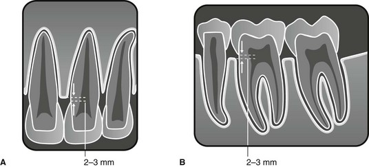
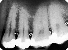
 (slightly reduced exposure) showing the radiographic features of a healthy periodontium (arrowed) before the onset of periodontitis.
• Thin, smooth, evenly corticated margins to the interdental crestal bone in the posterior regions.
• Thin, even, pointed margins to the interdental crestal bone in the anterior regions.
• Cortication at the top of the crest is not always evident, owing mainly to the small amount of bone between the teeth anteriorly.
• The interdental crestal bone is continuous with the lamina dura of the adjacent teeth. The junction of the two forms a sharp angle.
• Thin even width to the mesial and distal periodontal ligament spaces.
Important points to note
• Although these are the usual features of a healthy periodontium, they are not always evident.
• Their absence from radiographs does not necessarily mean that periodontal disease is present.
• Failure to see these features may be due to:
• Following successful treatment, the periodontal tissues may appear healthy clinically, but radiographs may show evidence of earlier bone loss when the disease was active. Bone loss observed on radiographs is therefore not an indicator of the presence of inflammation.
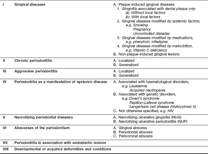
Classification of periodontal disease
Various classifications of periodontal disease have been put forward over the years. The most comprehensive, although not universally agreed, was produced by the International Workshop of the American Academy of Periodontology and the European Federation of Periodontology in 1999. A simplified version is shown in Table 19.2.
Stay updated, free dental videos. Join our Telegram channel
VIDEdental - Online dental courses


