9: Dental Caries

Dental Caries

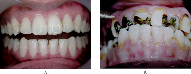
Dental caries is an irreversible microbial disease of the calcified tissues of the teeth, characterized by demineralization of the inorganic portion and destruction of the organic substance of the tooth, which often leads to cavitations. The word caries is derived from the Latin word meaning ‘rot’ or ‘decay’. It is a complex and dynamic process where a multitude of factors initiate and influence the progression of disease. Although effective methods are known for prevention and management of dental caries, it is a major health problem with manifestations persisting throughout life despite treatment. It is seen in all geographic areas in the world and affects persons of both genders in all races, all socioeconomic strata, and every age group. Some stay ‘caries-free’ for unknown reasons (Fig. 9-1). Despite extensive studies for more than a century, many aspects of etiology are still obscure, and efforts at prevention have been partially successful.
Epidemiology of Dental Caries

Caries Incidence in Modern Societies

By about the 17th century, there was a significant increase in the total caries experience and a smaller increase in the number of carious lesions involving the interproximal contact areas of teeth, characteristic of the pattern and occurrence of caries in modern population.

Extensive studies on the incidence of dental caries from various geographic areas have illustrated the apparent influence of civilization on dental disease. Mellanby in 1934 reviewed the literature on caries in existing primitive races and noted that the incidence was invariably less than that in modern man suggesting isolated populations that have not acquired the dietary habits of modern, industrialized man retain a relative freedom from dental caries. Native population living in the North West territories of Canada, Alaska and Greenland who consumed native food, had a lower evidence of carious lesion (0.1%) compared to those living at trading posts (13%). A comparable effect of diet upon caries was demonstrated by Mellanby in studies on natives of Southern Rhodesia. The determinants of the carious process are essentially local and limited to the oral cavity. Although there may be a certain degree of racial resistance to dental caries, dietary factor appears to be more significant, especially since caries incidence is increased by contact with ‘civilized’ food.

Comprehensive Assessment of Dental Caries Prevalence in Modern-day Population

While dental caries is all pervading in highly industrialized societies, the caries experience varies greatly among countries and even within a country. The difference in caries rates noted in different parts of the world are extreme from rates fewer than one decayed, missing and filled (DMF) tooth per person at all ages up to 39 years in Ethiopia (Littleton, 1963) to 60 times greater in Alaska-Aleuts (Russel et al, 1961). Findings from the Interdepartmental Committee on Nutrition for National Defence (ICNND) and WHO studies (Barmes, 1981) indicate that caries prevalence follows definite regional patterns. It is generally lowest (0.5–1.7 DMF) in Asian and African countries and highest (12–18 DMF) in America and other Western countries. Consistently, low to moderate caries rates were found in populations of the Indo-Chinese peninsula, Malaysia, central and southern Thailand, Burma, South Vietnam, mainland China, Taiwan, India and New Guinea.

Generally, highly industrialized countries have the highest caries indices with decayed, missing, and filled teeth (DMFT) of approximately 4.5. However, within this large group of countries a very high caries pattern of over 5.6 DMFT occurs in New Zealand, Australia, Brazil, and Argentina.

DMF and def Index

The most commonly employed method to measure the extent of previous damage to permanent dentition is by a measure known as the DMF index. The designation DMF (T) is used to denote decayed, missing, and filled teeth; DMF(S) denotes decayed, missing and filled surfaces in permanent teeth and therefore takes into account the number of surfaces attacked on each tooth. A similar index def (t) or def (s) index is used for primary dentition. The DMF/def index can be used to quantify both caries prevalence and caries incidence in a given population. It is an arithmetic index of the cumulative caries attack in a population.

A commonly used modified form of this test is the caries increment, which refers to the number of new carious lesions occurring in a specified time interval, either for an individual or averaged over a population. The assessment of the caries increment involves at least two examinations—one at the beginning and one at the end of the period in question. In children, primary teeth may be lost due to natural exfoliation and, for the purpose of the def index, it is essential that the examiner designates as missing only those teeth that are lost due to caries.

Factors Affecting Caries Prevalence

Age

Carious lesions that result in cavitation are irreversible and therefore, cumulative with age. There is a strong correlation between age and DMF indices. Several studies have shown that by the age of 6 years, about 20% of children have experienced dental caries in their dentition and a DMFT of 0.5 can be expected. By the age of 12 years, 90% of children would have experienced a DMFT of approximately 5.5. The decayed, missing and filled surface (DMFS) accelerates at a greater rate than the DMFT beyond the age of eight years. By the age 12, an average DMFS of 7.5 is seen in most populations. In general, other reports of caries prevalence among children in various parts of the world show rates that seem to be comparable to those cited here. Another common element is that children from families in lower socioeconomic groups consistently have greater caries prevalence than their peers from families at a higher socioeconomic level.

Current Trends in Caries Incidence

Significant data have been presented since the National Caries Program, USA in 1979–80 to substantiate numerous observations that there has been marked improvement in dental health as measured by prevalence of dental caries, especially in children and young adults, throughout the ‘civilized Western world’. Especially impressive was the increase in the percentage of children classified as caries-free in their permanent dentition. These changes had occurred in the absence of both fluoridation and organized preventive programs. This decrease in caries prevalence is also seen in England, Denmark, Ireland, the Netherlands, New Zealand, Norway, Scotland, and Sweden. A substantial decrease in the prevalence of dental caries has been reported from less developed countries. The cause for this widespread decline in the prevalence of dental caries is multifactorial. In some instances, communal water fluoridation has been present in the areas studied and in other cases organized preventive dentistry programs were available.

However, the time period involved in most of these studies coincides with the introduction and increased utilization of fluoride dentifrices and dietary fluoride supplements, as well as an increased awareness of the importance of oral health. The very limited studies available give no evidence that there is any change; for example, in the pervasiveness of Streptococcus mutans or any changes in dominant serotypes. Emphasis on improved physical health through food, exercise, and decreased carbohydrate consumption all may be the factors that have led to this decline.

Etiology of Dental Ca ries

The etiology of dental caries is generally agreed to be a complex problem complicated by many indirect factors that obscure the direct cause or causes. There is no universally accepted opinion for the etiology of dental caries. Numerous references on dental caries, including early theories attempting to explain its etiology, have been found in recorded history of ancient people. However, many theories have evolved through years of investigation and observation; the acidogenic theory of Miller (Miller’s chemico-parasitic theory), the proteolytic theory and the proteolysis chelation theory, are among those which have stood the test of time.

The Early Theories

Parasitic Theory

The first to relate microorganisms to caries on a causative basis as early as 1843 was Erdl who described filamentous organisms in the membrane removed from teeth. Shortly thereafter, Ficnus in 1847, a German physician in Dresden, attributed dental caries to ‘denticolae’ the generic term he proposed for decay related microorganisms. Leber and Rottenstein, two German physicians, disseminated the idea that dental caries commenced as a chemical process but that living microorganisms continued the disintegration in both enamel and dentin. In addition to these observations, Clark (1871, 1879), Tomes (1873) and Magitot (1878) concurred that bacteria were essential to caries, although they suggested an exogenous source of the acids. In 1880, Underwood and Miller presented a septic theory with the hypothesis that acid capable of causing decalcification was produced by bacteria feeding on the organic fibrils of dentin. They reported sections of decayed dentin having micrococci as well as oval and rod shaped forms.

Miller’s Chemico-Parasitic Theory or The Acidogenic Theory

The chemico-parasitic theory is a blend of the above mentioned two theories. Willoughby D Miller, an American who was working at the University of Berlin, is probably the best known of the early investigators on dental caries. He published extensively on the results of his studies, beginning in 1882, which culminated in the hypothesis, “Dental decay is a chemico-parasitic process consisting of two stages, the decalcification of enamel, which results in its total destruction and the decalcification of dentin as a preliminary stage, followed by dissolution of the softened residue. In case of enamel; however, the second stage is practically wanting, the decalcification of enamel signifying its total destruction”. The acid, which affects this primary decalcification, is derived from the fermentation of starches and sugar lodged in the retaining centers of the teeth. Miller found that bread, meat and sugar incubated in vitro with saliva at body temperature, produced enough acid within 48 hours to decalcify sound dentin. Subsequently, he isolated numerous microorganisms from the oral cavity, many of which were acidogenic and some were proteolytic. Since a number of these bacterial forms were capable of forming lactic acid, Miller believed that caries was not caused by any single organism, but rather by a variety of microorganisms. He assigned an essential role to three factors in the caries process: the oral microorganisms in acid production and proteolysis; the carbohydrate substrate; and the acid which causes dissolution of tooth minerals. Miller’s chemico-parasitic theory is the backbone of current knowledge and understanding of the etiology of dental caries.

However, Miller’s chemico-parasitic theory could not explain the predilection of specific sites on a tooth to dental caries and the initiation of smooth surfaces. Also, why some populations are caries-free and the phenomenon of arrested caries. The concept of dental plaque adhering to teeth and serving to localize bacterial enzymatic activity was proposed later in 1897 by Williams. This theory has been accepted by majority of investigators in a form essentially unchanged since its inception. The bulk of scientific evidence does implicate carbohydrates, oral microorganisms and acids, and for this reason, these deserve further consideration.

Role of Carbohydrates

Reference has been made previously to the finding that members of isolated primitive societies who had a relatively low caries index manifested a remarkable increase in caries incidence after exposure to refined diets. The presence of readily fermentable carbohydrates has been thought to be responsible for their loss of caries resistance.

The early studies of Miller showed that when teeth were incubated in mixtures of saliva and bread or sugar, decalcification occurred. There was no effect on the teeth when meat or fat was used in place of the carbohydrate. Both cane sugar and cooked starches produced acid, but little acid was formed when raw starches were substituted. Volker and Pinkerton reported the production of similar quantities of acid from mixtures of either sucrose or starch incubated with saliva with no difference in acid production between raw and refined sugarcane. The etiology of dental caries involves interplay between oral bacteria, local carbohydrates and the tooth surface that may be shown as follows: Bacteria + sugars + teeth → organic acids → caries.

The cariogenic carbohydrates are dietary in origin, since uncontaminated human saliva contains only negligible amounts regardless of the blood sugar level. Salivary carbohydrates are bound to proteins and other compounds, and are not readily available for microbial degradation. The cariogenicity of a dietary carbohydrate varies with the frequency of ingestion, physical form, chemical composition, route of administration and presence of other food constituents. Sticky, solid carbohydrates, soft retentive foods those that are cleared slowly, monosaccharides and disaccharides are more caries-producing. Plaque organisms produce little acid from the sugar alcohols, sorbitol, and mannitol. Glucose or sucrose fed entirely by stomach tube or intravenously, does not contribute to decay as they are unavailable for microbial breakdown. Meals high in fat, protein or salt reduce the oral retentiveness of carbohydrates.

Role of Microorganisms

Miller demonstrated the presence of microorganisms within the tubules of decayed teeth. These were mainly cocci and leptothrix, as he called them, and laid the foundation for the role of acids elaborated by bacteria in caries production. In 1900, Goadby isolated a gram-positive bacillus from carious dentin and termed it B. necrodentalis. These, he concluded, played a role in decalcification of both enamel and dentin. Later he changed his views, stating that certain streptococci were the active cause of caries. Later in 1922, McIntosh, James and Lazarus-Barlow were concerned with microorganisms capable of lowering the pH to the degree that the enamel was softened. From carious dentin they isolated bacteria which they called Bacillus acidophilus odontolyticus. Around the same time, Clarke in Great Britain isolated a streptococcus from teeth that was found to be in the early stages of the disease. In 1924, he described a new streptococcus species, S. mutans, which was almost always isolated from carious lesions in the teeth of British patients. Although the work was confirmed three years later by McLean, scientific interest in S. mutans lay dormant until its rediscovery in the mid 1960s.

Many of the earlier workers focused attention on L. acidophilus because it was found with such frequency in caries-susceptible persons that it came to be regarded as of etiologic importance. In 1925, Bunting and Palmerlee reported the bacillary forms in every initial lesion of caries similar to those described by McIntosh and they termed them B. acidophilus. Bunting stated in 1928, so definite is this correlation between B. acidophilus and dental caries that, in the opinion of this group, the presence or absence of B. acidophilus in the mouth constitutes a definite criterion of the activity of dental caries that is more accurate than any clinical estimation could be. Furthermore, it was noted that there was a spontaneous cessation of caries coincident with the disappearance of B. acidophilus from the mouth, either from prophylactic, therapeutic or dietetic control.

Bunting, Nickerson and Hard carried out extensive studies on B. acidophilus and reported that it was almost universally absent in the mouths of caries-immune persons, but was usually present in the mouths of caries-susceptible persons. Similar findings were reported in 1927 by Jay and Voorhees, who also found that the presence of L. acidophilus in persons without active caries was often a presage of the development of cavities some months later. Jay reported the isolation of 12 strains of Leptothrix in 1927, but doubted their importance in the carious process even though they produced acid from carbohydrates.

Between this period and the 1940s, numerous studies were carried out in attempts to confirm or deny the existence of a microorganism responsible for dental caries. Harrison observed streptococci to predominate on the surfaces of non-carious rat molars, about half of the strains being acidogenic. On the other hand, in rats with carious lesions, the surface flora consisted primarily of lactobacilli. Microorganisms isolated from the deeper carious cavities were mainly acidogenic streptococci and he thus concluded that there was an apparent relationship of lactobacilli with initial caries and of streptococci with more advanced lesions of dentin. Florestano, in 1942, cultured organisms from the saliva of carious and noncarious persons and studied their acidogenic potential. Aciduric streptococci and staphylococci were isolated from both the groups. Their acid production and presence in large numbers suggested a role in dental caries equal to that of lactobacilli. Bacteriologic studies in recent years have helped clarify the role of various organisms in the etiology of dental caries. Considerable emphasis has been placed on the various diet-bacterial interactions, which are involved in lesion development on different tooth surfaces. Specific microorganisms as well as combinations of microorganisms, including Lactobacillus, S. mutans, Actinomyces species and others, have been studied. Although there may be disagreement as to specifics, there is little doubt that bacteria are indispensable to the production of caries. One or more organisms are implicated in the initiation of caries, while other distinctly different organisms may influence the progression of the disease. Also, there is good evidence that different diet–bacterial interactions are involved in root surface and coronal caries, and they may represent two different diseases from the ecological and microbiological point of view.

In 1960, Keyes demonstrated that under certain laboratory conditions, dental caries in hamsters and rats could be considered an infectious and transmissible disease and therefore subject to those biologic principles which govern any infectious process. Fitzgerald and Keyes showed that even in a so-called caries-inactive strain of hamster, oral inoculation of certain pure cultures of streptococci isolated from hamster caries would induce the typical picture of active dental caries. The caries-inactive strain of hamster was found to have a noncariogenic microflora. These findings have led to interesting speculation about the importance of streptococci in the etiology of dental caries.

Microbial Flora and Dental Caries

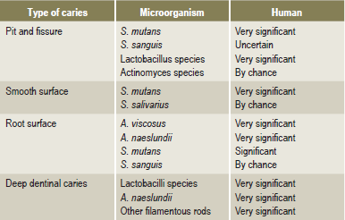
It is uniformly agreed that caries cannot occur without microorganisms. Several organisms have been found capable of inducing carious lesions when used as monocontaminants in gnotobiotic (germ-free) rats. These include the mutans group of streptococci, a Streptococcus salivarius strain, Streptococcus mitior, Streptococcus milleri, Streptococcus oralis, Streptococcus sanguis (different strains), Peptostreptococcus intermedius, Lactobacillus acidophilus, Lactobacillus casei, Actinomyces viscosus and Actinomyces naeslundii. In addition, in the same animal system, some streptococcis and lactobacilli such as Lactobacillus fermentum and Streptococcus lactis, were not able to induce caries, suggesting that not all organisms are cariogenic, at the same time caries will not occur even in the complete absence of microorganisms. Different organisms display certain selectivity for the tooth surface they localize and attack (Table 9-1).

A wide variety of organisms are able to initiate pit and fissure caries as they colonize in these retentive areas. A limited number of organisms have proved to colonize smooth surfaces and S. mutans is very significant in this respect. Some of the organisms involved in root caries are different from those in other smooth surface lesions because the initial lesion involves the cementum or dentin and not enamel. Bacteriological sampling of plaque covering caries of the root surfaces has yielded predominantly Actinomyces viscosus. However, other studies have found no difference in the prevalence of A. viscosus on carious versus intact root surface. Strains of Nocardia and S. sanguis, besides causing enamel caries may, at times, also cause root caries. While in the deep dentinal caries, the predominant organism is Lactobacillus, the exact extent to which these organisms participate in human disease is yet to be explored.

Studies in humans are largely based on the mathematical relationship between various streptococci, lactobacilli and dental caries. Available data strongly suggest an active involvement of S. mutans in caries initiation. Strains of S. mutans isolated from humans have proved to be cariogenic in animal studies and S. mutans can almost always be found in plaques over incipient lesions involving pits and fissures or smooth tooth surfaces. Not all studies support a unique or sole relationship between S. mutans and the initiation of caries in humans. The characteristics and properties of some known potentially cariogenic plaque microorganisms are discussed.

Lactobacilli

Lactobacilli are gram-positive, nonspore forming rods that grow best under microaerophilic conditions. The isolation of lactobacilli has been made possible by the use of a selective agar medium (Rogosa) which suppresses the growth of most other organisms by its low pH. The genus Lactobacillus includes many species and represents about 1% of oral flora. Among the homofermentative isolates, L. casei and L. acidophilus are the most common, while hetero fermentative members mostly include, L. fermentum and L. brevis. The idea that lactobacilli are important in the carious process was owing to the fact that they are both acidogenic as well as aciduric and could therefore multiply in the low pH of plaque and carious lesions. Lactobacilli as a universal etiologic agent in dental caries is; however, questioned because the amount of acid formed by lactobacilli present in plaque is insignificant in comparison to that produced by other acidogenic oral organisms. The occurrence of lactobacilli in carious lesions and their increased numbers in plaque and saliva does not necessarily establish their causative role although they could be secondary invaders. This possibility is supported by the observations that lactobacilli are not detectable in plaques covering white spot lesions on smooth surfaces and their predominant sites are in deep fissures and in deep dentinal lesions, favoring their retention.

Oral Streptococci

Of all the oral bacteria, streptococci have been studied most comprehensively. The most important species found in the oral cavity include: S. mutans, S. sanguis, S. mitior, S. salivarius, and S. milleri.

S. mutans

A Streptococcus that prevailed in many human carious lesions and first isolated in 1924 by Clarke was termed Streptococcus mutans. These bacteria are catalase negative, gram-positive cocci forming short to medium chains. On mitis salivarius agar, they grow as highly convex colonies. Unlike other oral streptococci, most strains of S.mutans can be selectively cultured in mitis salivarius agar containing 20% sucrose and 0.2% units/ml of bacitracin. Characteristically, S. mutans synthesizes insoluble polysaccharides from sucrose. It is homofermentative and is more aciduric than other oral streptococci.

Cariogenic strains of S. mutans contain a lysogenic bacteriophage which has not been isolated from non-cariogenic strains. Non-cariogenic strains are unable to adhere to glass and have decreased ability to form insoluble polysaccharides. In the oral cavity, S. mutans does not colonize the mouths of infants prior to the eruption of teeth. Likewise, it disappears from the mouth following the extraction of all teeth. Infants most likely become infected from their parents or from other individuals with whom they have frequent contact since these organisms are not found free living in nature and have only been isolated from humans and certain animals.

S. mutans forms a homogeneous group based on several phenotypic characteristics. However, based on nucleic acid base content and hybridization, S. mutans has been divided into five genotypes as S. mutans, S. rattus, S. sobrinus, S. cricetus, and S. ferus. Among, these, S. mutans and S. sobrinus are most commonly found in human plaque. S. mutans strains have also been divided into eight serotypes designated ‘a’ through ‘h’. The specific antigen for each serotype represents cell-wall constituents which have been isolated and chemically characterized as polysaccharides. Theoretical possibilities exist for inhibiting glucosyltransferase of several serotypes by an antiserum against purified glucose transferase of one single serotype.

Metabolism of S. mutans

The most important substrate for the involvement of S. mutans in the caries process is the disaccharide sucrose. Different pathways by which S. mutans may dissimilate sucrose, are by conversion of sucrose to adhesive extracellular carbohydrate polymers by cell bound and extracellular enzymes. The transport of sucrose into the cell interior is accompanied by direct phosphorylation for energy utilization through the glycolytic pathway, leading to lactic acid production and degradation of sucrose to free glucose and fructose by invertase. The intermediary metabolites from sucrose enter the glycolytic cycle or may be utilized in intracellular polymer synthesis in order to provide a reservoir for energy.

Most of the sucrose metabolized by S. mutans is utilized for its energy requirements and results in the production of lactic acid. Sucrose, which does not enter the cell, may be used for the extracellular synthesis of carbohydrate polymers. The ability of S. mutans to form adhesive plaques could explain its specific dependence on sucrose rather than other dietary carbohydrates.

It must be emphasized that S. mutans polymerizes the glucose and the fructose moieties of sucrose to synthesize glucans and fructans, which are two types of extracellular polymers. The enzymes responsible for the synthesis of extracellular glucans and fructans are called glucosyl- and fructosyltransferases, respectively. Synthesis of glucans from sucrose has been considered for several years to be the essential glue in S. mutans attachment to enamel and subsequent plaque formation. Two of the homopolymers of glucans are dextran and mutan. Mutan is an important constituent of fibrillar plaque matrix and is less soluble and more resistant to enzymatic attack than dextran.

Besides functioning as a resistant structural matrix, insoluble extracellular polysaccharides can act as a diffusion barrier. The transport of metabolites and salivary buffers into the plaque and the diffusion of acid out of the plaque may be affected by glucan. Besides producing glucan, certain oral bacteria can degrade this polymer and utilize it as a carbon source.

Fructans, on the other hand, unlike the mutan homopolymer of glucan, are generally soluble and can be degraded by plaque bacteria, thus serving as a reservoir of fermentable sugars for oral bacteria. A group of fructans produced by bacteria or created by breaking down other kinds of plant fructans are called levan beta 2, 6. Levans are both more soluble and more readily catabolized than glucans. Since levan hydrolysis is rapid, it may function as a short-term reservoir for the sustenance of bacterial anaerobic glycolysis in times of relative unavailability of dietary carbohydrate. Current opinion holds that this plaque component plays only a small role in the cariogenic potential of plaque because of the rapidity of its hydrolysis and the fact that it is purportedly not sticky.

Electron microscopic observation of the plaque formed by S. mutans reveals two types of extracellular products: a globular component, representing the water soluble, and a fibrillar component, the water insoluble glucan.

Lipoteichoic acid is another extracellular polymer that is found in cultures of S. mutans. These highly negatively charged compounds might contribute to the adhesiveness of bacteria. In addition to this, S. mutans strains have an ability to store intracellular glycogen amylopectin type polysaccharide, which provides a reservoir of substrate and enables prolonged periods of increased metabolic activity. Intracellular glycogen and extracellular polysaccharides serve as substrate reservoirs, which the organism may utilize for energy production, as the exogenous supplies of readily metabolized carbohydrate are depleted. In this fashion, both types of polysaccharides may play a role in the survival of organisms and in their potential to prolong acid production via glycolysis well beyond meal time.

It is known that sucrose-adapted S. mutans strains possess significant levels of invertase activity, and this enzyme is known to hydrolyze sucrose intracellularly to free glucose and fructose. Invertase is activated by inorganic phosphate and since phosphate accumulation is coupled with acid production, it is probable that one of the several mechanisms by which sucrose degradation is regulated in S. mutans is the activation of invertase by inorganic phosphate.

S. mitior

This is one of the most commonly isolated bacteria in the oral cavity. It produces soft, round and black-brown colonies on mitis salivarius agar. Along with S. sanguis it forms the most predominant organisms in dental plaque. However, its significance in human caries is assumed to be very minor.

Based on these observations from earlier studies, it is certain that bacteria, principally the gram-positive cocci and gram-positive pleomorphic rods, are essential for the development of caries. There is also a significant variation in the microbial flora associated with pit and fissure caries, smooth surface caries, root caries and deep dentinal caries. Since several factors may influence the formation, composition and metabolism of dental plaque, human dental caries may also be considered to be a diverse microbiologic disease. In the past, the total plaque was viewed as a pathogenic structure which had to be eliminated or reduced if caries was to be prevented. The present available data indicates that the qualitative nature of the flora in plaque determines the metabolism and the potential for caries production. This view is termed the Specific Plaque Hypothesis (Loesche, 1982), and according to this hypothesis, most but not necessarily all carious lesions are due to specific bacterial species. This concept suggests that cariogenesis is a specific bacterial infection and methods implemented for its elimination are more than just reduction of total plaque.

Role of Acids

The exact mechanism of carbohydrate degradation to form acids in the oral cavity by bacterial action is not known. It probably occurs through enzymatic breakdown of the sugar, and the acids formed are chiefly lactic acid, although others such as butyric acid are also formed. Since acid production is dependent upon a series of enzyme systems, methods of decreasing this acid formation by interference with certain enzymes could be an effective strategy to decrease caries.

The presence of acids in the oral cavity is of less significance than the localization of acids upon the tooth surface. This suggests a mechanism for holding acids, at a given point, for relatively long periods. Dental plaque fulfils this function. Acid production from carbohydrates have been extensively studied in human plaque. Generally, monosaccharides and disaccharides result in the greatest fall in plaque pH. On the other hand, acid formation is slower upon application of cooked starch. This is possibly because of the slower diffusion of larger starch molecules and acid production that occurs from the comparatively low concentration of maltose released from starch. Fermentation or glycolysis are ways of anaerobic catabolism of carbohydrates, which predominates in plaque and leads to acid production. The end products of glycolysis have the same empirical formulas as the starting substrate in that one molecule of glucose breaks into two molecules of lactic acid.

Organisms such as streptococci and lactobacilli ferment sugars, which produce 90% or more lactic acid as the end product; such bacteria are called homofermentative. Heterofermentatives produce a mixture of metabolites including other organic acids such as propionic, butyric, succinic, and ethanol using divergent metabolic pathways. For example, pyruvic acid, an intermediate metabolite in glycolysis, may be rendered to lactic acid by the enzyme lactic acid dehydrogenase or split into formic acid and acetyl coA by the enzyme pyruvate formate lyase. The acetyl coA is then converted into acetate and ethanol. The proportion of lactic acid or other organic acids formed by plaque may be markedly affected by growth conditions and by the bacterial types present. For example acid accumulation by S. mutans is substantially greater than by S. sanguis or S. mitis and Actinomyces are homolactic in anaerobic conditions but in the presence of carbon dioxide the fermentation is heterolactic with formate, acetate, lactate and succinate as their products.

Role of the Dental Plaque

Dental plaque (microbial plaque or bacterial plaque) is a structure of vital significance demonstrated for the first time in histologic preparations by Williams in 1897. It has been recognized for many years as a contributory factor to at least the initiation of the carious lesion.

Although Miller emphasized the role of foods and the acids produced by their bacterial degradation, he thought that the plaque protected enamel against attack by a carious process. In contrast, GV Black in 1899 regarded plaque to be important in the caries process and described it as “The gelatinous plaque of the caries fungus is a thin, transparent film that usually escapes observation, and which is revealed only by careful search. Neither it is the thick mass of material alba so frequently found upon the teeth, nor is the whitish gummy material known as sordes, which is often prominent in fevers and often present in the mouth in smaller quantities in the absence of fever.”

Plaque is the soft, nonmineralized, bacterial deposit which forms on teeth and dental prostheses that are not adequately cleaned. It characteristically forms on tooth surfaces which are not constantly cleansed, and appears as a tenacious, thin film, which may accumulate to a perceptible degree in 24–48 hours. A characteristic of plaque is that it resists removal by physiologic and oral cleansing forces such as saliva and tongue movement but is removable by toothbrushing. An important component of the dental plaque is acquired pellicle, which forms just prior to or concomitantly with bacterial colonization and may facilitate plaque formation. The pellicle is a glycoprotein that is derived from the saliva and is adsorbed on tooth surfaces. It is not dependent on bacteria but may serve as a nutrient for plaque microorganisms.

Dental plaque, or microcosm, as denoted by Arnim, is variable in both chemical and physical composition. It consists of salivary components such as mucin, desquamated epithelial cells and microorganisms. Plaque is composed of about 80% water and 20% solids. These are rich in bacteria with studies showing approximately 2×10¹¹ bacteria per gram. Bacterial and salivary proteins comprise about one half of the dry weight of plaque. Plaque also contains carbohydrates and lipids, which account for approximately 25% of the plaque’s dry weight. Most of the carbohydrates in the matrix consist of polymers, glucans, fructans, and heterosaccharides synthesized by the bacteria. Some of these polymers are thought to play a role in bacterial attachment and cohesion, and others are more important as a reservoir of fermentable substrates metabolized by bacteria when other more readily utilized carbohydrates in plaque become depleted.

Inorganic components of plaque account for approximately 5–10% of the dry weight of plaque. The concentration of calcium and phosphate in dental plaque is several magnitudes higher than in saliva. This is thought to be due, in part, to the infiltration of salivary proteins containing these constituents in the bound form. These probably include statherin, the salivary protein which, by adsorbing onto early crystal nuclei and preventing crystal growth, maintains super saturation of the fluid phase of plaque with apatite. In addition, bacteria may accumulate polyphosphates, which bind to calcium. Most of the calcium found in plaque is non-ionic and solubilisation occurs as pH drops. Dental calculus (q.v.) is plaque in which mineralization has involved both the plaque matrix and the microorganisms. However, the free surface of calculus usually harbors living bacteria.

There is a general agreement that enamel caries begins beneath the dental plaque. The presence of a plaque; however, does not necessarily mean that a carious lesion will develop at that point. Variations in caries formation have been attributed to the nature of the plaque itself, to the saliva or to the tooth. Extensive study of the bacterial flora of the dental plaque has indicated a heterogeneous nature of the structure. Most workers have stressed the presence of filamentous microorganisms, which grow in long interlacing threads and have the property of adhering to smooth enamel surfaces. Smaller bacilli and cocci then become entrapped in this reticular meshwork. Aciduric and acidogenic streptococci and lactobacilli are particularly numerous in this setup. Occasionally, strains of the filamentous organisms are actively acidogenic through carbohydrate fermentation, but this does not appear to be a general feature of this group.

Bibby (1940) studied the characteristics of different strains of filamentous organisms isolated from dental plaques and noted their ability to adhere to smooth surfaces. Blayney and his associates (1942) pointed out that the time required for the development of definite cavitation representing early caries in an intact enamel surface was several months. Hemmens and his coworkers (1946) believed that dental plaque was the most likely starting point for investigations aimed at understanding the earliest stage of enamel caries. They examined numerous plaques from areas of children’s teeth which became carious during the course of investigation. Aciduric streptococci were the organisms most commonly isolated from plaques during the period of caries activity, being present in varying numbers in 86% of the plaques. α-streptococci were isolated from slightly over 50% of the plaques from carious surfaces and from 75% of those from noncarious surfaces. The greatest incidence of occurrence of lactobacilli in plaque was 57%, but these organisms increased in incidence during that period in which the carious lesions were developing.

Most investigations of the microbiology of the dental plaque have concluded that three basic groups of microorganisms predominate: streptococci, actinomyces and veillonellae. The major strains of streptococci present in plaque are S.mutans, S. sanguis, S. mitior, S. milleri and S. salivarius (uncommonly). Major actinomyces strains include A. viscosus, A. naeslundii, A. israelii, and Rothia dentocariosa. The veillonellae group are the anaerobic gram-negative cocci organisms, chiefly V. parvula and V. alcalescens. Of all these, Streptococcus mutans is considered to be the chief etiologic agent in human dental caries today.

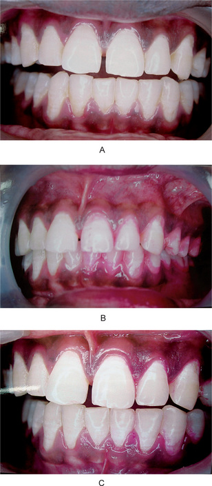
Plaques are classified as supra or subgingival, according to the anatomical area in which they form. Supragingival plaques play an essential role in the pathogenesis of dental caries while marginal and subgingival plaques are responsible for the initiation of periodontal diseases.The amount of plaque can be assessed directly by clinical examination (Fig. 9-2). Often dye solutions, referred to as disclosing agents, are used to stain plaques for visual scoring. Although carious lesions will not develop without plaque, it should be emphasized that plaques can often be relatively innocuous, have buffering capacity and protecting the teeth from exposure to acids present in many foods. However, when plaques contain appreciable proportions of highly acidogenic bacteria such as S. mutans and are exposed to readily fermentable dietary sucrose, they produce sufficient concentrations of acids to demineralise the enamel.
Mechanism of Plaque Formation

It is now known that the formation of dental plaque requires two types of specific bacterial adherent interactions. Firstly, bacteria attach selectively to the acquired pellicle, and secondly, bacteria accumulate via specific adhesive and cohesive interactions involving components of the plaque matrix and direct bacterial cell contact.

The pellicle appears as three distinct components. The subsurface component, below the surface of enamel having a dendritic configuration, the 1μm thick surface component closely associated with the surface of the tooth, and a suprasurface portion of 10 μm thickness which has a scalloped appearance. These amorphous organic films on the enamel surface may influence caries formation and bacterial adhesion. The acquired pellicle, like most proteinaceous adsorbed layers, is a membrane that may impart semipermeable properties to the enamel surface.

The role of the salivary pellicle in modifying plaque formation has been extensively studied because bacteria attached to salivary proteins adsorbed to the enamel rather than to the inorganic tooth surface. Some organisms such as S. salivarius, which are prominent on the dorsum of tongue and in saliva, do not adsorb well to teeth. Other organisms such as S. sanguis and A. viscosus, which are not as numerous in saliva, adsorb avidly to the pellicle and are prominent in developing new plaque. The important point is that organisms are not passively entrapped but rather selectively attached because of specific interactions involving their cell surface constituents and the macromolecules of salivary pellicle.

Stay updated, free dental videos. Join our Telegram channel

Jan 12, 2015 | Posted by in Oral and Maxillofacial Pathology | Comments Off on 9: Dental Caries

VIDEdental - Online dental courses

Get VIDEdental app for watching clinical videos