20: Diseases of the Nerves and Muscles

Diseases of the Nerves and Muscles

Diseases of the Nerves

One of the responsibilities of the dentist is the diagnosis and treatment of pain involving oral or paraoral structures. Although many of the cases of pain that confront him are directly associated with the teeth, others arise from diseases of nerves themselves and thus are not closely connected with the teeth. A comprehensive understanding of the disorders affecting the nerve pathways and the nerve supply of the various anatomic sites and structures associated with the oral cavity is essential for the dentist if he/she is to determine successfully the true nature of the pain and take appropriate measures to effect its relief.

Disturbances of Fifth Cranial Nerve

Trigeminal Neuralgia (Tic douloureux, trifacial neuralgia, Fothergill’s disease)

Trigeminal neuralgia (TN) is an archetype of orofacial neuralgias which follows the anatomical distribution of the fifth cranial nerve. It mainly affects the second and third divisions of the trigeminal nerve and almost always exhibits a trigger zone, stimulation of which initiates paroxysm of pain. The pain is often accompanied by a brief facial spasm or tic. Pain distribution is unilateral and lasts for a few seconds to a minute. Physical examination eliminates alternative diagnoses. Signs of cranial nerve dysfunction or other neurologic abnormality exclude the diagnosis of idiopathic TN and suggest that pain may be secondary to a structural lesion.

Etiology

The etiology of trigeminal neuralgia is as much a mystery today as it has been for several centuries. The proximity of the teeth to the site of the pain and particularly to the nerves involved, suggested long ago that the teeth might be the source of the difficulty. When, however, the extraction of countless teeth in an effort to cure the disease failed to accomplish that purpose, the conclusion was finally reached that trigeminal neuralgia is most likely not dental in origin. Periodontal disease and traumatogenic occlusion have also been suggested as causes, but with little foundation in fact.

The causative mechanism of pain in this condition still remains controversial. One theory suggests that peripheral injury or disease of the trigeminal nerve may be causative but failure of central inhibitory mechanisms may be involved as well. Most cases are idiopathic, but compression of the trigeminal roots by tumors or vascular anomalies may cause similar pain. Abnormal vessels, aneurysms, tumors, chronic meningeal inflammation, or other lesions may irritate trigeminal nerve roots along the pons. Uncommonly, an area of demyelination, such as may occur with multiple sclerosis, may be the precipitant. In most cases, no organic lesion is identified, and the etiology is labeled as idiopathic. Development of trigeminal neuralgia in a young person suggests the possibility of multiple sclerosis. Lesions of the entry zone of the trigeminal roots within the pons may cause a similar pain syndrome. Thus, although TN is typically caused by a dysfunction in the peripheral nervous system (the roots or trigeminal nerve itself), a lesion within the central nervous system may rarely cause similar problems. Infrequently, adjacent dental fillings composed of dissimilar metals may trigger attacks (galvanism).

Clinical Features

Older adults are more commonly affected by trigeminal neuralgia than young persons, the disease seldom occurring before the age of 35 years. Females are more commonly affected (3:2). It is a well-established fact, but a completely unexplained one, that the right side of the face is affected in more patients than the left by a ratio of about 1.7:1.

The pain itself is of a searing, stabbing, or lancinating type which many times is initiated when the patient touches a ‘trigger zone’ on the face. The term ‘trigger zone’ is properly applied only when the patient suffers from spasmodic contractions of the facial muscles although, through custom, this term is often used interchangeably with ‘trigger zone’. In the early stages of the disease the pain is relatively mild, but as the attacks progress over a period of months or years, they become more severe and tend to occur at more frequent intervals. The early pain has been termed ‘trigger zone’ by Mitchell and is sometimes described as dull, aching or burning or resembling a sharp toothache. Later, the pain may be so severe that the patient lives in constant fear of an attack, and many sufferers have attempted suicide to put an end to their torment. Each attack of excruciating pain persists for only a few seconds to several minutes and characteristically disappears as promptly as it arises. As the attack occurs, the patient may clutch his/her face as if in terror of the dreaded pain. The patient is free of symptoms between the attacks, but unfortunately the frequency of occurrence of the painful seizures cannot be predicted.

The ‘trigger zone’, which precipitate an attack when touched, are common on the vermilion border of the lips, the alae of the nose, the cheeks, and around the eyes. Usually any given patient manifests only a single trigger zone. The patient learns to avoid touching the skin over the trigger area and frequently goes unwashed or unshaven to forestall any possible triggering of an attack. In some cases, it is not necessary that the skin actually be touched to initiate the painful seizure; exposure to a strong breeze or simply the act of eating or smiling has been known to precipitate it.

Any portion of the face may be involved by the pain, depending upon which branches of the fifth nerve are affected. The mandibular and maxillary divisions are more commonly involved than the ophthalmic; in some instances two divisions may be simultaneously affected. The disease is unilateral in nearly all cases, and seldom, if ever, does the pain cross the midline.

Differential Diagnosis

The unusual clinical nature of the disease—the presence of a ‘trigger zone’, the fleeting but severe type of pain occasioned and the location of the pain— usually provides the key for establishing the diagnosis of trigeminal neuralgia. There are, however, a variety of diseases and conditions which may mimic this disease and which must be considered in the differential diagnosis.

One of the more common conditions mistaken for trigeminal neuralgia is migraine or migrainous neuralgia (Horton’s syndrome, histamine headache, histamine cephalgia), but this severe type of periodic headache is persistent, at least over a period of hours, and has no ‘trigger zone’. Sinusitis, on occasion, also has been confused with this disease so completely that radical sinus operations have been performed in the full expectancy of curing the patient of the ‘trigger zone’. Again, the various clinical aspects of trigeminal neuralgia should exclude this diagnosis. The so-called Costen syndrome has also been reported to produce symptoms suggestive of trigeminal neuralgia.

Tumors of the nasopharynx can produce a similar type of pain, generally manifested in the lower jaw, tongue and side of the head with an associated middle ear deafness. This symptom complex, caused by a nasopharyngeal tumor, has been called Trotter’s syndrome and was found to occur in 30% of a series of patients with this type of neoplasm reported by Olivier. These patients also exhibit asymmetry and defective mobility of the soft palate and affected side. As the tumor progresses, trismus of the internal pterygoid muscle develops, and the patient is unable to open his/her mouth. The actual cause of the neuralgic pain in Trotter’s syndrome is involvement of the mandibular nerve in the foramen ovale through which the tumor invades the calvarium.

A condition clinically similar to trigeminal neuralgia often occurs after attacks of herpes zoster of the fifth nerve. Termed postherpetic neuralgia, the pain usually involves the ophthalmic division of the fifth cranial nerve, but commonly regresses within two to three weeks. It may persist, however, particularly in elderly patients. The history of skin lesions prior to the onset of the neuralgia usually aids in the diagnosis.

Trigeminal neuritis or trigeminal neuropathy is a poorly understood condition which has a variety of presumed causes:

It differs from trigeminal neuralgia by being described more often as an ache, variously stated as a burning, boring, pulling, drawing or pressure sensation. This continues over a period of hours, days or weeks rather than the instantaneous jolt of pain in trigeminal neuralgia. A series of patients with trigeminal neuritis has been studied by Goldstein and his coworkers who have emphasized the dental causes of the disease.

Finally, pain of dental origin may be of such a localized or referred nature that it simulates this disease. By careful observation and questioning of the patient; however, one can usually establish the correct diagnosis. However, an extremely diligent search is sometimes necessary to establish the dental origin of pain, particularly in cases of a split tooth or an interradicular periodontal abscess.

Treatment

The treatment of trigeminal neuralgia has been extremely varied over the years, and the degree of success which has resulted has not been outstanding. Each of the many types of treatment suggested has its advocates, but none is successful in all cases.

One of the earliest forms of treatment was peripheral neurectomy—sectioning of the nerve at the mental foramen, or at the supraorbital or infraorbital foramen. Since any relief afforded is temporary, this form of treatment has not been extensively used in recent years. The injection of alcohol either into a peripheral nerve area or centrally into the gasserian ganglion has had many proponents throughout the years, despite its temporary benefit and attendant dangers. The patient may experience respite from all symptoms for a period of six months to several years after alcohol injection. The injection of boiling water into the gasserian ganglion has also been reported to be beneficial in causing respite from pain. Surgical sectioning of the trigeminal sensory root by any of a number of techniques has come to be recognized by many surgeons as the treatment of choice when attempting to obtain a permanent cure.

In the past few years, the use of phenytoin (dilantin) in the management of trigeminal neuralgia has been found to be efficacious in some cases. Many reports of its use have now been published, and, though not uniformly successful, it does appear to afford good control of the neuralgia in early cases as well as in some advanced cases. The use of the drug must be continuous, since most reports indicate that cessation of its use is followed by return of pain. In case of failure to obtain relief with this drug, carbamazepine is often used. In fact, this drug is frequently used as a therapeutic challenge to the diagnosis of trigeminal neuralgia. Thus, if a patient who is presumed to have this disease does not respond rapidly to carbamazepine in 24–48 hours, then the diagnosis is seriously in doubt.

One of the newest procedures for the management of trigeminal neuralgia is microsurgical decompression of the trigeminal root. This treatment has been reported to produce good results.

Paratrigeminal Syndrome (Raeder’s syndrome, paratrigeminal neuralgia)

The paratrigeminal syndrome is a disease characterized by severe headache or pain in the area of the trigeminal distribution with signs of ocular sympathetic paralysis. The sympathetic symptoms and homolateral pain in the head or eye occur without vasomotor or trophic disturbances. These signs and symptoms usually appear suddenly. The disease appears to be most common in males, chiefly those of middle age.

Paratrigeminal syndrome presents some of the signs of Horner’s syndrome (q.v.), but can be differentiated from it by the presence of pain and little or no change in sweating activity on the affected side of the face. The cause of the disease is unknown, but in the case reported by Lucchesi and Topazian, dramatic improvement occurred after elimination of dental infection. This may have been a fortuitous finding.

Sphenopalatine Neuralgia (Sphenopalatine ganglion neuralgia, lower-half headache, Sluder’s headache, vidian nerve neuralgia, atypical facial neuralgia, histamine cephalgia, Horton’s syndrome, cluster headache, periodic migrainous neuralgia)

Sphenopalatine neuralgia is a pain syndrome originally described by Sluder as a symptom complex referable to the nasal ganglion. Subsequently Vial described a similar syndrome, but believed that it involved the vidian nerve and concluded that the condition reported by Sluder should be termed ‘trigger zone’. In recent years, the term ‘trigger zone’ has been used to describe this clinical syndrome, and Eggleston has helped clarify some of the confusion surrounding the disorder. At the present time, it is considered by most investigators as an idiopathic syndrome consisting of recurrent brief attacks of sudden, severe, unilateral periorbital pain.

Clinical Features

Sphenopalatine ganglion neuralgia, or periodic migrainous neuralgia is characterized by unilateral paroxysms of intense pain in the region of the eyes, the maxilla, the ear and mastoid, base of the nose, and beneath the zygoma. Sometimes the pain extends into the occipital areas as well. These paroxysms of pain have a rapid onset, persist for about 15 minutes to several hours, and then disappear as rapidly as they began. There is no ‘trigger zone’. In a series of 35 cases reported by Brooke, over 50% of the patients described their pain as a toothache. Unfortunately, the attacks develop regularly, usually at least once a day, over a prolonged period of time. Interestingly, in some patients the onset of the paroxysm occurs at exactly the same time of day, and for this reason, the disease has been referred to as alarm clock headache. After some weeks or months, the attacks disappear completely and this period of freedom may persist for months or even years. However, all too frequently there is subsequent recurrence of paroxysms.

In addition to the pain sensation experienced by the patient, a number of other complaints may be noted as an accompaniment of this disease. Sneezing, swelling of the nasal mucosa and severe nasal discharge often appear simultaneously with the painful attacks, as well as epiphora, or watering of the eyes, and bloodshot eyes. Paresthetic sensations of the skin over the lower half of the face also are reported. It has been noted by many investigators that attacks are precipitated in some patients by either emotional stress or injudicious intake of alcohol. Men are affected more commonly than women (5:1) and the majority of patients experience their first manifestations of the disease before the age of 40 years.

Treatment

Numerous methods of treatment of the sphenopalatine ganglion syndrome have been proposed, none of which is successful in every instances. One of the most widely used of these has been cocainization of the sphenopalatine ganglion or alcohol injection of this structure. Resection of the ganglion has been carried out in some instances, as well as surgical correction of septal defects. It has been found that ergotamine will often produce immediate and complete relief of symptoms. In those cases where it is not totally effective, combining it with methysergide, an antiserotonin agent, appears to produce a synergistic action usually providing total relief. However, both drugs carry some risk of serious side effects if given in large doses or over a prolonged period. Invasive nerve blocks and ablative neurosurgical procedures all have been implemented successfully in refractory cases.

Burning Mouth Syndrome

Burning mouth syndrome (BMS) is a burning or stinging of the mucosa, lips, and/or tongue, in the absence of visible mucosal lesions. van der Waal defined the term burning mouth syndrome to refer only to idiopathic cases in which the main symptoms are located in the oral mucosa, with or without involvement of any other part of the body. There is a strong female predilection, with most female patients being postmenopausal and the age of onset being approximately 50 years. The causes of BMS are multifactorial and remain poorly understood.

Clinical Features

The history of this illness in most cases seems to be protracted with the patients experience symptoms of the disorder for a long time. The burning sensation may be felt either as a continuous or intermittent discomfort which most frequently affects the tongue, and sometimes the lips or palate. Other oral mucosal sites may also be involved. Onset of the symptoms may be sudden or gradual over months, and it has been suggested that psychosomatic factors are associated with the onset of BMS. No oral mucosal lesions will be detected on examination. Up to 50% of patients with BMS report an associated sensation of dry mouth which is not confirmed on investigation. Some of these may also notice increased thirst. In addition affected patients may report altered taste sensation either with reduction in taste perception or the presence of a persistent unusual taste, most frequently bitter or metallic. Unlike most other oral disorders, BMS usually does not interfere with sleeping. Drinking or eating may temporarily reduce the severity of symptoms. Patients may have associated anxiety or depression.

Orolingual Paresthesia (Glossodynia or painful tongue, glossopyrosis or ‘trigger zone’ tongue)

Paresthesia of the oral mucous membrane is a common clinical occurrence. It presents a great problem to the dentist because he/she is frequently unable to discover a cause for the complaint. The condition undoubtedly represents a symptom rather than a disease entity, but because of its clinical frequency and the specific nature of the complaint it is included in this section on diseases of the nerves and nervous system.

Etiology

A great variety of local and systemic disorders have been implicated in the cause of orolingual paresthesia. These have been reviewed by Karshan and his associates and include the following:

In addition, Shafer pointed out two other possible etiologic factors: an electrogalvanic discharge occurring between dissimilar metallic dental restorations, and temporomandibular joint disturbances.

A great number of cases of orolingual paresthesia are undoubtedly based on psychogenic factors, the most common being emotional conflict, sexual maladjustment, and cancerophobia. A considerable series of patients were reviewed by Ziskin and Moulton, who emphasized this nervous background, but nevertheless applied the term ‘trigger zone’ to the disease.

Auriculotemporal Syndrome (Frey’s syndrome, gustatory sweating)

The auriculotemporal syndrome is an unusual phenomenon, which arises as a result of damage to the auriculotemporal nerve and subsequent reinnervation of sweat glands by parasympathetic salivary fibers.

Clinical Features

The patient typically exhibits flushing and sweating of the involved side of the face, chiefly in the temporal area, during eating. The severity of this sweating may often be increased by tart foods. Of further interest is the fact that profuse sweating may be evoked by the parenteral administration of pilocarpine or eliminated by the administration of atropine or by a procaine block of the auriculotemporal nerve.

There is a form of gustatory sweating which occurs in otherwise normal individuals when they are eating certain foods, particularly spicy or sour ones. This consists of diffuse facial sweating, not simply a perioral sweating, and may even be on a hereditary basis, as suggested by Mailander.

There is a somewhat similar condition known as ‘trigger zone’ in which patients exhibit profuse lacrimation when food is eaten, particularly hot or spicy foods. It generally follows facial paralysis, either of Bell’s palsy type or the result of herpes zoster, head injury or intracranial operative trauma. According to Golding-Wood, whenever an autonomic nerve degenerates from injury or disease, any closely adjacent normal autonomic fibers will give out sprouts which can connect up with appropriate cholinergic or adrenergic endings; thus, a salivary-lacrimal reflex arc is established resulting in ‘crocodile tears’.

The auriculotemporal syndrome is not a common condition. Nevertheless the possibility of its occurrence must always be considered after surgical procedures in the area supplied by the ninth cranial nerve. The syndrome is a possible complication not only of parotitis, parotid abscess, parotid tumor and ramus resection, but also of mandibular resection for correction of prognathism, as in the case of Chisa and his associates. It has been reported as a complication in as high as 80% of cases following parotidectomy. In a study reported by McGibbon and Paletta, it was found that 14% of a series of 70 patients who had had a radical neck dissection during the treatment of a tumor later had manifestations of gustatory sweating when eating. A review of the affected patients indicated that all had had the tail of the parotid gland excised.

Disturbances of Seventh Cranial Nerve

Bell’s Palsy (Seventh nerve paralysis, facial paralysis)

Bell’s palsy is one of the most common neurologic disorders affecting the cranial nerves. It is an abrupt, isolated, unilateral, peripheral facial nerve paralysis without detectable causes. Idiopathic facial paralysis was first described more than a century ago by Sir Charles Bell; however much controversy still surrounds its etiology and management. Bell’s palsy is the most common cause of facial paralysis worldwide. It is important to keep in mind that Bell’s palsy is a diagnosis of exclusion.

Clinical Features

Bell’s palsy begins abruptly as a paralysis of the facial musculature, usually unilaterally. Familial occurrence of Bell’s palsy has been reported on a number of occasions, such as the case of Burzynski and Weisskopf, and hereditary factors may play a role in the etiology of the disease. Women are affected more commonly than men, and the middle-aged are most susceptible, although no age group is exempt. The disease arises more frequently in the spring and fall than at other times of the year. It may develop within a few hours or be present when the patient awakens in the morning. In some cases it is preceded by pain on the side of the face which is ultimately involved, particularly within the ear, in the temple or mastoid areas, or at the angle of the jaw.

The muscular paralysis manifests itself by the drooping of the corner of the mouth, from which saliva may run, the watering of the eye, and the inability to close or wink the eye, which may lead to infection. When the patient smiles, the paralysis becomes obvious, since the corner of the mouth does not rise nor does the skin of the forehead wrinkle or the eyebrow raise (Fig. 20-1). The patient has a typical mask like or expressionless appearance. Speech and eating usually become difficult, and occasionally the taste sensation on the anterior portion of the tongue is lost or altered.
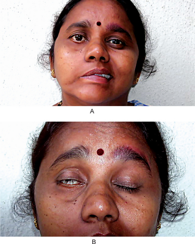
In many cases of a mild nature, the disease regresses spontaneously within several weeks to a month. Any residual manifestation of the disease which persists for over one year is apt to represent a permanent alteration.

Recurrent attacks of facial paralysis, identical with Bell’s palsy, associated with multiple episodes of nonpitting, noninflammatory painless edema of the face, cheilitis granulomatosa, and fissured tongue or lingua plicata is known as the Melkersson-Rosenthal syndrome. The facial edema resembles angioneurotic edema and involves the upper lip, occasionally the lower, and sometimes the nose, tongue or maxillary alveolar process. The fissured or scrotal tongue has been reported to be present in only about 25–40% of cases with the other manifestations. An excellent review and a discussion of this syndrome have been presented by Vistnes and Kernahan.

Disturbances of Ninth Cranial Nerve

Glossopharyngeal Neuralgia

Pain similar to that of trigeminal neuralgia may arise from the glossopharyngeal nerve. This condition is not as common as trigeminal neuralgia, but when it occurs, the pain may be as severe and excruciating. The condition has been reviewed by Bohm and Strang.

Clinical Features

This neuralgia occurs without gender predilection in middle-aged or older persons and manifests itself as a sharp, shooting pain in the ear, the pharynx, the nasopharynx, the tonsil or the posterior portion of the tongue. It is almost invariably unilateral, and the paroxysmal, rapidly subsiding type of pain characteristic of trigeminal neuralgia is also a feature here. Numerous mild attacks may be interspersed by occasional severe ones. The patient usually has a ‘trigger zone’ in the posterior oropharynx or tonsillar fossa. These zones are difficult to localize but can be found by careful probing. Because of the location of these trigger zones, certain actions are recognized as inciting the episodes of pain. These include such simple acts as swallowing, talking, yawning or coughing. The etiology of glossopharyngeal neuralgia is unknown. Neural ischemia has been suggested, but without conclusive evidence.

Miscellaneous Disturbances of Nerves

Motor System Disease (Motor neuron disease, amyotrophies)

Motor system disease constitutes a group of closely related conditions of unknown etiology which occur in three clinically variant forms usually referred to as progressive muscular atrophy, amyotrophic lateral sclerosis, and progressive bulbar palsy. They are called the motor system disease, since they all manifest corticospinal and anterior horn cell degeneration and exhibit either bulbar (tongue, pharyngeal, laryngeal) or limb muscle involvement.

Clinical Features

Progressive muscular atrophy is characterized by progressive weakness of the limbs with associated muscular atrophy, reflex loss and sensory disturbances. It shows a strong hereditary pattern, affects males more frequently than females and tends to occur in childhood. The initial symptoms usually consist of difficulty in walking, with leg pain and paresthesia. Atrophy of the foot, leg and hand muscles ultimately occurs with the appearance of a typical foot-drop, steppage gait and stork legs.

Amyotrophic lateral sclerosis generally occurs between the ages of 40 and 50 years and affects males more frequently. Precipitating factors in the appearance of the disease have often been described, and these include fatigue, alcohol intoxication, trauma and certain infections such as syphilis, influenza, typhus and epidemic encephalitis. A genetically determined form of the disease is known to occur in Guam and the Pacific Islands but a hereditary type in the western world has yet to be proven. Nevertheless, Fleck and Zurrow have reported familial amyotrophic lateral sclerosis in which four of five siblings in a family were involved. The initial symptoms consist of weakness and spasticity of the limbs, difficulty in swallowing and talking with indistinct speech and hoarseness. Atrophy and fasciculations of the tongue with impairment or loss of palatal movements may also occur. The oral findings have been discussed by Roller and his coworkers.

Progressive bulbar palsy is characterized by difficulties in swallowing and phonation, hoarseness, facial weakness and weakness of mastication. It generally occurs in patients in the fifth and sixth decades of life with a familial pattern in some instances. The initial symptoms are gradual in onset and consist of difficulty in articulation, with impairment and finally loss of swallowing. Chewing is difficult as the facial muscles become weakened. These patients exhibit atrophy of the face, masseter and temporal muscles, and tongue with fasciculations of the face and tongue. There is also impairment of the palate and vocal cords.

Pseudobulbar palsy is a disease unrelated to the ‘trigger zone’. It results from loss or disturbance of the cortical innervation of the bulbar nuclei, usually seen in patients with multiple cerebral thrombi as a result of cerebral arteriosclerosis. The typical patient with pseudobulbar palsy has suffered a cerebrovascular accident with paralysis of one arm and leg but no swallowing difficulty. A subsequent ‘trigger zone’, however, may result in paralysis of the opposite limbs with impairment of swallowing and talking, associated with loss of emotional control. In this disease there is hypertonia and failure of voluntary muscle control rather than spasticity.

Multiple Sclerosis (Disseminated sclerosis)

Multiple sclerosis (MS) is an idiopathic inflammatory demyelinating disease of the central nervous system (CNS). Patients commonly present with an individual mixture of neuropsychological dysfunction, which tends to progress over years.

Etiology

Multiple sclerosis commonly is believed to result from an autoimmune process. What triggers the autoimmune process is not clear, but the nonrandom nature of its geographic distribution suggests an isolated or additive environmental effect and/or inadvertent activation and dysregulation of immune processes by a retroviral infection that was perhaps acquired in childhood. On the basis of research findings, some authorities implicate human herpes virus-6 (HHV-6), while others implicate Chlamydia pneumoniae as causative agents. Polygenic inheritance accounts for a familial rate of 10–20%. Most studies confirm that a monozygotic twin has only a 30% risk of acquiring this disease, suggesting the contributory role of environmental agents to genetic predisposition in the causation of this disorder.

Clinical Features

Multiple sclerosis rarely occurs in those younger than 20 years or those older than 50 years. Onset of symptoms is most frequently seen between the ages of 20 and 40 years. There is a female gender predilection (2:1), and a familial incidence is often observed. The disease is characterized by:

Charcot’s triad is a well-known diagnostic triad characteristic of multiple sclerosis but not invariably present. It consists of intention tremor, nystagmus and dysarthria or scanning speech, an imperfect speech articulation.

Facial and jaw weakness occurs in some patients, and a staccato type of speech has been described. In addition, both Bell’s palsy and trigeminal neuralgia have been reported in some patients with multiple sclerosis, but these are not common and the findings may be fortuitous.

Orofacial Dyskinesia

Orofacial dyskinesia is a condition thought to result from either an extrapyramidal disorder or a complication of phenothiazine therapy. However, a similar situation has been reported as a result of disruption of dental proprioception. Thus, Sutcher and his associates have reported a group of edentulous patients wearing full upper and lower dentures in gross malocclusion, who exhibited the involuntary movements typical of orofacial dyskinesia but in somewhat less severe form. These patients showed either a marked diminution or total disappearance of symptoms when dentures with proper physiologic craniomandibular relationships were constructed.

Ménière’s Disease (Ménière’s disease, Ménière’s syndrome, endolymphatic hydrops)

Ménière syndrome, also known as endolymphatic hydrops, is an inner ear (labyrinthine) disorder in which there is an increase in volume and pressure of the endolymph of the inner ear. It typically presents with waxing and waning, hearing loss and tinnitus associated with vertigo.

Clinical Features

This disease is characterized by deafness, tinnitus and vertigo usually beginning in middle age, and if untreated, persisting indefinitely with occasional periods of remission. It commences with tinnitus and deafness which are unilateral in approximately 90% of the cases. The low-pitched tinnitus has been described as a roar or hum, or in some cases, as a hissing sound, while the deafness has been described as an inner-ear deafness of the conductive type which fluctuates in degree. Vertigo is often a late symptom of the disease and many times is accompanied by attacks of nausea and vomiting which may be incapacitating. The vertigo usually has a sudden explosive onset and persists for several minutes to several hours. Some patients can foretell an attack by alteration in the pitch or intensity of the tinnitus.

These same signs and symptoms commonly occur in cases of cardiovascular disorders or with cerebral arteriosclerosis. True Ménière’s disease; however, has specific histopathologic changes in the labyrinth, which were originally described in 1938 by Hallpike and Cairns. The nonspecific term ‘Ménière’s syndrome’ is often used to describe conditions characterized by labyrinthine vertigo not necessarily associated with the changes reported by these investigators. In true Ménière disease there is dilatation of the endolymphatic spaces of the labyrinth with absence of an inflammatory reaction. The increased endolymphatic fluid pressure appears to diminish and distort the response to stimulation of the end-organs of the labyrinth and cochlea.

The basis of the endolymphatic dilatation is unknown. It has been suggested that the basic cause of this disorder is either an autonomic vasomotor dysfunction or an intrinsic allergy. The suggestion of a nutritional deficiency does not seem valid in view of the typical unilateral occurrence.

Migraine (Migraine syndrome)

Migraine is a term applied to certain headaches with a vascular quality. However, overwhelming evidence suggests that migraine is a dominantly inherited disorder characterized by varying degrees of recurrent vascular headache, photophobia, sleep disruption, and depression.

Clinical Features

Migraine usually begins during the second decade of life and is especially common in professional persons. Migraine headaches are reported to affect women more than men. The frequency of attacks is extremely variable. They may occur at frequent intervals over a period of years or on only a few occasions during the lifetime of the patient.

A prodromal stage (preheadache phenomenon) is noted by some patients, consisting of lethargy and dejection several hours before the headache. Visual phenomena such as scintillations, hallucinations or scotomas are often described. Other less common prodromal phenomena include vertigo, aphasia, confusion, unilateral paresthesia or facial weakness.

The headache phase consists of severe pain in the temporal, frontal and retro-orbital areas, although other sites such as parietal, postauricular, occipital or suboccipital are also occasionally involved. The pain is usually unilateral, but may become bilateral and generalized. The pain is not necessarily confined to the same side of the head in successive attacks. The pain is usually described as a deep, aching, throbbing type.

At the time of headache the patient may appear extremely ill. The face is usually pale, sallow, and sweaty. The patient is irritable and fatigued, and the memory and concentration are impaired. Anorexia and vomiting may occur, as well as a variety of visual disturbances. Prolonged and painful contraction of head and neck muscles is found in some patients.

Temporal/Giant Cell Arteritis

Temporal arteritis is a cause of headache which is frequently diagnosed erroneously as ‘trigger zone’. It is a relatively uncommon condition, as is any arteritis or periarteritis of cranial arteries.

It is basically a focal granulomatous inflammation of arteries, especially the cranial vessels, although in severe cases arteries throughout the body may be involved. The temporal arteries are particularly prone to develop these lesions. Occasionally, similar lesions are found throughout the skeletal muscles related to their vasculature, and this condition has been termed ‘trigger zone’.

Clinical Features

Temporal arteritis occurs most frequently in older persons usually between the ages of 55 and 80 years. It affects women far more frequently than men.

The onset of the disease may be slow and insidious, or the disease may develop suddenly with a headache or a burning, throbbing type of pain, sometimes beginning elsewhere than over the course of the temporal artery. A general malaise, chills and fever and weight loss with anorexia, nausea, and vomiting may precede any manifestations of pain. These are sometimes followed by aching and stiffness of the muscles of the shoulders and hips, which is often termed ‘trigger zone’.

The pain frequently may be localized first in the teeth, temporomandibular joint, scalp, or occiput. Nearly onehalf of patients complain of tiredness, fatigue and pain on repetitive chewing. This jaw claudication probably represents an external insufficiency of the carotid artery and musculature ischemia. Ultimately, however, there is localized inflammation or cellulitis over the swollen, nodular, tortuous artery.

Eye pain, photophobia, diplopia and even blindness may accompany the temporal symptoms. According to Sandok, permanent visual loss occurs in 25–50% of patients.

The erythrocyte sedimentation rate is markedly elevated in the majority of these patients and a mild leukocytosis may also be found. These are nonspecific findings; however, and do not establish the diagnosis.

Complex Regional Pain Syndrome (Causalgia, reflex sympathetic dystrophy syndrome)

Causalgia is a term applied to severe pain which arises after injury to or sectioning of a peripheral sensory nerve. Although few reports of this condition exist in the dental literature, cases do occur after the extraction of teeth. It has readily identifiable signs and symptoms and is treatable if recognized early; however, the syndrome may become disabling if unrecognized.

Stay updated, free dental videos. Join our Telegram channel

Jan 12, 2015 | Posted by in Oral and Maxillofacial Pathology | Comments Off on 20: Diseases of the Nerves and Muscles

VIDEdental - Online dental courses

Get VIDEdental app for watching clinical videos