Chapter 8
Pedicle Grafts: Coronally Advanced Flaps
HISTORY
Bernimoulin et al. (1975) first reported the coronally positioned graft succeeding grafting with a free gingival autograft. This was a two-stage procedure. In the first stage, a free gingival graft was placed apical to the margins of the recession to be treated. The second stage occurred a few months later, when the graft was coronally positioned over the denuded root surfaces.
In 1986, Tarnow described the semilunar coronally positioned flap. This was a one-stage, no-suture, coronally repositioned flap aimed at correcting mild gingival recessions. In 1989, Allen and Miller reported the use of a one-stage, coronally positioned flap associated with citric acid root conditioning aimed at correcting shallow marginal recessions (2.5–4.0 mm).
INDICATIONS
A pedicle graft is used to cover gingival recessions affecting natural teeth. In the case of mild recessions, the one-stage, coronally positioned flap technique is sufficient. When the recession is moderate to severe (≥4 mm), a two-stage procedure will bring more predictable, long-term results.
Maynard (1977) outlined the following requirements as criteria for success when using coronally positioned flaps:
- The presence of shallow crevicular depths on proximal surfaces
- Normal interproximal bone heights
- Tissue height within 1 mm of the cemento-enamel junction of adjacent teeth
- Six-week healing of the free gingival graft prior to coronal positioning
- Reduction in root prominence
- Adequate release of the flap during the second-stage surgery to prevent retraction during healing
ARM AMENTARIUM
This includes the basic surgical kit plus the following:
- Gelfoam (Pharmacia-Upjohn, Kalamazoo, MI, USA) or Surgicel (Johnson & Johnson, New Brunswick, NJ, USA) (for the two-stage procedure)
- PeriAcryl (GluStitch; Delta, BC, Canada) (for the two-stage procedure)
- Citric acid, pH 1 (40%), or 1 capsule of tetracycline hydrochloride (HCl), 250 mg
CORONALLY POSITIONED FLAP:TWO STAGES
Technique
Fiststage
The first part of a two-stage, coronally positioned flap is a free gingival autograft (see Chapter 5) (Figs. 8.1 & 8.2).
Second stage
After a minimum 3 months of healing, the gingival graft is coronally positioned to cover the root, which should be carefully planned and conditioned with citric acid or tetracycline HCl (50–100 mg/ml) for 3–5 min. If the root has a proeminent concavity or convexity, it should be eliminated or reduced with a back-action chisel or a burr to graft on a flat surface.
Figure 8.1. Tooth 20 has gingival recession and no attached gingiva.
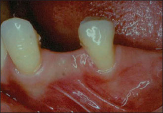
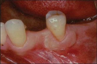
Figure 8.2. Tooth 20 at 2 months after receiving a free-gingival graft, first-step procedure.
The second stage requires a split-thickness dissection with mesial and distal vertical releasing incisions (Fig. 8.3). The vertical incisions are made at the line angles of the adjacen/>
Stay updated, free dental videos. Join our Telegram channel
VIDEdental - Online dental courses


