Chapter 15
Improving Patients’ Smiles: Aesthetic Crown-Lengthening Procedure
HISTORY
There are two aspects to the crown lengthening procedure: aesthetic and functional. In both cases, the surgical procedure is aimed at reestablishing the biological width apically while exposing more tooth structure. The biological width is defined as the sum of the junctional epithelium and supracrestal connective tissue attachment (Cohen 1962).
Gargiulo et al. (1961), who measured the human dentogingival junction, found that the average space occupied by the sum of the junctional epithelium and the supracrestal connective tissue fibers is 2.04 mm. Violation of that space by restorations impinging on the biological width has been associated with gingival inflammation, discomfort, gingival recession, alveolar bone loss, pocket formation, and the like (Parma-Benfenati et al. 1985; Tarnow et al. 1986; Tal et al. 1989).
To have a harmonious and successful long-term restoration, Ingber et al. (1977) advocated 3 mm of sound supracrestal tooth structure between bone and prosthetic margins, which allows for the reformation of the biological width plus sulcus depth. This can be achieved surgically (crown lengthening) or orthodontically (forced eruption) or by a combination of both (Ingber 1976; Pontoriero et al. 1987; De Waal and Castellucci 1994).
INDICATIONS
- To improve the gummy smile of a patient with a high smile line
- To rehabilitate dentition that is compromised by the presence of extensive caries, short clinical crowns, traumatic injuries, or severe parafunctional habits
- To restore gingival health when the biological width has been violated by a prosthetic restoration that is too close to the alveolar bone crest
Crown lengthening can be limited to the soft tissues when there is enough gingiva coronal to the alveolar bone, allowing for surgical modification of the gingival margins without the need for osseous recontouring (that is, pseudopockets in cases of gingival hyperplasia). An external or internal bevel gingivectomy (gingivoplasty) is the procedure of choice in these cases.
The biological width has not been compromised, and, as a result, the soft tissue pocket is eliminated and the teeth exposed without the need for osseous resection. Unfortunately, the majority of cases will involve bone recontouring as well as gingival resection to accommodate aesthetics and function. This is a more delicate procedure that requires exposing root surface, positioning gingival margins at the desired height, and apically reestablishing the biological width.
The crown-lengthening procedure enables restorative dentists to develop an adequate zone for crown retention without extending the crown margins deep into periodontal tissues. After the procedure, it is customary to wait 6–8 weeks before cementing the final restoration. In the aesthetic zone, a waiting period of at least 6 months is recommended before final impression (Pontoriero & Carnevale 2001). This reduces the chances of gingival recession following prosthetic crown insertion, specifically if there is a thin biotype.
A FEW WORDS ABOUT AESTHETICS
As the saying goes, “Beauty is in the eye of the beholder.” Oral aesthetics is part art and part science. The enhancement of a person’s smile culminates in the individualization of the general rules governing dental aesthetics for that person. Every patient is different, and yet a nice smile is the result of an orderly combination of several components. Knowing the general guidelines that make a smile appealing and tailoring them to an individual patient gives that smile uniqueness.
The aesthetic zone has been defined as the area encompassed by the upper and lower lips (Saadoun & LeGall 1998). It is the harmonious relationship among the dentition (premolar to premolar), the periodontium (gingival line), and the lips that will make or break a smile.
In 1984, Tjan et al., after observing several hundred dental and hygiene students, defined a standard of normalcy in an aesthetic smile (Figs. 15.1 & 15.2).
ARMAMENTARIUM
This includes the basic kit plus crown-lengthening burrs and bone chisels.
Figure 15.1. A pleasant smile line reveals 75%-100% of the maxillary anterior teeth and the interproximal gingiva only (68.94% of the subjects). The gingival margins of the central incisors and canines are located horizontally at the same level, whereas the gingival margins of the laterals are 2 mm below. The maxillary incisal curve is parallel with the lower lip (84.8% of the subjects). From Tjan et al. (1984).
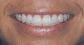
Figure 15.2. The position of the anterior contact point progressing from incisal to cervical and from central incisors to canines (horizontal lines). The location of the gingival zenith (blackarrows), the most apical point of the gingival tissue, referencing the tooth axis, is distal on the maxillary central incisors and canines, and coincidental on lateral incisors (Rufenacht 2000).The golden percentage (25%, 15%, and 10%) is considered a starting point in designing the relative width of teeth in a beautiful smile. With all of these width ratios added together, the total canine-to-canine width becomes the golden percentage
Stay updated, free dental videos. Join our Telegram channel
VIDEdental - Online dental courses


