Chapter 10
Acellular Dermal Matrix Graft (AlloDerm)
HISTORY
Acellular dermal matrix allograft, originally intended to cover burn wounds (Wainwright 1995), has been introduced as a less invasive alternative to soft tissue grafting (Silverstein & Callan 1997). This allograft is a freeze-dried, cellfree, dermal matrix comprised of a structurally integrated basement-membrane complex and extracellular matrix in which collagen bundles and elastic fibers are the main components (Wei et al. 2000).
INDICATIONS
- Soft tissue augmentation
- Multiple adjacent gingival recessions
- Lack of graftable palatal tissue
- Patient reluctant to have a second surgical site
- Correction of gingival/mucosal amalgam tattoos
ARMAMENTARIUM
This includes the basic surgical kit, AlloDerm, sterile saline, and two sterile dishes.
TECHNIQUE
After scaling and root planning, the root surfaces are conditioned. A partial thickness flap creating a pouch is formed using a no. 15 blade (Figs. 10.1 & 10.2). All these steps are similar to those described for the subepithelial connective tissue graft.
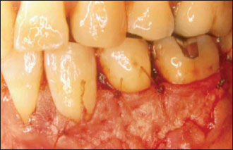
The AlloDerm is rehydrated in two consecutive 10- to 15-min sterile saline baths (depending on size and thickness of the piece used). The graft is inserted into the pouch with the connective tissue side—the bloody side—against the recipient bed. The papillae are de-epithelialized, and the graft is immobilized with resorbable sutures at the level of the cemento-enamel junction (Fig. 10.3). The buccal flap is then sutured over the AlloDerm to cover the graft as much as possible. It is important to not leave any AlloDerm exposed, if possible (Fig. 10.4).
POSTOPERATIVE INSTRUCTIONS
Postoperative instructions include systemic antibiotherapy for 7 days after the surgery. This is helpful to avoid complications. Nonsteroidal or steroidal anti-inflammatory drugs should be prescribed to keep the pain and swelling down (Greenwell et al. 2004).
Figure 10.1. Envelope incision: a pouch is created.
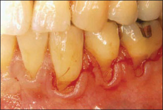
Figure 10.2. The roots are scaled, and the pouch is ready to accommodate the graft.
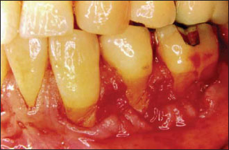
Figure 10.3. The AlloDerm is trimmed to fit the pouch, cover the roots, and suture to the papillae.These have been de-epithelialized.
Stay updated, free dental videos. Join our Telegram channel
VIDEdental - Online dental courses


