8: Craniomaxillofacial Trauma Surgery

Craniomaxillofacial Trauma Surgery

Oral and maxillofacial surgeons are the only specialists who have the training to provide complete craniomaxillofacial trauma care that includes medical and dental management and the capability to address reconstruction of all elements of the face. However, only with continued training, participation in trauma care, and observation of one’s own “personal residency” beyond formal training can surgeons maintain current skills for management of facial trauma. As specialists, we should endorse and support this essential component of oral and maxillofacial surgery (OMFS), because it has had a crucial impact on the advancement of the profession and has contributed to our gaining our rightful seat among other surgical disciplines.

The modern management of maxillofacial trauma has evolved with the advent of new biomaterials, improved diagnostic imaging, and refined instrumentation with associated techniques. The approach to maxillofacial trauma is in part related to the surgeon’s training and the available facilities; however, the basis of evaluation of the trauma patient who has sustained maxillofacial injuries remains unchanged. The importance of adherence to advanced trauma life support (ATLS) protocols and a comprehensive physical examination cannot be overemphasized. The use of bone grafts and the placement of dental implants put our specialty in the unique position of being able to offer complete rehabilitation beyond the immediate surgical repair of the fractured segments. An understanding of occlusion and the related musculoskeletal apparatus is essential for correct management of facial fractures, many of which involve the dentate segments. The goal of maxillofacial trauma surgery is restoration of the preinjury level of function and optimal cosmetic outcome.

In this chapter we present a series of cases representing the spectrum of maxillofacial injuries as they are encountered in the most classic way. Although most of the cases presented (except for the panfacial trauma case) represent isolated injury patterns, it is important to recognize that injuries can present in any combination.

It is not our intent to provide an exclusive approach to the evaluation and management of these injuries, but rather to emphasize common patterns of presentation, treatment options, and complications and to discuss other pertinent factors. In clinical practice three interdependent factors are related to a successful outcome; the patient, the injury, and the surgeon. These are different every time.

The Facial Injury Severity Scale (FISS) is a tool recently devised for designation of the severity of facial injuries (Box 8-1). The result is a numeric value that is the sum of all facial fractures and fracture patterns. The FISS is a predictor of the severity of facial injury as measured by the operating room charges required for treatment and the length of hospital stay.

Box 8-1   Facial Injury Severity Scale (FISS)

image

*Each midfacial fracture is assigned 1 point unless it is part of a complex.

Unilateral Le Fort fractures are assigned half the value shown.

From Bagheri SC, Dierks EJ, Kademani D, et al: Application of a facial injury severity scale in craniomaxillofacial trauma, J Oral Maxillofac Surg 64:404-414, 2006.

The Glasgow Coma Score is a universally used system for evaluation of neurologic status (Box 8-2).

Dentoalveolar Trauma

HPI

The patient reports that he was riding his mountain bike when an abrupt bump resulted in him striking his lower face against the handlebars. He dismounted the bicycle without other injuries and drove himself to an outside emergency department. He denies loss of consciousness, nausea, vomiting, visual disturbances, or headache (indicative of head trauma with intracranial injury). He further denies stridor, dyspnea, or increased work of breathing (suggestive of foreign body aspiration resulting from dislodged teeth, dental restorations, or orthodontic appliances). He notes several dental fractures, profound mobility of the lower teeth, and gingival bleeding. He undergoes primary and secondary surveys, according to the ATLS protocol, that are found to be negative. A computed tomography (CT) scan is obtained, and the patient is given instructions to report by private car to your office for evaluation.

Examination

General. The patient is a well-developed, well-nourished adult male in mild distress secondary to pain and oral bleeding. He is neurologically intact.

Maxillofacial. There are no lacerations, contusions, or abrasions of the scalp, midface, or chin. The pupils are equal at 4 mm, round, and reactive to light and accommodation. The nasal dorsum is midline and stable. There is no rhinorrhea (a concern for violation of the cribriform plate with cerebrospinal fluid [CSF] leakage) and no septal hematoma on speculum examination. The ears are symmetric and without injury to the pinnae. Examination of the external auditory meatus reveals no otorrhea (also a concern for CSF leakage) or disruption of the tympanic membrane. There is no mastoid ecchymosis noted (Battle’s sign, significant for occult skull base fracture). The orbits, midface, and mandible are without step deformity or crepitus on palpation. Cranial nerves II through XII are intact.

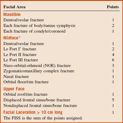
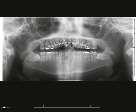
Intraoral. There is a 1-cm abrasion of the upper lip skin without significant laceration. Intraorally, there is profound ecchymosis of the upper lip mucosa with a laceration extending into the sublabial vestibule (Figure 8-1). Teeth #7 and #8 are mobile as a single unit with displacement of the buccal cortical plate on manipulation. Tooth #9 is grossly mobile and subluxed several millimeters. It also demonstrates an oblique coronal fracture with pinpoint pulp exposure (Ellis class III). In addition, tooth #9 is sensitive to mechanical stimulation with a cotton-tipped applicator and tender to percussion. There is occlusal prematurity with interference of the maxillary anterior teeth on attempted intercuspation.

Imaging

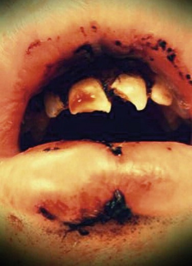
A maxillofacial CT scan demonstrates an alveolar segment fracture involving teeth #7 and #8, with subluxation of tooth #9 and fracture of the alveolar plate (Figure 8-2). There are otherwise no injuries to the maxillofacial skeleton, cervical spine, brain, or cranium. The minimum radiographic study necessary for diagnosis of dentoalveolar fractures is a periapical radiograph, although the diagnosis can often be made with a physical examination alone. Based on availability, other radiographic studies may include:

Treatment

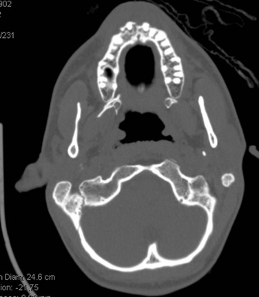
Initial stabilization includes reduction of the teeth and splinting (Figure 8-3). With the current patient, bonded flexible wire splinting was not available, so the teeth were splinted with an arch bar. The occlusion was checked, and the teeth were not in occlusion during maximum intercuspation. The patient’s tetnus status was up to date. He was given a prescription for amoxicillin and chlorhexidine and discharged home. The teeth were splinted for 8 weeks because of the alveolar segment fracture. Root canal therapy was initiated on day 10 with calcium hydroxide therapy.

Complications

In the past, only about 25% to 40% of replanted, avulsed teeth show periodontal ligament (PDL) healing. This has been attributed to poor handling of the tooth. Three different types of posttraumatic external root resorption have been distinguished in the literature: surface resorption (repair-related root resorption), inflammatory resorption (infection-related root resorption) and replacement resorption (ankylosis-related root resorption). Surface resorption has no significant clinical consequences and can be observed. However, the other types of resorption can ultimately result in tooth loss. In the avulsed tooth, if the PDL that is still attached to the tooth does not dry out, the cells can remain viable for an extended period, depending on the storage medium. Once the tooth has been reimplanted and stabilized, the viable PDL cells reattach to the PDL within the socket. When the injury to the cementum of the root is localized, there is minimal destructive inflammation, allowing for new cementum to be laid down after the inflammation resolves. When there is poor handling of the avulsed tooth (e.g., drying or storage in nonphysiologic solutions), damage and necrosis of the PDL occur. Subsequently, there is a large area of inflammation to remove the damaged PDL and cementum. This must be replaced by new tissue. The slower moving cementoblasts compete with the osteoblasts in the replacement process, resulting in some areas of the root surface being replaced by bone. Over time, through osseous remodeling, this can result in osseous or replacement resorption. Internal root resorption can occur through persistent inflammation or metaplastic replacement of normal pulp tissue. This can result in late tooth fractures. Root surface treatments and root canal therapy are directed toward prevention of this complication. Ideal management of dentoalveolar trauma may have to be delayed due to life-threatening injuries that must be managed first (Figure 8-4). This may result in resorptive complications.

Discusson

Appropriate diagnosis is critical in identifying and treating dentoalveolar injuries, which are known to affect one fourth of all children and one third of all adults. Depending on the mechanism of injury, a number of maxillofacial injuries may present with concomitant intracranial or cervical spine injuries, despite normal neurologic findings on physical examination. After a thorough physical examination that follows the ATLS protocol, attention is directed to the head and neck. Contaminated facial wounds should be irrigated with normal saline if available, although tap water has been shown to be as effective as saline. Patients with grossly contaminated wounds or facial injuries caused by dog or human bites should be considered for tetanus immunization based on their vaccination history. If an adult has an ambiguous immunization history or has received fewer than three prior doses of tetanus toxoid, he or she should receive tetanus immune globulin (TIG) and the tetanus-diphtheria (Td) or tetanus-diphtheria-acellular pertussis (Tdap) vaccine. Prior tetanus disease is inadequate at providing immunity, because a small amount of the highly potent toxin is sufficient to cause clinical neuromuscular weakness and airway compromise. Antibiotic coverage must be based on the mechanism and extent of injury. It is indicated in contaminated wounds with significant soft tissue injury, luxated teeth, avulsed teeth, pulp exposures, root fractures and alveolar fractures. Amoxicillin is usually chosen unless the patient is allergic to penicillin; in such cases, clindamycin can be substituted. Chlorhexidine oral rinse is an excellent choice for most oral injuries to help prevent infection.

The cause of dentoalveolar trauma varies among different demographics, but it generally results from falls, playground accidents, domestic violence, bicycle accidents, motor vehicle accidents, assaults, altercations, and sports injuries. Gassner and colleagues reported an incidence of 48.25% in all facial injuries, 57.8% in play and household accidents, 50.1% in sports accidents, 38.6% in accidents at work, 35.8% in acts of violence, 34.2% in traffic accidents, and 31% in unspecified accidents. Falling is the primary cause of dentoalveolar trauma in early childhood. Andreasen reported a bimodal trend in the peak incidence of dentoalveolar trauma in children aged 2 to 4 and 8 to 10 years.

Dentoalveolar injuries have been classified by the International Association of Dental Traumatology, which regularly reviews and updates its guidelines and publishes them online at www.dentaltraumaguide.org. Broadly, the discrete categories of dentoalveolar injury include:

Injuries to the periodontium resulting from forces directed through the tooth and to the surrounding bone and periodontal attachment are the most common types of dental trauma in the primary dentition.

Periodontal Injuries

Periodontal injuries may be classified according to the following system.

Concussion. No visible trauma to tooth or alveolar structures but pain on percussion. Treatment is conservative, with a no-chew diet only and surveillance of pulpal vitality.

Subluxation. Increased mobility of the tooth without dislocation. Treatment is conservative, although a flexible splint may be applied for the patient’s comfort for up to 2 weeks (Box 8-3).

Extrusion. Coronal dislocation of the tooth due to separation of the periodontal ligament without alveolar bone disruption. Treatment involves repositioning the tooth into the socket, stabilizing the tooth for 2 weeks with a nonrigid, flexible splint, and performing root canal therapy in teeth with closed apices. If the marginal alveolar bone demonstrates radiographic signs of breakdown at follow-up, prolonged splinting is recommended, for up to 6 weeks after the injury.

Lateral luxation. Tooth displacement with fracture of the alveolar process. Treatment includes flexible splinting for 4 weeks; root canal therapy is indicated for cases of pulpal necrosis to prevent root resorption.

Intrusion. Apical dislocation of the tooth, with crushing injury of supporting alveolar bone. Treatment depends on the status of the root apex. Incomplete root formation is treated conservatively, allowing several weeks for passive eruption. If no spontaneous movement is appreciated, orthodontic repositioning may be attempted after several weeks of conservative treatment. Teeth with complete apical development undergo immediate orthodontic repositioning if intruded more than 3 mm and conservative observation if intruded less than 3 mm, with application of orthodontic forces after 2 to 4 weeks. Teeth with 7 mm or greater of intrusive displacement should undergo immediate surgical repositioning regardless of the root apex development. Repositioned teeth must then undergo stabilization using a flexible splint for 4 to 8 weeks. Teeth with complete root formation that are intruded will likely develop pulpal necrosis and must undergo root canal therapy 2 to 3 weeks after injury. Formal obturation may be preceded by calcium hydroxide canal treatment if the tooth is actively being repositioned.

Avulsion/extrarticulation. The complete loss of tooth from alveolar supporting bone. The most commonly avulsed tooth is the maxillary central incisor, and the condition most often affects children 7 to 10 years of age. In most cases, replantation of avulsed permanent teeth should be attempted. Contraindications to replantation include an immunosuppressed patient after transplant surgery and patients with cardiac valve replacement. When possible, teeth should be positioned back into the socket immediately and stabilized. If this is not possible, the prognosis of the avulsed tooth depends on how it was handled. The prognosis is improved if there is no dry time, the tooth is stored in physiologic solution, and replantation is performed within 1 hour. Organ transport solution allows the PDL cells to survive for 1 week, Hank’s Balanced Salt Solution (HBSS) allows cells to survive for 24 hours, but milk allows only 6 hours of survival. Water is a poor storage medium for teeth. Because it is hypotonic, it results in rapid lysis of the PDL cells. For teeth with a closed apex and no dry time, stored in HBSS for less than 24 hours, or in milk or saliva for less than 6 hours, the tooth should be placed in doxycycline (0.05 mg/ml) for 5 minutes and then replanted. In animal studies, Cvek and colleagues and Yanpiset and Trope demonstrated that the use of doxycycline in this fashion significantly enhanced revascularization. Tetracycline has antiresorptive and antimicrobial properties. Tetracycline has a direct inhibitory effect on collagenase activity and osteoclasts. Its antimicrobial effects help to eliminate bacteria that have contaminated the alveolus, PDL, and pulpal tissues. The tooth is stabilized with flexible wire and composite for 7 to 10 days. At the 7- to 10-day follow-up visit, root canal therapy is started. The pulp is extirpated, and calcium hydroxide therapy is started. Calcium hydroxide is an effective antimicrobial agent that decreases resorption and promotes healing. The more alkaline environment in the dentin slows the resorptive cells and promotes hard tissue formation. Therapy continues usually until a viable PDL is radiographically demonstrated (6 to 24 months). The root canal can then be obturated with the final filling material, such as gutta percha. For teeth with an open apex and dry time less than 1 hour, the goal is to encourage revascularization, continued root formation, and apex closure. For these teeth, soaking in doxycycline (0.05 mg/ml) for 5 minutes is recommended. The tooth is stabilized with a flexible wire and composite and monitored for signs of pulpal necrosis. Apexification therapy should be performed with calcium hydroxide treatment if pulpal necrosis develops. Teeth that have been out of the mouth for longer than 1 hour and have not been kept in a storage medium will have a necrotic PDL and a poorer prognosis, with a greater risk of root resorption. To improve the prognosis, the PDL is debrided by placing the tooth in a sodium hypochlorite solution for 30 minutes. Extraoral root canal therapy is completed with gutta percha. The tooth is then placed sequentially in citric acid solution for 3 minutes, 1% stannous fluoride solution for 5 minutes, and 0.005% doxycycline solution for 5 minutes. The tooth is then replanted and splinted for 7 to 10 days. A study using Emdogain (Straumann; Basel, Switzerland) has shown some beneficial effects for teeth with extended dry times. Emdogain is an enamel matrix protein that has been shown to make the root more resistant to resorption and also to stimulate new PDL formation from the socket. The prognosis for teeth with an open apex and longer than 1 hour dry time is poor. No attempt should be made at revascularization. Instead, calcium hydroxide therapy for apexification can be initiated. An alternative treatment is to perform root canal therapy prior to replantation of the tooth; this allows for better sealing of the open apex. Treatment of the root surface is the same as for the closed apex. The tooth will likely undergo resorption, but it can allow for maintenance of alveolar width and height until the patient is old enough for implant placement.

Prominent maxillary central incisors that protrude beyond the confines of the upper lip are associated with a higher incidence of dental trauma in these children. Children are more challenging to examine and treat, and the parents’ cooperation is required. Injuries in the primary dentition, especially intrusion, can result in crown deformation or enamel hypoplasia of the underlying permanent teeth. For these reasons, primary teeth, if avulsed, should not be replanted for fear of injury to the underlying permanent teeth. Luxated and intruded teeth should likewise be removed.

Bonded composite with flexible wire is the treatment of choice for injuries to the periodontium and root fractures. This technique permits flexible stabilization that allows some movement of the tooth in relation to the alveolus. This in turn allows for healing of the PDL and reduces the risk of ankylosis or resorption. The recommended fixation time for an injury to the periodontium is 7 to 10 days. Arch bars are more rigid and provide better stabilization for alveolar segment fractures; they also may be less technically challenging to place. The disadvantages of the arch bar technique are that it car produce an eruptive or extrusive force because of the placement of the wire beneath the height of contour of the tooth; also, the rigid nature of this technique can facilitate ankylosis and resorption.

Dental Crown and Root Injuries

Injuries to the dental crown and root are classified as follows:

Crown fractures are common, and many times treatment is delayed to allow management of more severe injuries. Treatment is based on the extent of the crown-root involvement and/or pulpal involvement. Crown fractures that extend longitudinally onto the root below the level of the bone require extraction. Crown lengthening or orthodontic extrusion can be used to salvage some teeth. Treatment of root fractures depends mainly on the location of the fracture. Horizontal root fractures in the apical one third have the best prognosis. If the tooth is stable, it may not require treatment. Teeth that are mobile must be splinted for 12 weeks or extracted. Fractures in the cervical one third are usually extracted. Crown lengthening or orthodontic extrusion may also be performed.

The two types of treatment for pulpal injuries are direct pulp cap and root canal therapy. Direct pulp cap therapy is indicated for small, pinpoint exposures treated within 24 hours for mature teeth with a closed apex, and also for small and large pulp exposures in teeth with an open apex, to encourage apexification. Calcium hydroxide is used to seal small exposures. For teeth with an open apex and a large exposure, or exposure for longer than 24 hours, or deciduous teeth, a pulpotomy is performed first and then the calcium hydroxide therapy. Root canal therapy, with pulp extirpation, instrumentation of one or more canals, and sealing of the root, is recommended for pulpal injuries in mature teeth for large exposures or exposures exceeding 24 hours.

Injuries to the Alveolar Bone

Injuries to the supporting alveolar bone are classified as follows:

Alveolar fractures associated with intrusion or luxation are managed by immediate closed reduction of the fracture to realign the segments, reduce the teeth, and set the teeth into the best occlusion. Splinting with a rigid splint using acid-etched resin or orthodontic brackets and wire on either side of the fractured alveolus for 4 to 6 weeks is an option. In isolated alveolar segment fractures with no associated luxation injury, closed reduction is performed, followed by fixation with a single arch bar and 24- or 26-gauge wire for 4 weeks. Rigid fixation with titanium mini-plates and screws is generally reserved for alveolar fractures associated with fractures involving the basal bone and requiring open repair.

Bibliography

Andersson, LJ, Friskopp, J, Blomlof, L. Fiber-glass splinting of traumatized teeth. ASDC J Dent Child. 1983; 50(1):21–24.

Andreasen, JO. Effect of extra-alveolar period and storage media upon periodontal and pulpal healing after replantation of mature permanent incisors in monkeys. Int J Oral Surg. 1981; 10(1):43–53.

Andreasen, JO. Traumatic dental injuries: a manual, ed 3. Chichester, West Sussex, UK: Wiley-Blackwell; 2011.

Andreasen, JO, Borum, MK, Jacobsen, HL, et al. Replantation of 400 avulsed permanent incisors. 4. Factors related to periodontal ligament healing. Endod Dent Traumatol. 1995; 11(2):76–89.

Andreasen, JO, Andreasen, FM, Andersson, L, et al. Textbook and color atlas of traumatic injuries to the teeth, ed 4. Oxford, UK: Blackwell Munksgaard; 2007.

Andreasen, JO, et al. The dental trauma guide. www. dentaltraumaguide. org, 2012.

Blomlof, L. Milk and saliva as possible storage media for traumatically exarticulated teeth prior to replantation. Swed Dent J Suppl. 1981; 8:1–26.

Bystrom, AR, Claesson, R, Sundqvist, G. The antibacterial effect of camphorated paramonochlorophenol, camphorated phenol and calcium hydroxide in the treatment of infected root canals. Endod Dent Traumatol. 1985; 1(5):170–175.

Centers for Disease Control and PreventionAtkinson W, Wolfe S, Hamborsky J, eds. Epidemiology and prevention of vaccine-preventable diseases, ed 12, Washington DC: Public Health Foundation, 2012.

Chen, JW. Cervical spine injuries. Oral Maxillofac Surg Clin North Am. 2008; 20(3):381–391.

Cohenca, N, Simon, JH, Roges, R, et al. Clinical indications for digital imaging in dento-alveolar trauma. Part 1. Traumatic injuries. Dent Traumatol. 2007; 23(2):95–104.

Cvek, M, Cleaton-Jones, P, Austin, J, et al. Pulp revascularization in reimplanted immature monkey incisors: predictability and the effect of antibiotic systemic prophylaxis. Endod Dent Traumatol. 1990; 6(4):157–169.

Cvek, M, Cleaton-Jones, P, Austin, J, et al. Effect of topical application of doxycycline on pulp revascularization and periodontal healing in reimplanted monkey incisors. Endod Dent Traumatol. 1990; 6(4):170–176.

Diangelis, AJ, Andreasen, JO, Ebeleseder, KA, et al. International Association of Dental Traumatology guidelines for the management of traumatic dental injuries. Part 1. Fractures and luxations of permanent teeth. Dent Traumatol. 2012; 28(1):2–12.

Elias, H, Baur, DA. Management of trauma to supporting dental structures. Dent Clin North Am. 2009; 53(4):675–689.

Filippi, A, Pohl, Y, von Arx, T. Treatment of replacement resorption with Emdogain: preliminary results after 10 months. Dent Traumatol. 2001; 17(3):134–138.

Fuss, Z, Tsesis, I, Lin, S. Root resorption: diagnosis, classification and treatment choices based on stimulation factors. Dent Traumatol. 2003; 19(4):175–182.

Gassner, R, Bösch, R, Tuli, T, et al. Prevalence of dental trauma in 6,000 patients with facial injuries: implications for prevention. Oral Surg Oral Med Oral Pathol Oral Radiol Endod. 1999; 87(1):27–33.

Gazelius, B, Olgart, L, Edwall, L. Non-invasive recordings of blood flow in human dental pulp. Endod Dent Traumatol. 1986; 2:219–221.

Gonda, F, Nagase, M, Chen, RB, et al. Replantation: an analysis of 29 teeth. Oral Surg Oral Med Oral Pathol. 1990; 70(5):650–655.

Hiltz, J, Trope, M. Vitality of human lip fibroblasts in milk, Hanks balanced salt solution, and Viaspan storage media. Endod Dent Traumatol. 1991; 7(2):69–72.

Iqbal, MK, Bamaas, N. Effect of enamel matrix derivative (EMDOGAIN) upon periodontal healing after replantation of permanent incisors in beagle dogs. Dent Traumatol. 2001; 17(1):36–45.

Jamal, BT, Diecidue, R, Qutob, A, et al. The pattern of combined maxillofacial and cervical spine fractures. J Oral Maxillofac Surg. 2009; 67(3):559–562.

Karayilmaz, H, Kirzioimagelu, Z. Comparison of the reliability of laser Doppler flowmetry, pulse oximetry and electric pulp tester in assessing the pulp vitality of human teeth. J Oral Rehabil. 2011; 38(5):340–347.

Kehoe, JC. Splinting and replantation after traumatic avulsion. J Am Dent Assoc. 1986; 112(2):224–230.

Kloss, F, Laimer, K, Hohlrieder, M, et al. Traumatic intracranial haemorrhage in conscious patients with facial fractures: a review of 1,959 cases. J Craniomaxillofac Surg. 2008; 36(7):372–377.

Malmgren, B, Andreasen, JO, Flores, MT, et al. International Association of Dental Traumatology guidelines for the management of traumatic dental injuries. Part 3. Injuries in the primary dentition. Dent Traumatol. 2012; 28(3):174–182.

Mithani, SK, St. -Hilaire, H, Brooke, BS, et al. Predictable patterns of intracranial and cervical spine injury in craniomaxillofacial trauma: analysis of 4,786 patients. Plast Reconstr Surg. 2009; 123(4):1293–1301.

Needleman, HL. The art and science of managing traumatic injuries to primary teeth. Dent Traumatol. 2011; 27(4):295–299.

Reynolds, JS, Reynolds, MT, Powers, MP. Diagnosis and management of dentoalveolar injuries. In: Fonseca RJ, Walker RV, Barber HD, eds. Oral and maxillofacial trauma. ed 4. St Louis: Saunders; 2013:248–292.

Sjögren, U, Figdor, D, Spangberg, L, et al. The antimicrobial effect of calcium hydroxide as a short-term intracanal dressing. Int Endod J. 1991; 24(3):119–125.

Soder, PO, Otteskog, P, Andreasen, JO, et al. Effect of drying on viability of periodontal membrane. Scand J Dent Res. 1977; 85(3):164–168.

Tronstad, L, Andreasen, JO, Hasselgren, G, et al. pH changes in dental tissues after root canal filling with calcium hydroxide. J Endod. 1981; 7(1):17–21.

Trope, M. Root resorption of dental and traumatic origin: classification based on etiology. Pract Periodontics Aesthet Dent. 1998; 10(4):515–522.

Trope, M. Avulsion and replantation. Refuat Hapeh Vehashinayim. 2002; 19(2):6–15.

Trope, M, Friedman, S. Periodontal healing of replanted dog teeth stored in Viaspan, milk and Hanks balanced salt solution. Endod Dent Traumatol. 1992; 8(5):183–188.

Valente, JH, Forti, RJ, Freundlich, LF, et al. Wound irrigation in children: saline solution or tap water? Ann Emerg Med. 2003; 41(5):609–616.

Wilder-Smith, PEEB. A new method for the non-invasive measurement of pupal blood flow. Int Endod J. 1988; 21:307–312.

Wolfe, RM. Update on adult immunizations. J Am Board Fam Med. 2012; 25(4):496–510.

Yanpiset, K, Trope, M. Pulp revascularization of replanted immature dog teeth after different treatment methods. Endod Dent Traumatol. 2000; 16(5):211–217.

Subcondylar Mandibular Fracture

HPI

The patient reports that during routine daily exercise, he jumped over a fence, tripped, and landed on his chin and right hand. He was seen at a local emergency department for the deep laceration on his chin. He also complains of inability to open wide and pain in front of his right ear. The patient did not lose consciousness, and no bleeding from the ear canal, auditory dysfunction, dizziness, tinnitus, nausea/vomiting, or visual changes are present. There is no dyspnea, stridor, or inability to manage secretions. The patient states that he is unable to get the teeth to interdigitate, he has reduced ability to open the mouth, and he feels intense pain when attempting excursive movements. There are no neurosensory changes in the lip, chin, tongue, or midface.

A thorough review of systems is essential when evaluating these patients, as is ensuring appropriate advanced trauma life support (ATLS) evaluation. Traumatic force to the mandible is transmitted to the skull base. A review of symptoms related to intracranial injury and closed head injury, as mentioned previously, allows the surgeon to determine additional studies, evaluations, or referrals. The surgeon must also be diligent about investigating signs of cervical injury; the association between mandibular fractures and cervical spine injuries is well established (although these two types of injuries infrequently occur together), and any neck pain warrants further evaluation. In addition, the surgeon must evaluate for signs of concomitant mandibular fractures, because more than half of fractures are associated with contralateral parasymphysis or body/angle fracture. A significant impact to the chin, as occurred in the current patient, raises concern for bilateral subcondylar fracture with the potential for airway compromise; therefore, a review of symptoms related to airway obstruction must be performed.

Examination

General. The patient is a well-developed and well-nourished man in no apparent distress.

Maxillofacial. There is a 3-cm hemostatic laceration at the submental region with no foreign body or signs of obvious fracture. Upon opening, the mandible deviates to the right (due to the unopposed contralateral lateral pterygoid muscle and impaired rotation and translation on the affected side). The maximal interincisal opening is limited to 20 mm, with associated pain. There is edema of the right preauricular region, no deformity of the ear, no blood at the external auditory canal (EAC), no hemotympanum, and normal auditory acuity (hemotympanum and blood at the EAC may indicate perforation of the anterior tympanic plate). There is no otorrhea or Battle’s sign (which may indicate basilar skull fracture and CSF leakage). Palpation of the right preauricular region also elicits pain (pain in the preauricular area with a history of trauma to the symphysis is highly suggestive of a subcondylar fracture).

Intraoral. Left lateral excursive movement is limited to 2 mm (excursive movement of the mandible to the left requires the function of the right lateral pterygoid against an intact condylar neck). There are no associated intraoral lacerations and no dental trauma (fractures of the teeth are not uncommon with forceful closure of the mandible at the time of trauma). Occlusal examination shows premature contacts on the right side, with a posterior left open bite (secondary to collapse of the vertical height of the mandible on the right). The airway is patent with no obstruction or reduction in airflow.

Extremities. There is pain to passive range of motion (ROM) of the right wrist. A palpable radial pulse and normal capillary refill in the nail beds are present (vascular compromise from a distal radial fracture or a carpal bone fracture, or in a compartment system, is a surgical emergency).

Imaging

Depending on the facility, initial imaging for evaluation of the mandible may include a computed tomography (CT) scan, cone-beam CT scan, panoramic radiograph, or plain view mandibular series that includes lateral and posteroanterior cephalometric films, a reverse Towne’s view, and oblique views of the mandible. Many rural hospitals still use a plain view series of the mandible. Most hospitals use a CT scan, which has become the gold standard imaging modality. A CT scan allows the entire face to be evaluated in one study. The mandible can also be evaluated in several different anatomic planes. The axial and coronal planes are the two most commonly used views. The coronal plane can be very helpful for condylar process fractures and for determining dislocation and orientation; the axial planes are useful for intracapsular fractures and the remainder of the mandible. Direct coronal imaging requires hyperextension of the neck and should not be obtained in patients with a suspicion of cervical spine injury. Three-dimensional reconstructions are extremely valuable and allow preoperative planning in a more sophisticated manner for complex cases such as gunshot wounds or severely comminuted fractures. A panoramic film is the single best plain film for evaluating the entire mandible at once. In combination with a reverse Towne’s view, the sensitivity for detecting a condylar process fracture increases. However, all modalities have limitations, and surgeons should use imaging studies based on individual cases and available resources.

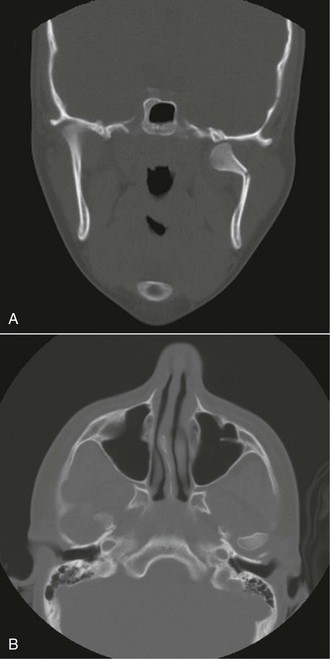
For the current patient, a CT scan was obtained as the initial study. It demonstrated a right subcondylar fracture on coronal and axial views (Figure 8-5). A plain wrist film was also obtained, which revealed a right-sided fracture of the distal radius (Colles fracture).

Treatment

The treatment of fractures of the mandibular condyle is one of the most widely debated topics in the maxillofacial literature. Several variables should be considered when determining treatment and predicting the prognosis, including the level of fracture, degree and direction of displacement, age and medical status of the patient, concomitant injuries, and status of the dentition. Assael has developed a comprehensive list of considerations affecting treatment selection and outcome, all of which should be included in the evaluation of the patient prior to the institution of therapy. Although comprehensive discussion of these considerations is beyond the scope of this chapter, the variables can be divided into patient, surgeon, and third-party categories. Age, gender, medical status, compliance, associated injuries, and fracture type are a few of the patient-specific variables. The surgeon’s ability and resources, in addition to resources to cover the expense of treatment, are also pertinent to successful treatment.

The primary goal in the treatment of any fracture is adequate stabilization that allows for fracture healing and primary osseous union. In the treatment of mandibular condyle fractures, the goals of treatment are:

Preinjury alignment of the mandibular condyle within the glenoid fossa is not essential for adequate rehabilitation after mandibular condyle fractures. The pull of the lateral pterygoid muscle characteristically displaces the condyle anteriorly and medially; therefore, closed reduction (more correctly termed “closed treatment”) typically does not reduce the condyle into its original position.

The treatment options are categorized into surgical and nonsurgical modalities. Surgical treatment includes open reduction with or without internal fixation; however, most agree that if an open approach is taken, fixation should be applied. Endoscopic reduction and fixation of condylar fractures has gained popularity during the past decade. The use of this technique requires familiarity with the endoscope and the ability to convert the procedure to an open method if endoscopic reduction fails to successfully complete the procedure. The options for nonsurgical treatment include closed reduction (closed treatment) with maxillomandibular fixation (CR-MMF) and dietary modification with ROM exercises. In the treatment of facial fractures, patients older than 10 years are treated in a manner similar to that for adults; however, it is rarely advocated that children and teenagers undergo open reduction of condylar fractures. A soft diet with mobilization is the treatment of choice in patients 15 years old or younger. If the occlusion is unstable and not reproducible, a short period of intermaxillary fixation (2 weeks) can be advocated.

For the current patient, the occlusion was reestablished easily with minimal manipulation, and after extensive discussion of procedures, alternatives, risks, and benefits, the patient was placed in maxillomandibular fixation (MMF) for 4 weeks, After the 4 weeks, an aggressive post-treatment physiotherapy program was instituted, with active and passive range of motion exercises. Return to full function occurred within 4 weeks of release from MMF. There were no postoperative complications and, the patient returned to full function with stable and repeatable occlusion.

Complications

The complications of treating fractures of the mandibular condyle are well described in the literature and are often used as the basis of comparison for surgical and nonsurgical treatment. One of the most severe late complications can be temporomandibular joint (TMJ) ankylosis (fusion between the mandibular condyle and the glenoid fossa). Patients with TMJ ankylosis often have a history of facial trauma. Prevention of ankylosis was discussed by Zide and Kent in 1983. They advocated appropriate physiotherapy early in the phase of nonsurgical treatment. Other types of late mandibular dysfunction have been cited as complications of closed reduction, including chronic pain, malocclusion, internal derangement, asymmetry, limited mobility, and gross radiographic abnormalities (however, radiographic abnormalities in the absence of pain or functional impairment have no clinical significance). Long-term complications of open reduction and internal fixation (ORIF) are scar perception, facial nerve palsy/paralysis, loss or failure of fixation, Frey’s syndrome, avascular necrosis, TMJ dysfunction, and facial asymmetry. The early complications are few and can include early failure of fixation, malocclusion, pain, and infection.

Stay updated, free dental videos. Join our Telegram channel

Jan 12, 2015 | Posted by in Oral and Maxillofacial Surgery | Comments Off on 8: Craniomaxillofacial Trauma Surgery

VIDEdental - Online dental courses

Get VIDEdental app for watching clinical videos