7: Vesiculobullous Disorders

Vesiculobullous Disorders

There are a variety of oral lesions which clinically present as vesiculobullous (VB) lesions. Although the lesions start as vesicles or bullae they rupture early and appear as ulcerated or erosive areas. As a result they are better called ulcerovesiculobullous diseases. In this chapter common oral VB lesions will be discussed.

A vesicle is defined as a fluid-filled elevated lesion, less than 1 cm in diameter. A bulla is a fluid-filled elevated lesion greater than 1 cm in diameter.

Classification of Vesiculobullous Lesions

Acute and chronic vesiculobullous lesions (Table 1)

Table 1

Comparison between acute and chronic vesiculobul-lous lesions

  Acute–VB lesions Chronic–VB lesions
Duration Short Long
Age Young Middle aged–older
Etiology Allergy, burns, viruses Autoimmune
Examples Herpes simplex infections Pemphigus
  Chicken pox Bullous pemphigoid
  Herpes zoster Cicatricial pemphigoid
  Herpangina Bullous lichen planus
  Hand, foot and mouth disease Chronic herpes simplex
  Erythema multiforme Linear IgA disease

II Based on the clinical presentation


III Histopathological classification

    Intraepithelial vesiculobullous lesions

    HSV infection

    Varicella infection

    Herpangina

    Hand, foot and mouth disease

    Pemphigus

    Familial benign chronic pemphigus

    Epidermolysis bullosa

    Mucosal erythema multiforme

    Subepithelial vesiculobullous lesions

    Bullous pemphigoid

    Cicatricial pemphigoid

    Epidermolysis Bullosa

    Dermal Erythema multiforme

    Dermatitis herpetiformis

    Linear IgA disease

IV Based on whether the lesions are infectious or non-infectious

    Infectious VB lesions

    Herpes simplex infections

    Varicella infections

    Herpangina

    Hand, foot and mouth disease

    Non-infectious VB lesions

    Pemphigus

    Paraneoplastic pemphigus

    Bullous pemphigoid

    Cicatricial pemphigoid

    Erythema multiforme

    Dermatitis herpetiformis

    Epidermolysis bullosa acquisita

    Linear IgA disease

Predominantly Vesicular Lesions

Herpes Viruses

The word herpes is derived from the Greek word ‘herpein’ which literally means to creep. The word signifies the creeping or spreading nature of the skin lesions caused by many of the herpes viruses.

It is estimated that out of the 80 known herpes viruses, at least eight are known to infect human beings. The herpes viruses that are known to cause infection in humans are herpes simplex virus (HSV 1 and 2), varicella zoster virus (VZV), cytomegalovirus (CMV), Epstein-Barr virus (EBV), human herpes virus 6 (HHV-6), human herpes virus 7 (HHV-7) and human herpes virus 8 (HHV-8). These viruses are usually transmitted from host to host by direct contact or through saliva and genital secretions. Herpes viruses are shed in the saliva of asymptomatic hosts.

When an individual comes in contact with the virus, primary infection is seen. Subsequently these viruses establish latency in the host. The site of latency for each form of herpes virus is different (Table 2).

Table 2

Site of latency of viruses

Name of virus Site of latency
HSV 1 & 2, VZV Sensory nerve ganglia
CMV Lymphocytes, salivary gland tissue (rarely)
EBV B lymphocytes and salivary gland tissue
HHV-6, HHV-7 CD4 lymphocytes
HHV-8 Still unknown but believed to be associated with B lymphocytes circulating in hematopoietic system

Herpes Simplex Virus (HSV) Infections

Etiology and pathogenesis

Herpes simplex virus infection is caused by two types of DNA viruses namely HSV 1 and HSV 2. Characteristically, HSV 1 is responsible for infections involving the oropharyngeal regions, dermatitis and meningoencephalitis (infections above the waist) and HSV 2 is responsible for anogenital infections (infections below the waist). However, changing sexual practices have shown that these viruses may not necessarily be responsible for infections of specific sites.

In contrast to other viruses HSV needs physical contact to transfer infection. The incubation period ranges from 2 days to 3 weeks. After the primary infection the virus hides within the trigeminal ganglia and becomes activated when the environment is conducive for viral replication. Approximately 90% of USA population is seropositive for antibodies to HSV. Of this group only 1% exhibit primary herpetic infections and 30–40% demonstrate recurrent infections. Primary HSV infections are usually seen after 6 months of age and peak at about 2–3 years of age.

Other specific HSV infections include herpetic whitlow, herpetic gladiatorum, herpetic meningoencephalitis, herpetic conjunctivitis, herpetic eczema (Kaposi’s varicelliform eruption) and disseminated herpes simplex of newborn.

Clinical features

Primary HSV infection

Only 1% of the population in USA exhibit all signs and symptoms of primary infection. It usually affects young children and adolescents, and occasionally young adults. The primary HSV infection seldom occurs in the first 6 months of life as the infant is protected by the maternal antibodies that are still circulating in the newborn.

Primary herpetic gingivostomatitis is characterized by fever, malaise, anorexia, irritability and regional lymphadenopathy (especially, submandibular and superficial cervical group of nodes are involved). Subsequently the mouth becomes sore and the individual may complain of burning sensation in the mouth and difficulty in swallowing. Apart from the gingiva, the buccal mucosa, palate, tongue, tonsillar and pharyngeal region may be affected.

The gingiva is erythematous, boggy and bleeds spontaneously or on the slightest of provocation. As the disease advances multiple tiny to moderately large (few mm to almost a centimeter in size) yellow colored vesicles develop that rupture to form shallow painful ulcers that are usually covered by a grayish colored membrane. These ulcers are bound by an erythematous halo.

Generally in about 2 weeks the lesion heals without scarring. Immediately after the resolution of the primary lesion, the virus travels along the nerve pathway and lies dormant in the nerve ganglia regional to the site of the primary infection. Within the regional ganglion the virus incorporates its DNA into the host’s DNA thereby establishing the beginning of a life-long affliction. The trigeminal ganglion is usually the site for dormancy for HSV-1 and HSV-2.

Herpetic Whitlow and Herpes Gladiatorum

Herpetic whitlow is an intense painful infection affecting the fingers or toes of children, adults and healthcare workers. It typically involves the terminal phalanx of the index and thumb fingers of children and healthcare workers. It is believed that HSV-1 is responsible for 60% of the known cases of herpetic whitlow and 40% are caused by HSV-2. In children, most cases can be attributed to autoinoculation of HSV-1 (infants suffering from acute herpetic gingivo-stomatitis who engage in thumb/finger sucking). In young adults and elderly, herpetic whitlow is caused by autoinoculation of HSV-2, usually due to digital-genital contact. Literature review reveals few reports describing herpetic whitlow affecting the toe. Ozawa et al (2004) reported an elderly patient who exhibited herpetic whitlow of the toe.

The lesions of herpetic whitlow are usually preceded by prodromal symptoms of burning or tingling sensation and pruritus of the affected digit and sometimes the whole limb. As the lesion progresses, the involved digit reveals multiple tiny vesicles which are extremely tender. The surrounding area appears edematous and erythematous. The lesion may rupture to form shallow ulcers and subsequently heal in about 10 days time.

Similar vesicular lesions seen on the bodies of sportsmen (especially contact sports) and wrestlers is referred to as herpes gladiatorum.

Investigations

Typical clinical features will usually suffice to make a clinical diagnosis. However for definitive diagnosis cytological smears, antibody titers and viral isolation can be used.

Cytological smear requires deroofing a fresh vesicle, followed by scraping of the base of the lesion and the smear made should be stained with Papanicolaou’s, Wright’s or Giemsa’s stain.

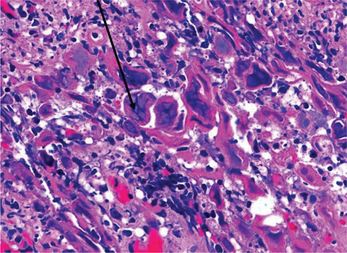
The characteristic findings include the presence of multinucleated giant cells and intranuclear viral inclusion bodies such as Lipschutz bodies or Cowdry Type A (ovoid, amorphous, eosinophilic bodies that exhibit peri inclusion halo that is caused by the peripheral displacement of the nucleolus and the nuclear chromatin). The cells exhibit ballooning degeneration of the nucleus.

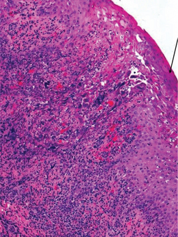
On biopsy the microscopic features include an ulcerated epithelium with large keratinocytes showing glassy giant marginated nuclei (Tzanck cells) (Figures 1 and 2).
Alternatively viral isolation can be done. It is one of the most definitive methods of identifying HSV.

Serological tests can be used to detect antibodies in patient suffering from primary HSV. For the test, blood sample should be taken in the initial 3 days of an acute infection. Alternatively a convalescent blood sample can be obtained 4 weeks after the primary infection. It is believed that the antibody titer should increase by four times in convalescent serum sample for the diagnosis of the primary infection.

Recurrent Herpes Infections

Almost 30–40% of the individuals exposed to HSV infection will exhibit at least one episode of recurrent herpes infections. Recurrent infections can present either as recurrent herpes labialis or recurrent intraoral herpes.

Various factors have been known to trigger these recurrent attacks such as stress, trauma, exposure to sunlight and menstruation. These recurrent infections usually have a milder course.

Herpetic Labialis

This is the most common secondary herpetic lesion, usually following exposure to UV light or extreme cold. Groups of vesicles appear at the vermillion border, and rupture may result in crusty lesions. The lesions are tingly and painful, normally healing within 2 weeks. The lesions are highly contagious at the weeping stage.

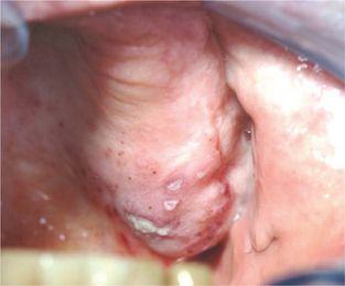
Intraoral herpetic lesions are evident as clusters of vesicles (Figure 3). These vesicles rupture, leaving ulcerations on bound mucosa, usually within 3 days of recent dental treatment. These are also present with prodromal symptoms such as paresthesia, swelling, burning and tingling.
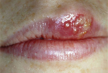
Secondary herpetic lesions recur, and the recurrence rate is > 6 times/year. Secondary intraoral herpetic lesions are seen in patients who are immunocompromised from HIV/AIDS, pre-transplant and post-transplantation surgery or post-chemotherapy. Intraorally ulcerations occur on bound and mobile mucosa.

Yeo et al (2002) reported the presence of herpes labialis in musicians who play woodwind instruments and brass instruments such as the flute, saxophone, trumpet and horn. They suggest that the herpetic lesions are precipitated by mechanical trauma of the lips and stress suffered by the musicians preparing for a performance. It is commonly seen that the woodwind players tend to have lesions on the lower lip and brass instrument players have herpetic lesions on the upper lip.

Treatment

The aims of managing HSV infections are to inhibit autoinoculation and transmission. The management is targeted at symptomatic relief. Topical and systemic antivirals like acyclovir can be used (Table 3).

Table 3

Treatment regimen for different kinds of HSV infection

  Immunocompetent Immunosuppressed
Primary herpetic gingivostomatitis—children Symptomatic—no aspirin or NSAIDs Acyclovir capsules 200 mg or valacyclovir caplets 500 mg
Primary herpetic gingivostomatitis—adult Symptomatic, aspirin and NSAIDs, magic mouthwash Acyclovir capsules 200 mg, 5 times daily for 10 days or valacyclovir caplets 500 mg, 2 caplets twice daily for 5 days
Recurrent herpetic labialis Abreva, Denavir 5%, valacyclovir 1,500 mg, single dose or valacyclovir 750 mg, twice daily for 1 day started within 1 hour of onset of prodromal signs Valacyclovir caplets 500 mg, 4 caplets taken at prodromal symptoms and 4 caplets 12 hours later
Recurrent intraoral herpes Acyclovir 200 mg 5 times a day for 10 days  

To be effective all treatment should be started within 72 hours of initial disease presentation. Patient compliance is shown to be better with fewer and shorter doses (Whitley, 2006).

Varicella Zoster Infections

The primary and secondary infections are caused by VZV belonging to Herpedes family. Chicken pox is the primary infection and herpes zoster is caused by reactivation of the VZV. This infection occurs due to inhalation and primary infection occurs in children. Reactivation occurs when the patient is stressed or immunosuppressed. It usually affects dermatomes in the trunk, head or neck.

Clinical features

Secondary infection (herpes zoster)

Herpes zoster is the reactivated form of the VZV. It is also referred to as shingles. The word shingles is reportedly derived from the Latin word ‘cingulum’ which means ‘girdle’. The term girdle is apt to describe the lesion of herpes zoster as it presents as a unilateral rash that can wrap around the waist or torso like a girdle. The word zoster is Greek word that refers to a belt like object that is used by warriors to hold the armor in place at the waist.

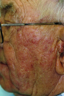
Secondary varicella zoster infection occurs in adults with altered immune status or under stress and is unilateral. The main distinguishing feature of VZV is unilateral and involves one of the branches of the trigeminal nerve. Clinically tingling and vesiculation similar to herpes simplex are seen. Clinical appearance of these lesions is very similar to herpes simplex except for the unilateral location (Figures 4 and 5). Patients may complain of mild to severe pain that may exacerbate on the slightest of touch. The dermal lesions usually resolve in about a week forming a scab.

Complications Associated with Herpes Zoster

The most common complication associated with HZ is the development of postherpetic neuralgia. In this condition the pain continues to persist even after the herpetic lesions have resolved. The pain is typically presented as sharp or burning pain. Postherpetic neuralgia is usually seen in individuals over the 5th decade of life.

Herpes zoster is generally unilateral. However in immunocompromised individuals or an associated malignancy, herpes zoster can exhibit a generalized involvement. Other relatively uncommon complications involve encephalitis, peripheral nerve palsies and myelitis.

Zoster ophthalmicus is seen when herpes zoster involves the ophthalmic ganglion of the trigeminal nerve. Patients present with painful inflammatory condition of the eye along with impaired vision or transient blindness.

Some individuals exhibit only the prodromal symptoms of pain and paresthesia without the development of visible cutaneous rash. This phenomenon is referred to as ‘zoster sine herpete’.

Ramsay Hunt syndrome is one of the rare manifestations of HZV infection where geniculate ganglion is involved. The facial and auditory nerves can be involved. The condition is characterized by facial paralysis, pain and vesicles in the external auditory canal and pinna of the ear and the oral cavity. Other associated findings are tinnitus, vertigo and ipsilateral hearing loss.

Hand, Foot and Mouth Disease

The term hand, foot and mouth (HFM) disease was used for the first time in 1960 following an outbreak of a relatively mild febrile condition associated with papular and vesicular lesions on the dermis and the oral cavity in Birmingham, England.

Etiology

Enteroviruses are known to cause HFM disease. The enteroviruses that have been implicated are Coxsackie virus serotype A 16, Coxsackie virus serotype A 4 to 7, 9, 10 and Coxsackie virus serotype B 1 to 3 and 5. Asia Pacific region including regions like Singapore, Japan, Indonesia, Malaysia and Taiwan in the 1970s reported severe forms of HFM disease resulting in a widespread epidemic. The cause for this epidemic was Enterovirus-71 A, B, C (EV 71). The EV-71 apart from causing HFM disease can also cause flaccid paralysis, myocarditis, pulmonary hemorrhage, encephalitis and meningitis.

Deshpande et al (2003) isolated a strain of EV-71 from the stool of a child suffering from acute flaccid paralysis following administration of oral polio vaccine. They termed this isolated strain as EV 71 D genotype.

Sasidharan et al (2005) studied 81 children (age group of 7 months to 8 years) who presented with papulovesicular exanthems in Calicut, India. In their study 19 children showed a significant rise in the titers of IgM antibody against EV-71.

Infection usually occurs by the fecal-oral route, leading to viremia and invasion of the skin and mucosa. The incubation period is approximately 3–7 days.

Herpangina

Herpangina was first described by John Zahorsky in 1920. Herpangina is primarily caused by coxsackie virus A 1–10, 16, or 22. Other viruses that may cause herpangina are coxsackie virus B 1–5, enterovirus 71 and echovirus 3, 6, 9, 11, 16, 17, 22, 25, and 30. Herpangina occurs in epidemics and usually occurs in the summer months. It usually affects young children and adolescents. The mode of spread is the fecal-oral route. The incubation period varies from 1 to 10 days and usually lasts 4 days.

Dermatitis Herpetiformis

Dermatitis herpetiformis (DH) is a rare, chronic, pruritic immunobullous disorder of the skin that is associated with gluten hypersensitivity. The condition is characterized by the subepidermal deposition of IgA antibody against the tissue transglutaminase. Antibody deposition leads to the formation of dense neutrophilic abscesses, subepidermal vesicles and bullae. Spurkland et al (1997) described the genetic basis for DH. They showed that DH and celiac disease are associated with class II HLA alleles A1*0501 and B1*02, which encode the HLA-DQ2 heterodimer. Salmela et al (2001) showed that there was an upregulation of metalloelastase in both intestinal and lesional skin.

Stay updated, free dental videos. Join our Telegram channel

Jan 12, 2015 | Posted by in Oral and Maxillofacial Radiology | Comments Off on 7: Vesiculobullous Disorders

VIDEdental - Online dental courses
Get VIDEdental app for watching clinical videos