Chapter 7
Endodontic Retreatment of Teeth Restored with Adhesive Techniques and Fibre Posts
Aim
To describe the procedure for the removal of a cemented fibre post from the root canal.
Outcome
At the end of this chapter, the reader should be able to identify the risks and be knowledgeable on methods for the removal of fibre posts from root canals.
Introduction
The term “post-treatment disease” includes all persistent, recurrent and emerged apical periodontitis associated with endodontically treated teeth (Fig 7-1a–c). This condition, caused by microbial infection of the root canal system, is frequently associated with a periapical radiolucency. It affects between 5–35% of root canal treated teeth.
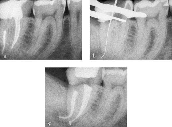
Fig 7-1 (a) A lower right second molar affected by post-endodontic disease: note the fibre post inserted in the distal root. (b) After removal of the fibre post, the canal is negotiated and the working length determined. (c) Awaiting final coronal restoration.
Coronal leakage associated with secondary caries, debond and loss of coronal restorations is often implicated in post-treatment endodontic disease. After removal of defective restorations and secondary caries, an endodontic retreatment is often attempted in order to eliminate infection from the root canal space.
Post removal is often considered a major challenge to endodontic retreatment, in particular if the post is bonded with adhesive cement. Added to this, few practitioners have experience of fibre post removal, and the challenge is often considered to be daunting.
Diagnosis
Radiographic identification of contemporary fibre posts is straightforward, but earlier, radiolucent posts may be difficult to identify. Under such a circumstance, an apparently empty canal may be surrounded by a thin, radiopaque line of resin cement.
A decision must often be made whether to remove the crown or work through it. Working through a crown may present special challenges, since instruments will need to be inserted along the long axis of the post to be removed, and this may be impossible through a conservative coronal access. Although the canal which contains the post may be readily identified, other canals which may also need retreatment may be difficult to identify within a mass of tooth-coloured composite, in a deep cavity.
The wise option is usually to remove the crown as part of the package of care.
Armamentarium
As with many procedures in dentistry, the correct tools are helpful. These may include:
-
instrumentation for crown and bridge removal
-
magnification
-
rotary instruments
-
ultrasonic equipment and tips.
Magnification aids include simple loupes, with or without supplementary lighting, and operating microscopes (Figs 7-2 to 7-5). An operating microscope greatly facilitates retreatments.
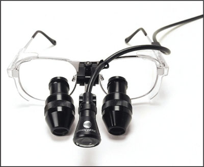
Fig 7-2 Loupes with an LED lamp.
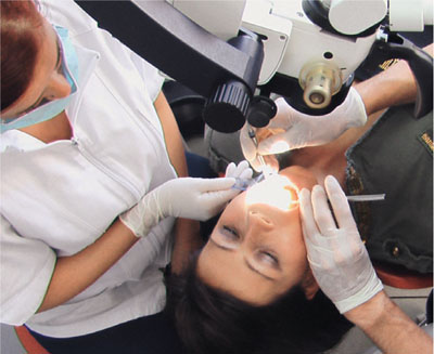
Fig 7-3 A typical session of endodontic microintervention.
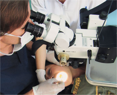
Fig 7-4 Optimal positioning of operator, assistant and patient while working on a lower tooth under magnification.
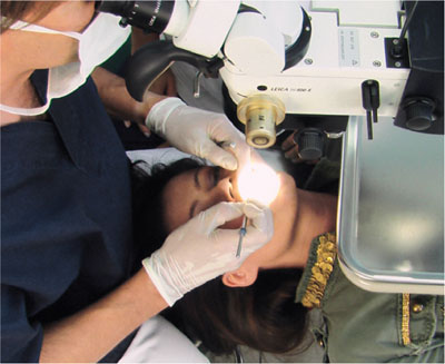
Fig 7-5 Positioning for a maxillary tooth.
Microscopes have not yet become established in mainstream general practice, owing largely to the capital cost, but also to the training and familiarisation required by both the dentist and the chair-side personnel. Loupes are, however, widely employed in practice.
Working with magnification
Operator position, patient position and role of the dental nurse may be altered in practices which adopt magnification.
Working with a microscope, the operator position of choice is usually behind the patient’s head, in the “12 o’clock” position. The patient may be supported in neck extension, in particular when working on lower teeth. The dental nurse needs her own binocular and should be trained in “four-handed work under magnification”.
Principles of Post Removal
Unlike metallic posts, fibre posts are not readily removed by simple lute-disruption: by means of ultrasonic and pulling techniques. Fibre posts must be cut out wi/>
Stay updated, free dental videos. Join our Telegram channel

VIDEdental - Online dental courses


