7: Connective Tissue Lesions

Connective Tissue Lesions

Connective tissue lesions comprise a large and diverse number of entities ranging from reactive conditions to neoplasms. Reactive conditions are derived from mesenchymal cells and are represented by fibrous hyperplasias or exuberant proliferations of granulation tissue. Tumors of connective tissue elements are heterogeneous and form a complex collection of diseases. Prediction of biological behavior from histology alone is problematic and is reflected in the difficulties in grading individual tumors. Traditionally, tumors of connective tissues have been classified on a model of presumed histogenetic lineage. Hence neoplasms are subdivided into tumors of fibrous, fibrohistiocytic, myofibroblastic, vascular, neural, muscular, adipose, and other types of tissue. Increasingly, it is becoming evident that many tumors do not arise from their mature, differentiated counterparts, because soft tissue tumors can arise in sites that are devoid of their mature tissue counterpart. For example, liposarcomas often arise at sites where no adipose tissue is present, and rhabdomyosarcomas often arise at sites that contain no striated muscle. It is likely that soft tissue tumors arise from primitive progenitor cells that can develop along any differentiation pathway, which is dictated by the expression of specific differentiation genes. However, for the purposes of describing these entities, a histogenetic classification has been maintained here.

Fibrous Lesions

Reactive Hyperplasias

Reactive hyperplasias comprise a group of fibrous connective tissue lesions that commonly occur in oral mucosa secondary to injury. They represent a chronic process in which exuberant repair (granulation tissue and scar) follows injury. As a group, these conditions present as submucosal masses that may become secondarily ulcerated when traumatized during mastication. Their color ranges from lighter than the surrounding tissue (because of a relative increase in collagen) to red (because of an abundance of well-vascularized granulation tissue). Because nerve tissue does not proliferate with reactive hyperplastic tissue, these lesions are painless. The reason for the exuberant repair is unknown. Treatment generally consists of surgical excision and removal of the irritating factor(s).

Although these lesions are all pathogenically related, different names or subdivisions have been devised because of variations in the anatomic site, clinical appearance, or microscopic picture. Those lesions that present as prominent red masses are discussed in Chapter 4.

Peripheral Fibroma

Clinical Features

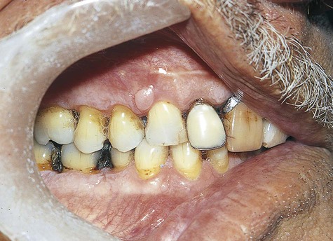
By definition, peripheral fibroma is a reactive hyperplastic mass that occurs on the gingiva and is believed to be derived from connective tissue of the submucosa or periodontal ligament (Figure 7-1). It may occur at any age, although it does have a predilection for young adults. Females develop these lesions more commonly than do males, and the gingiva anterior to the permanent molars is most often affected.

Peripheral fibroma may present clinically as a stalked (pedunculated) or a broad-based (sessile) mass that is similar in color to surrounding connective tissue. Ulceration may be noted over the summit of the lesion. It rarely causes erosion of subjacent alveolar bone.

Histopathology

Peripheral fibroma is a form of fibrous hyperplasia that may also be called hyperplastic scar. It is highly collagenous and relatively avascular, and it may contain a mild to moderate chronic inflammatory cell infiltrate. This lesion is basically the gingival counterpart to traumatic fibroma occurring in other mucosal regions.

Microscopically, several subtypes of this lesion have been identified. These are essentially of academic interest because the biological behavior and treatment of these microscopic variants are the same.

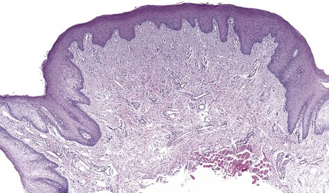
Peripheral ossifying fibroma is a gingival mass in which islands of woven (immature) bone and osteoid are seen. The bone is found within a lobular proliferation of plump, benign fibroblasts. Chronic inflammatory cells tend to be seen around the periphery of the lesion (Figure 7-2). The surface is typically ulcerated.
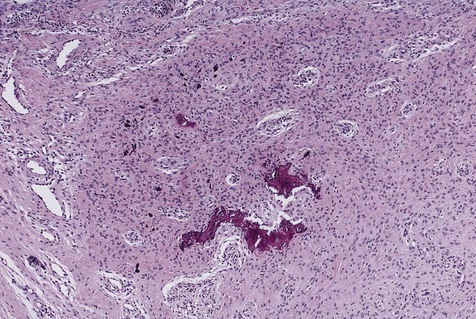
Peripheral odontogenic fibroma is a gingival mass composed of well-vascularized, nonencapsulated fibrous connective tissue. The distinguishing feature of this variant is the presence of strands of odontogenic epithelium, often abundant, throughout the connective tissue. Amorphous hard tissue resembling tertiary (reactive) dentin, so-called dentinoid, may also be present. The lesion is usually nonulcerated.

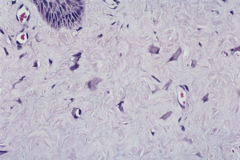
Giant cell fibroma is a fibrous hyperplasia in which many of the mesenchymal cells are relatively larger than normal fibroblasts (giant cells) and assume a stellate shape. Immunohistochemical studies have shown that most of these stellate cells are fibroblasts (a few factor XIIIa–positive dendritic cells are also typically present) (Figure 7-3). These same peculiar stellate cells can be found in focal fibrous hyperplastic lesions throughout the oral mucosa and occasionally on the skin (fibrous papule). One form of this lesion is known as retrocuspid papilla of the mandible.

Focal Fibrous Hyperplasia

Clinical Features

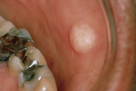
No gender or racial predilection for the development of this intraoral lesion has been noted. It is a very common reactive hyperplasia that is typically found in frequently traumatized areas, such as the buccal mucosa, the lateral border of the tongue, and the lower lip (Figure 7-4). It is a painless, broad-based swelling that is paler in color than the surrounding tissue because of its relative lack of vascular channels. The surface may occasionally be traumatically ulcerated, particularly in larger lesions. Lesions have limited growth potential and do not exceed 1 to 2 cm in diameter.

Multiple fibromas may be part of a rare autosomal-dominant syndrome known as Cowden’s syndrome or multiple hamartoma syndrome. Many organ systems, such as the mucosa, skin, breast, thyroid, and colon, may be affected. Frequently encountered abnormalities include numerous oral fibromas and papillomas; cutaneous papules, keratoses, and trichilemmomas; benign and malignant neoplasms of the breast and thyroid; and colonic polyps. The underlying genetic problem appears to be related to germline mutations of the tumor suppressor gene PTEN found on chromosome 10q23.

Denture-Induced Fibrous Hyperplasia

Clinical Features

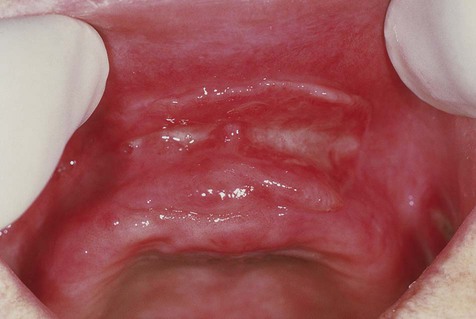
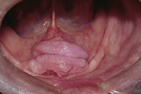
Denture-induced fibrous hyperplasia is a common lesion that occurs in the vestibular mucosa and less commonly along the mandibular lingual sulcus where the denture flange contacts tissue (Figures 7-6 and 7-7). As the bony ridges of the mandible and the maxilla resorb with long-term denture use, the flanges gradually extend farther into the vestibule. There, chronic irritation and trauma may incite an overexuberant fibrous connective tissue reparative response. The result is the appearance of painless folds of fibrous tissue surrounding the overextended denture flange.

Generalized Gingival Hyperplasia

Etiology

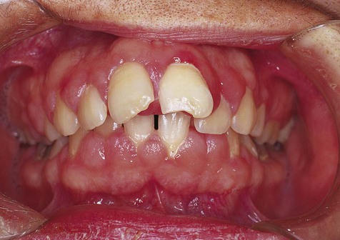
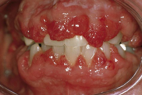
In generalized gingival hyperplasia, overgrowth of the gingiva may vary from mild enlargement of the interdental papillae to such severe uniform enlargement that the crowns of the teeth may be covered by hyperplastic tissue (Box 7-2). Uniform or generalized gingival fibrous connective tissue hyperplasia may be due to one of several etiologic factors. Most cases are nonspecific and are the result of an unusual hyperplastic tissue response to chronic inflammation associated with local factors such as plaque, calculus, or bacteria. Why only some patients have a propensity for the development of connective tissue hyperplasia in response to local factors is unknown. Recent studies have reported a possible role for keratinocyte growth factor (a member of the fibroblast growth factor family) in this condition.

Other conditions such as hormonal changes and drugs can significantly potentiate or exaggerate the effects of local factors on gingival connective tissue. Hormonal changes that occur during pregnancy and puberty have long been known to be associated with generalized gingival hyperplasia. This hyperresponsiveness during pregnancy has led to the infrequently used and inappropriate term pregnancy gingivitis. Altered hormonal conditions act in concert with local irritants to produce the hyperplastic response. It is questionable whether significant gingival enlargement during periods of hormonal imbalance would occur in individuals with scrupulous oral hygiene.

Phenytoin (Dilantin), a drug used in the control of seizure disorders, is a well-known etiologic factor in generalized gingival enlargement. It is thought that phenytoin causes impaired collagen degradation through suppression of matrix metalloproteinases (MMPs)/tissue inhibitor of metalloproteinase-1 (TIMP-1) and α2β1-integrin–mediated endocytosis. The extent or severity of so-called Dilantin hyperplasia is dependent on the presence of local factors such as plaque and calculus. The effects of time and dose of the drug on gingival tissue are not clear. Reported prevalence has ranged from 0% to 80%, depending on the investigator’s clinical criteria and the number of patients observed. A value of 50% is generally accepted as the probable prevalence. In any event, the fact that not all patients taking phenytoin develop gingival hyperplasia indicates that some patients are predisposed to the development of this condition. It has only rarely been described in edentulous patients and in children before tooth eruption.

Cyclosporine, the immunosuppressant drug that is used to modulate T-lymphocyte function in transplant recipients and in patients with various autoimmune diseases, has also been linked to fibrous hyperplasia of the gingiva. The cause of this condition is not known, but edema secondary to increased sulfated-glycosaminoglycan synthesis by fibroblasts may play an important role. Not all patients are affected (10% to 70%), and local factors have a synergistic role. Unlike phenytoin-related hyperplasia, cyclosporine-induced hyperplasia has been reported to be a reversible process following cessation of drug use.

Nifedipine and other calcium channel blockers used in the treatment of cardiac angina, arrhythmias, and hypertension are known to contribute to gingival hyperplasia. The process mimics phenytoin-related hyperplasia but, similar to cyclosporine-induced gingival hyperplasia, appears to be reversible.

Gingival enlargement is also known to occur in patients with leukemia, especially those with the chronic monocytic form. This is believed to be a result of infiltration of the gingival soft tissues by malignant white blood cells. It may be modulated by local factors such as plaque and calculus; because of the bleeding tendency associated with leukemia, patients may be unable or reluctant to practice correct oral hygiene, resulting in the accumulation of plaque and debris. This accumulation may provide the inflammatory stimulus for connective tissue hyperplasia.

Some rare types of gingival hyperplasia that occur in early childhood have a hereditary basis. The best recognized is hereditary gingival fibromatosis, which clinically can resemble Dilantin-induced gingival hyperplasia. Patients with other rare syndromes such as Zimmerman-Laband, Cross’, Rutherfurd’s, Murray-Puretic-Drescher (juvenile hyaline fibromatosis), and Cowden’s syndromes can develop varying degrees of fibrous gingival hyperplasia.

Clinical Features

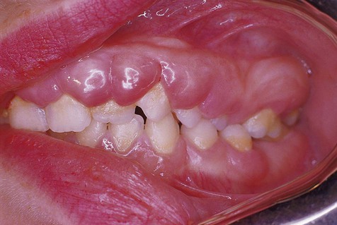
The clinical feature common to the variously caused gingival hyperplasias is an increase in bulk of the free and attached gingiva, especially the interdental papillae (Figures 7-8 to 7-10). Stippling is lost, and gingival margins become rolled and blunted. The consistency of the gingiva ranges from soft and spongy to firm and dense, depending directly on the density and amount of fibrosis. A range of color from red-blue to lighter than surrounding tissue is also seen; this varies with the severity of the inflammatory response as well. Generally, hyperplasias associated with nonspecific local factors and hormonal changes appear more inflamed clinically than drug-induced and idiopathic forms. The idiopathic type is particularly dense and fibrous, with relatively little inflammatory change.

Neoplasms

Solitary Fibrous Tumor

Solitary fibrous tumor is a benign proliferation of spindle cells of disputed but probable fibroblastic origin (Box 7-3). This lesion was first described as a tumor of the pleura and has subsequently been described at many other sites. Oral lesions are seen in adults and present as submucosal masses predominantly in the buccal mucosa (Box 7-4).

Microscopically, lesions are circumscribed and are composed of a “patternless” proliferation of spindle cells (Figure 7-11). Some areas may suggest neurofibroma or Schwannoma, whereas others may suggest hemangiopericytoma or leiomyoma. Tumor cells characteristically stain positive for CD34 (90% to 95% of cases), CD99 (70%), and Bcl-2 (20% to 35%) by immunohistochemistry. Many factor XIIIa–positive cells may be found. Immunohistochemistry has permitted a better understanding of this entity and more reliable identification; therefore many oral tumors previously diagnosed by light microscopy as other soft tissue neoplasms such as leiomyoma, hemangiopericytoma, and benign fibrous histiocytoma probably represent solitary fibrous tumor.
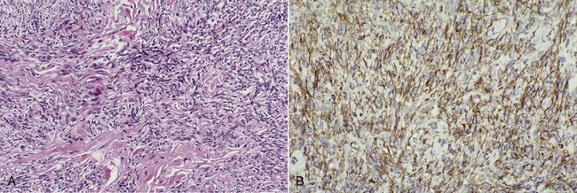
Treatment consists of surgical excision. Although most cases are benign, the behavior of solitary fibrous tumors is unpredictable. Approximately 10% to 15% behave aggressively, warranting long-term follow-up. It is important to note that correlation between histologic features and overall behavior is poor.

Giant Cell Angiofibroma

Myxoma

Histopathology

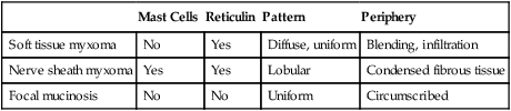
Oral myxomas are not encapsulated and may exhibit infiltration into surrounding soft tissue. Dispersed stellate and spindle-shaped fibroblasts are found in a loose myxoid stroma. Soft tissue myxomas may be confused with other myxoid lesions, such as nerve sheath myxoma and oral focal mucinosis (Table 7-1).

TABLE 7-1

MUCOSAL MYXOID LESIONS: MICROSCOPIC DIFFERENTIATION

  Mast Cells Reticulin Pattern Periphery
Soft tissue myxoma No Yes Diffuse, uniform Blending, infiltration
Nerve sheath myxoma Yes Yes Lobular Condensed fibrous tissue
Focal mucinosis No No Uniform Circumscribed

image

Nerve sheath myxoma arises from the endoneurium of a peripheral nerve. This lesion typically exhibits lobulated mucoid tissue containing stellate and spindle-shaped cells. Condensed connective tissue, representing perineurium, surrounds the lesion. With special stains, a fine reticulin network is seen throughout. Mast cells are characteristically present in this lesion and neural markers such as S-100 are expressed by the tumor.

Oral focal mucinosis represents the mucosal counterpart of cutaneous focal mucinosis. The lesion appears as a well-circumscribed area of myxomatous connective tissue in the submucosa. It contains no mast cells and no reticulin network, except that which surrounds supporting blood vessels. Unlike the nerve sheath myxoma, neural markers such as S-100 are negative.

Nasopharyngeal Angiofibroma

Clinical Features

Nasopharyngeal angiofibroma is also known as juvenile nasopharyngeal angiofibroma because of its almost exclusive occurrence in the second decade of life. For this uncommon to rare neoplasm that nearly always affects boys, it is thought that sex hormones variably play a role in its pathogenesis, with expression of androgen, testosterone, and dihydrotestosterone receptors. This lesion characteristically produces a mass in the nasopharynx that arises along the posterolateral wall of the nasal roof and over time leads to obstruction or epistaxis that may, on occasion, be severe. Rarely, this lesion may present intraorally, causing palatal expansion or inferior displacement of the soft palate, which appears blue because of the intense vascularity of the lesion. It generally can be described as benign and slow-growing but unencapsulated and locally invasive. On occasion, it may exhibit aggressive clinical behavior, characterized by direct extension into the bones of the midface and the skull base. The symptom triad includes recurrent epistaxis, nasal obstruction, and mass effect within the nasopharynx.

Nodular Fasciitis

Clinical Features

Nodular fasciitis, also known as pseudosarcomatous fasciitis, is a well-recognized entity representing a myofibroblastic proliferation. A closely related lesion known as proliferative myositis occurs in muscle. The cause of this proliferation is unknown. Although some patients with nodular fasciitis report a history of trauma at the site of the lesion, most patients do not. Traditionally considered a reactive condition, recent molecular evidence indicates that, at least in some cases, the cells in nodular fasciitis are clonal, suggesting a benign neoplasm. Expression analysis has shown that the proliferating cells in nodular fasciitis produce several molecules involved in inflammation and cytokine signaling. The condition typically presents as a firm mass in the dermis or the submucosa and exhibits such rapid growth clinically that malignancy may be suspected. Pain or tenderness often accompany the process. No gender predilection has been noted, and young adults and adults are usually affected. The trunk and the extremities are most commonly involved, with about 10% of cases reported to occur in the head and neck, usually in the skin of the face and the parotid sheath. Intraorally, the buccal mucosa is the most commonly affected site. All of these lesions are benign, and they often are managed by excision to remove the growing mass and to confirm the diagnosis. Ultimately if left untreated, regression will occur.

Histopathology

A nodular growth contains plump spindle cells with vesicular nuclei in a haphazard to storiform arrangement (Figure 7-12). Myxoid areas are usually found. Multinucleated giant cells are occasionally present and may originate from adjacent muscle or from fusion of macrophages. Mitotic figures may be frequent but are morphologically normal in appearance. Inflammatory cells and extravasated red blood cells are also microscopic features of nodular fasciitis. By immunohistochemistry, the cells in nodular fasciitis express smooth m/>

Only gold members can continue reading. Log In or Register to continue

Stay updated, free dental videos. Join our Telegram channel

Jan 12, 2015 | Posted by in Oral and Maxillofacial Pathology | Comments Off on 7: Connective Tissue Lesions

VIDEdental - Online dental courses

Get VIDEdental app for watching clinical videos