6: Verrucal-Papillary Lesions

Verrucal-Papillary Lesions

Reactive/Infectious Lesions

Squamous Papilloma/Oral Wart

Oral squamous papilloma is a generic term that is used to include papillary and verrucous growths composed of benign epithelium and minor amounts of supporting connective tissue.

Oral squamous papilloma (including the vermilion portion of the lip) is the most common papillary lesion of the oral mucosa and accounts for approximately 2.5% of all oral lesions. Similar to verruca vulgaris (warts) on the skin, many oral squamous papillomas have been shown to be associated with the human papillomavirus (HPV). The type of HPV varies, with some cases having the same subtype as cutaneous warts and others having different HPV strains. Whether all oral papillomas are of viral origin is unresolved. It has been shown that the class of HPVs is very large (more than 100 subtypes), and that individually these viruses are associated with many conditions of squamous epithelium. For example, HPV subtypes 2 and 4 have been demonstrated within cutaneous warts; flat warts of the skin have been associated with HPV subtypes 3 and 10. HPV subtype 11 has been found within papillomas of the sinonasal tract and the oral cavity. HPV subtypes 16 and 18 have been related to neoplastic changes of cervical squamous epithelium and to oropharyngeal squamous cell carcinoma (Table 6-1).

TABLE 6-1

LESIONS CAUSED BY HUMAN PAPILLOMAVIRUS SUBTYPES

Lesion HPV Subtype
Oral papilloma/wart 2, 6, 11, 57
Focal epithelial hyperplasia 13, 32
Dysplastic wart (HIV) 16, 18, others
Verruca vulgaris, skin 2, 4, 40, others
Flat wart 3, 10
Condyloma acuminatum 6, 11, others
Laryngeal papilloma 11
Conjunctival papilloma 11

HIV, Human immunodeficiency virus; HPV, human papillomavirus.

Etiology

HPV, the putative etiologic agent of papillomas of the upper aerodigestive tract, is a member of the papovavirus group. It is a DNA virus containing a single molecule of double-stranded DNA comprising approximately 8000 nucleotide base pairs. The viruses themselves are nonenveloped icosahedral particles ranging from 45 to 55 nm in diameter with 72 capsomeres in a skewed arrangement. Various species are antigenically distinct, sharing some common antigenic determinants. HPV specifically infects basal epithelial cells and establishes productive infection only in the stratified squamous epithelium of the skin and mucosa. Replication of HPV occurs within the nuclei of epithelial cells with the viral genome expressed in both early and late stages. In HPV-associated benign epithelial proliferations, the virus is episomal, but in HPV-associated cancers, the virus is integrated into the host cell DNA. If progeny production is blocked, persistent infection may result. However, if intact viruses are produced, new infective particles can be released with or without cell death.

Clinical Features

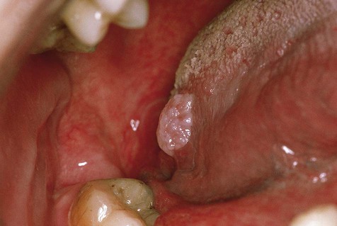
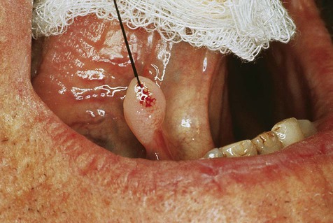
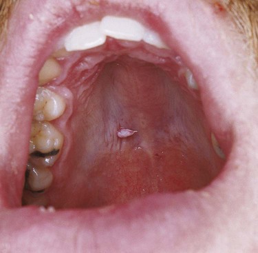
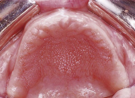
Oral squamous papillomas may be found on the vermilion portion of the lips and on any intraoral mucosal site, with predilection for the hard and soft palate and the uvula (Box 6-1; Figures 6-1 to 6-3). The latter three sites account for approximately one third of all lesions. The lesions generally measure less than 1 cm in greatest dimension and appear as pink to white exophytic granular or cauliflower-like surface alterations. They are generally asymptomatic and solitary in their presentation, although multiple lesions may occur.

image
FIGURE 6-3 Oral wart, palate.

The incidence of oral warts is increasing in patients with human immunodeficiency virus (HIV)/acquired immunodeficiency syndrome (AIDS) in contrast to other oral manifestations of the disease. This has been associated with the use of antiretroviral therapeutics, particularly highly active antiretroviral therapy (HAART). Lesions range from flat-topped plaques to verruciform or cauliflower-shaped exophytic masses. Lesions also tend to be multiple and recurrent.

Histopathology

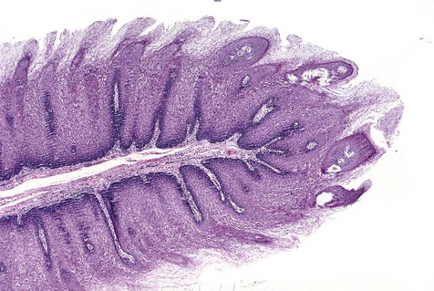
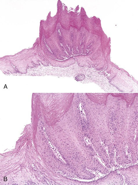
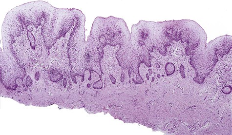
Oral squamous papillomas represent an exaggerated growth of normal squamous epithelium (Figures 6-4 to 6-6). The lesions are exophytic and are composed of finger-like extensions of epithelium, supported by a well-vascularized connective tissue core. The histologic architecture may mimic the pattern of the cutaneous wart. Upper level epithelial cells demonstrate nuclei that are pyknotic (condensed) and crenated, often surrounded by an edematous or optically clear zone, forming the so-called koilocytic cell. In the uterine cervix, this cytologic appearance represents HPV infection, which by extension is generally thought to present in the oral cavity.
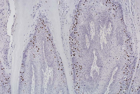
image
FIGURE 6-4 Papilloma.
image
FIGURE 6-5 A and B, Oral wart.

Dysplastic Oral Warts

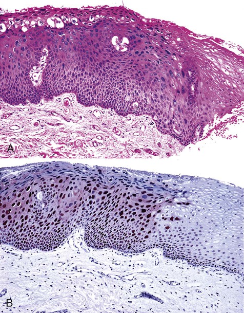
A small subset of HIV/AIDS patients develop oral warts that exhibit microscopic changes that are dysplastic in appearance (Box 6-2; Figure 6-7). The degree of dysplasia ranges from mild to severe. The outcome, or natural history, of these dysplastic warts is unknown, although invasive carcinoma has yet to be reported despite several years of follow-up. A wide variety of HPV subtypes, including 16 and 18, can be demonstrated in these lesions.

Papillary Hyperplasia

Etiology

Papillary hyperplasia, or palatal papillomatosis, appears almost exclusively on the hard palate and almost always in association with a removable prosthesis. The precise cause of papillary hyperplasia is not well understood, although it appears to be associated with an ill-fitting or loose denture that creates a potential space between the denture base and tissue, predisposing to or potentiating growth of Candida albicans organisms. Tissue hyperplasia has been related to the presence of the fungal organism in the setting of low-grade chronic trauma.

Histopathology

On perpendicular cross-section, papillary hyperplasia appears as numerous small fronds or papillary projections covered with intact parakeratotic stratified squamous epithelium (Figure 6-9). The epithelium is supported by hyperplastic central cores of well-vascularized stromal tissue. The epithelium is hyperplastic and often demonstrates pseudoepitheliomatous features, occasionally severe enough to mimic squamous cell carcinoma. No evidence of dysplasia is found in association with this lesion and risk of malignant transformation is not increased.

Differential Diagnosis

The range of possibilities in the differential diagnosis of papillary hyperplasia of the palate is rather narrow because this particular entity is seldom confused with other forms of pathology. The chief lesion to be separated from papillary hyperplasia is nicotine stomatitis involving the hard palate; however, nicotine stomatitis does not occur on the hard palate of those who wear complete maxillary removable appliances. Also, nicotine stomatitis tends to be more keratinized and usually demonstrates the presence of a small red dot or punctum in the center of each nodular excrescence, which represents the orifice of the subjacent minor salivary gland duct. Rarely, in Darier’s disease, the mucosa of the palate may demonstrate numerous papules. Numerous squamous papillomas may occur on the palate; however, these lesions tend to be more keratinized with more delicate projections. In the so-called malignant form of acanthosis nigricans, oral lesions are papillary in nature and may regress relative to the treatment response of the underlying distant malignancy. Finally, in the multiple hamartoma syndrome (Cowden’s syndrome), the oral mucosa may exhibit numerous papillary mucosal nodules. These nodules, composed of benign fibroepithelial proliferations, may impart a cobblestone appearance, usually to the tongue, buccal mucosa, and gingiva. Affected patients usually have other stigmata of the syndrome, including hamartomatous papules of the skin, benign breast lesions, and malignancies of the thyroid and kidney.

Treatment and Prognosis

Surgical removal is indicated before a denture is reconstructed for the patient. The actual surgical method is often a matter of individual preference and may include curettage, cryosurgery, electrosurgery, microabrasion, or laser ablation.

Removal of appliances at bedtime and soaking in a weak disinfecting or antifungal medium, as well as maintenance of good oral hygiene coupled with topical antifungal therapy, may significantly reduce the intensity of lesions. In mild cases, the use of soft tissue conditioning agents and liners, with frequent changes of the lining material, can produce sufficient resolution to preclude surgery. Topical antifungal ointment, alone or mixed with a corticosteroid ointment, may help reduce the size and intensity of the lesions, although it will not effect a complete cure when used alone.

Condyloma Latum

Condyloma latum is one of the many and variable expressions of secondary syphilis. As with all forms of syphilis, cutaneous, mucosal, and systemic lesions that mimic other conditions or diseases can be seen. Characteristic of condyloma latum is the presence of exophytic, sometimes friable, papillary to polypoid lesions within the oral cavity. Condyloma latum contains abundant microorganisms (Treponema pallidum), making it potentially infectious.

Condyloma latum usually appears on the skin, especially in the perianal and genital areas. Lesions may also be noted within the oral cavity. Here the tissue is formed into a soft, red, often mushroom-like mass with a generally smooth, lobulated surface.

Microscopically, the overlying epithelium demonstrates significant acanthosis, along with intracellular and intercellular edema and transmigration of neutrophils. A perivascular plasma cell infiltrate is common within the lamina propria in the absence of a true vasculi/>

Stay updated, free dental videos. Join our Telegram channel

Jan 12, 2015 | Posted by in Oral and Maxillofacial Pathology | Comments Off on 6: Verrucal-Papillary Lesions

VIDEdental - Online dental courses

Get VIDEdental app for watching clinical videos