Chapter 7 Color and Shade
Section A Understanding and Manipulating Color
Brief History of Clinical Development and Evolution of the Procedure
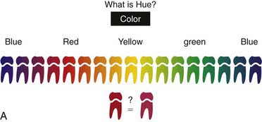
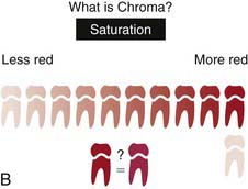
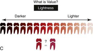
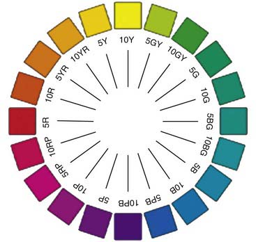
Although this created some organization of the color perception experience, it was only a beginning. At the time of its discovery, the color circle seemed to describe some basic law of physics, but it turns out that the organization of color into a color circle has more to do with the physiology of the eye and psychology of the observer. It does, however, form the basis for several present-day workable systems of color. In 1905, Albert Henry Munsell, an American artist and art teacher, further modified the color circle, devising a system of color organization that centered around three unique aspects of color: hue, chroma, and value (Figure 7-1). Using these three aspects, Munsell was able to construct a three-dimensional color wheel (Figure 7-2).
In the fifteenth century, Flemish painters such as Jan van Eyck worked with a method of painting known as the “wet-on-wet” technique, in which layers of wet paint are applied to previous layers of wet paint (Figure 7-3). Different details of the paintings are placed in different strata over the canvas, and the layers of paint toward the outside are usually increasingly translucent, creating an illusion of depth and vitality that would be impossible if only opaque paints were used (Figure 7-4). In some paintings as many as 30 layers are used to create maximum effect. This is not that different from the technique a master dental technician uses when different shades of porcelain along with flecks of inlaid color are employed to imitate the dentinal and enamel layers.

FIGURE 7-3 Two examples of Jan van Eyck’s work. A, Portrait of a Man (1433). B, The Arnolfini Portrait (1434).
(Copyright © The National Gallery, London.)
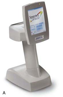
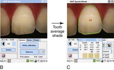
Over the last decade, computerized shade-matching systems have appeared on the market (Figure 7-5). This innovative technology offers better accuracy, improved efficiency, and esthetic benefits to patient, dentist, and technician. Based on technology imported from the painting industry, these systems analyze the color of the natural teeth; calculate the exact ratio of hue, chroma, and value for a multitude of points on the tooth surface; and display this information on the dentist’s computer screen. The process illuminates the guesswork often associated with reading the shade tabs and greatly facilitates the communication of information to the lab technician. This improved flow of information encourages the fabrication of predictably accurate, highly esthetic restorations. From the lab’s perspective, the frequency of remakes is reduced.
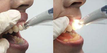
FIGURE 7-5 VITA Easyshade intraoral dental spectrophotometer in use.
(Courtesy Vident, Brea, California).
Computerized shade matching eliminates the subjectivity and frequent perception errors associated with traditional shade taking. It improves the accuracy and predictability of restorative procedures, particularly those difficult situations in which a single anterior tooth is being replaced (Figure 7-6). There is no requirement for standardizing the light environment of the dental operatory, and thus accurate shades can be taken anywhere, anytime.
Clinical Considerations
Clinical Options
Shade-Matching Techniques
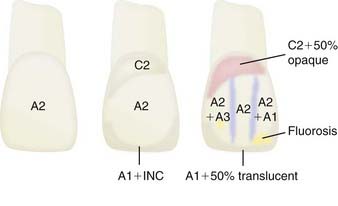
Traditional shade taking involves matching one or more selected colors from a range of shade tabs to the teeth adjacent or contralateral to the teeth to be restored. This serves as a guide to the lab technician fabricating the crown or the bridge. The more information (and accuracy) that the dentist can provide in the prescription, the more lifelike the technician’s output can become. Thus the dentist who provides a drawing of a tooth color map, indicating the various shades within the tooth and their borders, is more likely to have a positive result than the dentist who describes the shade as a single generic color (Figure 7-7).
Earlier shade guides (Figure 7-8, A) were developed haphazardly, with no infrastructural relationship among the various shades. Today’s advanced guides (Figure 7-8, B) have been developed logically, with incremental relationships among the various shade tabs, an organizationally structured series of family and color groupings, and a range that covers the entire tooth-visible color envelope. These modern guides are designed for ease of learning and ease of use. The entire shade guide system is available at less than $100.
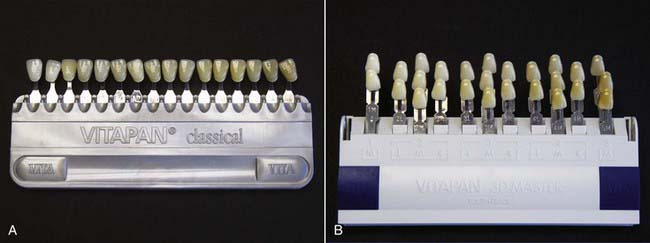
FIGURE 7-8 A, VITAPAN Classical Shade Guide. B, VITAPAN 3D-Master Shade System (Vident, Brea, California).
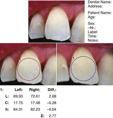
The current best approach to shade taking is the spectrophotometer. It provides the most accurate method for matching the coloration of the tooth. Some systems provide readings of translucence and reflectivity as well. Spectrophotometers provide consistent shade measurement regardless of the environment, lighting conditions, or other operatory variables including the dental team member who is conducting the shade-taking process. With some systems, a further comparative analysis can be undertaken on shade scans taken before and after treatment to provide the color difference between the two measurements. This is particularly useful for tooth-whitening procedures (Figure 7-9).
Other Considerations
The Additive System
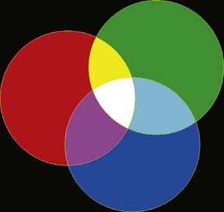
Knowledge of this system (the so-called “additive” system of color) (Figure 7-10) has enabled the creation of such devices as the color television. Using only three phosphors, one each of the three primary colors, the color television is able to produce a seemingly unlimited range of shades. One such television monitor boasts a palette of 16,777,216 colors that are available on the screen. In the additive system, white is the balanced mixture of all the colors, and black is the absence of color. Yellow is a balanced mixture of red and green. Because the additive system of color does such a laudable job of organizing color, it may seem that there is no need for any other approach.
The Subtractive System
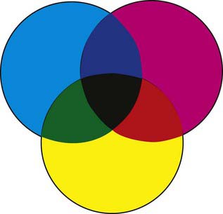
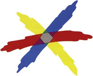
Those involved in art, however, tend to emphasize another arrangement. In this system, the so-called “subtractive” system, the three primary colors are red, yellow, and blue (Figure 7-11).
In the subtractive system, black is the result of a mixture of the three primaries, and white is the absence of color. This system is popular because it is perhaps the easiest to use when dealing with pigments (Figure 7-12).
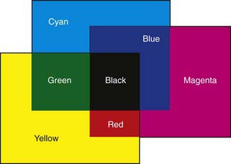
FIGURE 7-12 In the subtractive system, black is the result of a mixture of the three primary colors.
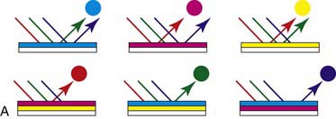
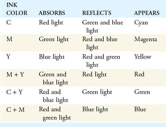
This system works the way it does because the pigments within the crayons absorb certain parts of the spectrum and reflect others. Thus the pigments are “light traps.” Red pigment, for instance, absorbs all parts of the light spectrum except red (Figure 7-13 and Table 7-1).
If pigments displayed perfect efficiency, the mixture of any two primary colors would result in the production of black. If in the crayon example one crayon absorbed all the spectrum except red, and the other one absorbed everything except blue, there would be nothing left over. Normal pigment concentrations, however, are notoriously inefficient in this regard and are almost always thinned out to create a relatively low saturation. Thus a red crayon selectively absorbs certain wavelengths and reflects those centering around the 700-nm (red) range (Figure 7-14).
In the subtractive system, when the three primary colors are arranged in the traditional color wheel, diametrically opposed colors are called complementary colors. Yellow and violet, for instance, are complementary colors. The mixture of two highly saturated complementary colors results in the elimination of color and the production of black. Because the pigments used in dentistry are poorly saturated and imperfect, the mixture of the stains usually produces some shade of grey instead of black (Figure 7-15).
Problems Inherent to Matching the Shades of Teeth
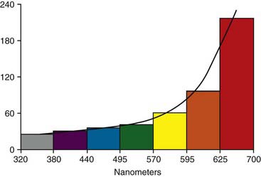
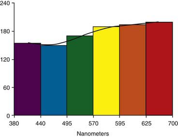
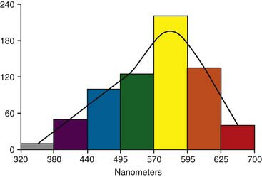
The apparent color of a tooth is affected by the color of the incident light. For example, in full-spectrum light a tooth might normally have a reflectance curve such as that shown in Figure 7-16.
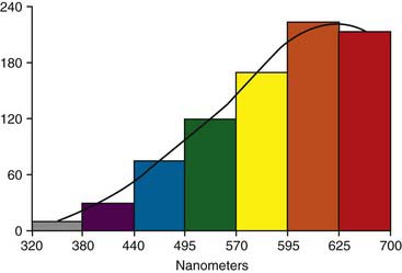
If the source of light changes, however, the apparent color can change dramatically. The normal transmission curve of a typical incandescent light bulb (Figure 7-17) contrasts with the normal transmission curve of a fluorescent light tube (Figure 7-18).
Figure 7-19 shows the two different reflectance curves a tooth would display under these two different sources of light. Even with a constant source of light, the light that actually reaches the tooth can be affected by the colors in the environment at the moment. A dark shade of lipstick or an intensely colored outfit can easily affect the available spectrum for reflection by the tooth.
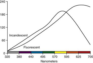
FIGURE 7-19 Reflectance curve of a tooth under two different sources of light, incandescent and fluorescent.
In addition, there are variations in operator sensitivity. Color blindness is no small problem. It is a fact that nearly one in 10 dentists in the United States has some degree of color deficiency in the red and green areas. If other color deficiencies are also included, the percentage of visually deficient operators goes up even further. The likelihood of a male dentist being color deficient is more than 10 times that of his female counterpart (Table 7-2).
TABLE 7-2 Color Blindness Distribution in the General Population and Various Groups
| INCIDENCE IN MALES | INCIDENCE IN FEMALES | |
|---|---|---|
| Caucasians | 8.08 ± 0.26% | 0.74 ± 0.11% |
| Northern European American Australian |
||
| Asiatics | 4.90 ± 0.18% | 0.64 ± 0.08% |
| Japanese Chinese Others (e.g., Korean, Filipino) |
||
| Other Racial Groups | 3.12 ± 0.40% | 0.69 ± 0.07% |
| American Indian Mexican African American Eskimo |
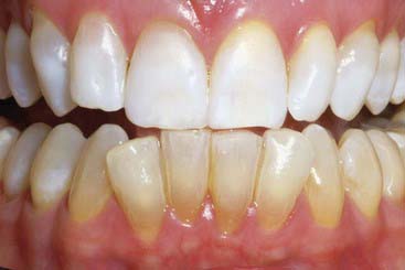
Even when the dentist’s innate visual color sensitivity is found to be optimal, however, there is still no guarantee of consistent color judgments. The eye can sustain a decrease in sensitivity from nerve fatigue, the same as any other sensory organ. For this reason, it is important to avoid staring at the tooth and shade guides when taking a shade. Instead, short glances should be employed, with the first reading considered the most accurate. One other important point: during the cementation step for veneers, the area is often isolated. If many laminates are involved, the isolated teeth have time to desiccate during the extended procedure. After only a few minutes of drying, the appearance of the teeth begins to change. The dried teeth are markedly whiter, and their surfaces more opaque. To demonstrate this, a patient with perfectly matched anterior teeth had a rubber dam placed, exposing the maxillary teeth to air for 20 minutes. When the rubber dam was removed, the difference in appearance was clearly evident (Figure 7-20).
Metamerism
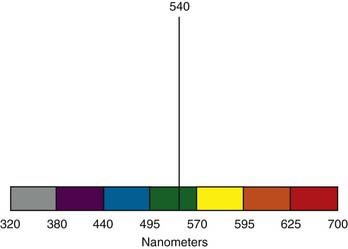
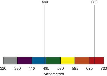
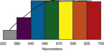
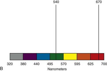
Simply put, the eye is incapable of distinguishing between certain combinations of light stimuli. Both of the spectral curves shown in Figure 7-21 are perceived as yellow-green. Under full-spectrum lighting, the surface that reflects light centered around 540 nm is indistinguishable in hue from one that reflects two loci of reflectance, one centered around 490 nm and the other around 650 nm.
When the lighting source changes, however, the perceived color of the objects also changes. Sunlight on an average day produces a spectral distribution similar to that seen in Figure 7-22. Contrast this with the curve shown in Figure 7-17 for typical tungsten light. As can be seen, when a surface is illuminated by a tungsten source, there is very little light in the 490-nm range available for reflection. In this situation, the samples no longer match.
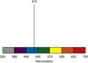
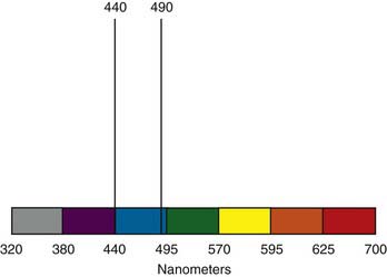
This example is by no means unique. A nearly infinite number of combinations can be computed that create metameric pairs. Figure 7-23 demonstrates one of the many pairs that would appear to be blue under full-spectrum light but that would not match under other light sources. The metameric pair in Figure 7-23 results from the fact that the eye cannot distinguish between a pure blue hue at 475 nm and the combination of 440 nm (reddish blue) and 490 nm (greenish blue), one of the many pairs that would appear to be blue under full-spectrum light.
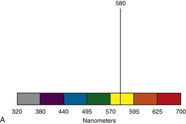
The pair shown in Figure 7-24 would be seen as yellow. Unfortunately, metamerism is a common illusion in the dental field. A factor that complicates this even further is the fact that human vision is most acute in the yellow range—a color range of particular importance in dentistry. In other words, not only is it impossible to accurately determine a nonmetameric color match, but human eyes are uniquely most sensitive to this error in the yellow range.
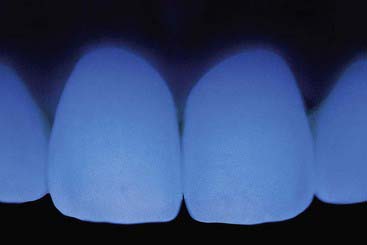
When a fluorescent porcelain is used in place of the nonfluorescent one, the shade match can even carry over to situations with intense ultraviolet light (Figure 7-25). Clearly there is a distinct advantage to creating the porcelain laminate veneer crown or bridge out of a variable fluorescing porcelain. There may be some advantage to using a luting agent that displays variable fluorescence as well.
Stay updated, free dental videos. Join our Telegram channel

VIDEdental - Online dental courses


