6
Respiratory disorders
Introduction
The essential function of the respiratory system is to absorb oxygen from the air into the blood and to excrete carbon dioxide. Ventilation is the process of drawing air containing 21% oxygen into the lungs, and it depends on the size of each breath, the respiratory rate, the resistance of the airways and the distensibility of the lungs. Perfusion of the lungs carries deoxygenated blood to the lungs from the venous system and oxygenated blood away from the lungs to the arterial system. The processes of ventilation and perfusion bring air from the atmosphere and blood from the circulation into close contact across the alveolar capillary membrane so that oxygen can be absorbed and carbon dioxide removed.
The respiratory centre in the brainstem controls breathing and may be affected by sedative drugs (e.g. midazolam and morphine) or brainstem disease (e.g. stroke). Respiratory diseases can affect the airways (e.g. chronic obstructive pulmonary disease (COPD) and asthma), the lung tissue (e.g. lung fibrosis and pneumonia), the pleura (e.g. pleural effusions) or the pulmonary vasculature (e.g. pulmonary embolism).
Two key features of respiratory disease are breathlessness and hypoxia. The lungs are a major interface between the person and the environment as 8 litres of air are inhaled each minute. This air contains noxious materials. Thus the lungs are vulnerable to infections from inhaled microbial pathogens, allergic reactions from antigens, cancer from carcinogens (e.g. tobacco smoke) and inflammation and scarring from dusts and toxins. The trachea is in direct continuity with the oropharynx, therefore the lungs are vulnerable to aspiration of material from the mouth. Periodontal infection can be a source of bacteria tracking down into the lungs, giving rise to pneumonia or lung abscess, and items of dental equipment or fragments of teeth can be inhaled, causing acute airway obstruction.
Clinical assessment
Respiratory disease is common and can impact on dentistry in several ways such that respiratory symptoms, signs and investigations are an important part of the overall medical assessment of the patient.
Symptoms
The main respiratory symptoms (defined later) are breathlessness, wheeze, stridor, cough, sputum production, haemoptysis and pleuritic pain (Table 6.1). Breathlessness (dyspnoea) can be graded according to the patient’s exercise capacity, indicated by breathlessness on climbing stairs, on walking short distances and at rest. It may be episodic, as in asthma, or persistent as in COPD.
Many patients with severe lung disease may be more breathless when lying flat (orthopnoea) and may have difficulty in tolerating equipment that blocks their mouth during dental treatment.
Wheeze is a whistling noise on expiration caused by air passing through narrowed airways. It is a particular feature of asthma and COPD. Stridor is a noise on inspiration and is a feature of obstruction of the trachea or central airways (e.g. by a tumour or an inhaled foreign body). Cough is a forceful expiratory blast and is a protective reflex to remove inhaled material from the lungs. It is a common symptom of many lung diseases. It may be productive of sputum, which is typically purulent and green in lung infections, e.g. chronic bronchitis, pneumonia, bronchiectasis and tuberculosis. Haemoptysis (coughing up blood) is an important symptom that always requires investigation. Patients with haemoptysis should be referred to a doctor for further assessment. Sometimes the blood may have arisen from an oropharyngeal source, but blood arising from the lower respiratory tract is a sinister symptom, which may indicate diseases such as bronchial or laryngeal carcinoma, tuberculosis, severe bronchitis, pneumonia or bronchiectasis. Pleuritic pain is a sharp stabbing pain on breathing. It indicates irritation of the pleura by inflammation (pleurisy), infection, pulmonary embolism, tumour or an air leak from the lung (pneumothorax). It may also arise from injury to the ribs and chest wall.
Table 6.1 Respiratory symptoms
|
|
|
|
|
|
|
Diseases of the lung can be associated with many general symptoms such as poor appetite and weight loss in lung cancer, fever and sweating in lung infections, lethargy, malaise and peripheral oedema from hypoxia, and headaches from carbon dioxide retention. Chronic hypoxia causes a rise in pulmonary artery pressure, which may result in right ventricular failure. This is referred to as cor pulmonale and manifests as an elevated jugular venous pressure and peripheral oedema.
Examination
Some signs of respiratory disease may be evident to the dental practitioner from general examination of the clothed patient. Patients with impaired lung function often have an increased respiratory rate and use accessory muscles of respiration (e.g. the sternocleidomastoids) to increase ventilation. Conversely, slow shallow breathing may be a sign of respiratory depression during use of sedative medication. The respiratory rate is most accurately assessed by counting it over a period of 30 sec. This is best done surreptitiously, perhaps after counting the pulse rate, as patients tend to breathe faster if they are aware that you are focusing on their breathing. Wheeze or stridor may be audible. Hoarseness of the voice may be an important clue to laryngeal disease (e.g. carcinoma) or to recurrent laryngeal nerve damage by a lung cancer.
Distension of the jugular veins may indicate an elevated venous pressure from failure of the right side of the heart (cor pulmonale) or from obstruction of the superior vena cava by a lung cancer. Enlargement of the cervical or supraclavicular lymph nodes can result from spread of disease from the chest (e.g. cancer or TB). Horner’s syndrome (Table 6.2) consists of drooping of the eyelid (ptosis), constriction of the pupil (miosis), reduced prominence of the eye (enophthalmos) and loss of sweating on one side of the face (anhidrosis). These appearances are due to damage to the sympathetic nerve supply, which may occur as a result of a tumour at the lung apex.
Table 6.2 The components of Horner’s syndrome
|
|
|
|
Pallor of the skin and mucous membranes may indicate anaemia, which can cause breathlessness. Cyanosis is a bluish discoloration of the skin and mucous membranes as a result of deoxygenated haemoglobin. It is the key sign of severe hypoxia and is best seen on the tip of the tongue. It is not usually detectable until the oxygen saturation is below 85% (normal level is 97%).
Oral candidiasis may be an adverse effect of inhaled or oral steroid therapy. Gingival overgrowth is an adverse effect of some drugs such as ciclosporin used after organ transplantation.
Tar staining of the fingers is a feature of heavy tobacco smoking, which is associated with COPD and lung cancer. Clubbing is characterised by an increased curvature of the nail with loss of the angle between the nail and nail bed such that the finger resembles a ‘club’ when viewed in profile (Chapter 1). It is an important sign that is associated with a number of diseases, including lung cancer, cystic fibrosis, asbestosis and fibrotic lung disease. A tremor may be due to high dose β-agonist medication such as nebulised salbutamol. Venous dilatation, a bounding pulse and a flapping twitching of the hand (asterixis) are signs of severe carbon dioxide retention.
General medical history
Certain features of the patient’s overall medical history are particularly important. Patients with major respiratory disease will often have had a full assessment by their general medical practitioner or hospital specialist. In complex cases it is useful to discuss the patient’s problems with their doctor so as to coordinate and optimise medical and dental care. For example, patients receiving chemotherapy for lung cancer may be at particular risk of developing infection or bleeding after oral surgery as a result of bone marrow suppression with reduced white cell and platelet counts.
Good dental care and hygiene are particularly important for patients receiving immunosuppressive drugs for inflammatory lung disease (e.g. the chronic granulomatous disease sarcoidosis) or after lung transplantation as the oropharynx is a potential source of infection which can spread to the lungs.
Particular care is needed when considering sedation for patients with hypoxia because of the risks of respiratory depression. It is important to have a complete list of the patient’s current medications and any drug allergies. Adverse effects of drug treatments may be apparent, e.g. oral candidiasis from inhaled or systemic corticosteroids, gingival overgrowth from ciclosporin, or mucositis and xerostomia from anticancer chemotherapy. Potential drug interactions need to be considered, for example macrolide antibiotics (e.g. erythromycin and clarithromycin) reduce the hepatic clearance of theophylline and can give rise to toxic adverse effects (e.g. vomiting or tachycardia). A small percentage of patients with asthma may develop wheeze if NSAIDs (e.g. naproxen or ibuprofen) are used for analgesia.
Aspects of the patient’s environment can be important in the causation of lung disease. Tobacco smoking predisposes to lung cancer and COPD. Occupational exposure to dusts such as asbestos, coal and silica can cause lung fibrosis. Contact with pet birds can cause lung inflammation and fibrosis in the form of bird fancier’s allergic alveolitis. The lungs can be involved by systemic diseases such as rheumatoid arthritis or their treatments (e.g. methotrexate alveolitis). The lungs are particularly vulnerable to infections when the patient is immunocompromised by HIV infection or by the use of immunosuppressive drugs (e.g. ciclosporin or azathioprine).
Investigating respiratory disease
Some simple investigations (e.g. peak expiratory flow rate or oximetry) can be performed in the dental surgery and are useful in assessing and monitoring the patient’s respiratory status. The peak expiratory flow rate is the maximum rate of airflow during a forced expiration. It is a measure of the airway calibre and is reduced in diseases such as asthma and COPD. Many patients will have a peak flow meter and use it to monitor the activity of their asthma.
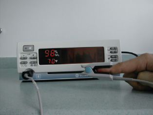
An oximeter (Fig. 6.1) measures the percentage of oxygenated haemoglobin using a probe placed on a finger or ear lobe, and comprises two light-emitting diodes and a detector. Oxygenated blood appears red whereas deoxygenated blood appears blue. Skin pigmentation or use of nail varnish can interfere with light transmission. Oximetry is particularly useful in monitoring the patient’s oxygenation continuously during procedures (e.g. bronchoscopy), anaesthesia or sedation, particularly in patients with respiratory disease. Oximetry does not provide information about the patient’s carbon dioxide level. In hospital practice, a sample of blood can be taken from an artery to measure oxygen and carbon dioxide levels directly.
Figure 6.1 A pulse oximeter. The probe (right of picture) can be placed on a finger and the oxygen saturation (upper figure on display as a percentage) and pulse rate (lower figure) are measured. No nail varnish is allowed.

Hospital laboratories can measure other aspects of lung function. For example, the total lung capacity is reduced in diseases which restrict the volume of the lung (e.g. fibrotic lung disease). The transfer factor measures the rate at which gas passes from the alveoli to the bloodstream, and this is reduced in diseases such as emphysema, lung fibrosis or pulmonary embolism. Clinical examination lacks sensitivity, and major disease of the lungs can be present without any detectable clinical signs. The chest X-ray, therefore, has a key role in the investigation of respiratory disease.
Computed tomography (CT) provides a much more detailed cross-sectional image, and various techniques can be used to display the lung parenchyma (e.g. lung fibrosis), the mediastinal structures (e.g. lymph node enlargement in lung cancer) and the vasculature (e.g. pulmonary embolism). Bronchoscopy allows direct visualisation of the main airways, biopsies may be taken for histopathological examination (e.g. lung cancer) and secretions can be aspirated for cytology and microbiology tests. Sputum microbiology is used routinely to detect microbial pathogens in diseases such as chronic bronchitis, pneumonia, bronchiectasis and tuberculosis.
Asthma
Asthma is characterised by bronchial inflammation, which results in mucosal oedema and irritability of the airways such that they are prone to constriction of the bronchial smooth muscle. This results in symptoms such as wheeze, cough and breathlessness, and airways obstruction, which is variable and reversible with treatment. About 7% of the adult population in the UK have asthma. Asthma is multifactorial in origin, arising from a complex interaction of genetic and environmental factors.
Atopy is a constitutional tendency to produce IgE on exposure to common antigens, and atopic individuals have a high prevalence of asthma, allergic rhinitis and eczema. The prevalence of asthma is increasing, and this seems to be related to a modern urban economically developed environment. Some patients with atopic asthma develop reactions to common antigens such as house dust mite, found in carpets, soft furnishings and bedding, and to pets and antigens in the environment, e.g. grass pollens, and in the workplace, e.g. isocyanates or hard wood dusts.
Attacks of asthma can be precipitated by co-factors such as respiratory tract infections, cigarette smoke, pollutants and cold air. β-Blocker drugs (e.g. atenolol) can induce bronchoconstriction, and a small percentage of asthmatics develop wheeze if given aspirin or NSAIDs (e.g. ibuprofen).
Symptoms may develop at any age and may be episodic or persistent. Episodic asthma is a common pattern in children and young adults. The patient is often asymptomatic between attacks, but episodes of bronchoconstriction are provoked by factors such as infection or contact with an inhaled antigen. Sometimes the pattern is of persistent asthma with chronic wheeze and dyspnoea. Airway remodelling in chronic severe asthma can result in permanent changes in the bronchial wall with fixed airways obstruction.
The variable nature of symptoms is a characteristic feature of asthma. Typically there is a diurnal pattern, with symptoms and peak expiratory flow rate (PEFR) being worse early in the morning (‘morning dipping’). Symptoms such as cough and wheeze often disturb sleep (‘nocturnal asthma’). Symptoms may be provoked by exercise (‘exercise asthma’), but many elite athletes can compete at the highest level despite having asthma. Patients with asthma usually have a dramatic improvement in their symptoms and PEFR when given a bronchodilator drug or corticosteroids.
The key feature of asthma is airways obstruction, which can be measured objectively using a PEFR meter. Patients will often have a PEFR meter that they use to monitor their asthma. They may be aware of their best achievable PEFR and they often have a self-management plan whereby they start a course of prednisolone and increase their bronchodilator medications if their PEFR falls below a set level.
Drugs used to treat asthma
In most patients, asthma can be controlled by a regular inhaled corticosteroid to reduce airway inflammation and an inhaled bronchodilator to be taken as required to reverse wheeze.
Bronchodilators (‘relievers’)
β2-Agonists (e.g. salbutamol and terbutaline) stimulate β-adrenoreceptors in the smooth muscle of the airway producing muscle relaxation and bronchodilatation. They have an onset of action within 15 min and last 4–6 hours. Adverse effects are rare but include tremor, palpitations and muscle cramps. Patients should carry their ‘reliever inhaler’ with them and it should be available when undertaking dental procedures so that it can be used if the patient becomes wheezy. Long-acting β2–agonists (e.g. salmeterol and formoterol) have a duration of action of >12 hours. Anticholinergic bronchodilators (e.g. ipratropium and tiotropium) produce bronchodilatation by blocking the bronchoconstriction effect of vagal nerve stimulation on bronchial smooth muscle. Adverse effects are rare, but nebulised ipratropium may be deposited in the eyes, aggravating glaucoma. Theophyllines increase cyclic adenosine monophosphate (cAMP) stimulation of β-adrenoreceptors by inhibiting the metabolism of cAMP by the enzyme phosphodiesterase. They are not available in inhaler form and are taken as tablets. Adverse effects include nausea, headache and tachycardia. Interactions with other drugs can cause toxicity. Hepatic clearance of theophyllines is reduced by drugs such as ciprofloxacin, erythromycin and cimetidine.
Anti-inflammatory drugs (‘preventers’)
Inhaled corticosteroids (e.g. beclometasone, budesonide and fluticasone) are the mainstay of asthma treatment as they control airway inflammation, which is the key underlying process in asthma. They need to be taken regularly, usually twice daily, to prevent asthma attacks. Adverse effects include oropharyngeal candidiasis and hoarseness of the voice, which can be reduced by using a spacer device and gargling the throat after inhalation. Higher dose inhaled steroids can cause some suppression of adrenal function, increased bone turnover and skin purpura. Sodium cromoglicate is a preventative inhaler that has anti-inflammatory effects in stabilising mast cells, but it is less effective than inhaled corticosteroids. Leukotriene receptor antagonists (e.g. montelukast and zafirlukast) are a new modality of anti-inflammatory therapy, given orally in tablet form. They block the effect of cysteinyl leukotrienes, which are metabolites of arachidonic acid with bronchoconstrictor and pro-inflammatory effects. Prednisolone (e.g. 30 mg/day for 7 days) is usually given as ‘rescue treatment’ for acute attacks of asthma. Pat/>
Stay updated, free dental videos. Join our Telegram channel

VIDEdental - Online dental courses


