6: Dental Implant Surgery

Dental Implant Surgery

The number of different dental professionals who place dental implants has increased dramatically during the past decade. This is likely to continue due to many changing healthcare and economic trends. Oral and maxillofacial surgeons remain the only specialists who can offer the full spectrum of dental implant surgery, encompassing complex implant reconstruction of the atrophic mandible and maxilla, restoration of the dentition after tumor resections, grafting for small and large defects, and esthetic implantology. Our specialty is also well positioned to provide safe and effective anesthesia, either in the office or in the operating room, for healthy patients and for those with complex medical comorbidities.

Although cone-beam computed tomography (CBCT) is not the standard of care for placement of dental implants, its use can have significant diagnostic and treatment planning implications. CBCT can improve the accuracy of implant placement and outline anatomic boundaries to reduce the risk of complications.

In this chapter we include eight cases that cover some of the contemporary issues related to dental implants. Two cases representing the routine placement of maxillary and mandibular implants are presented, followed by six cases that discuss sinus augmentation, zygoma implants, treatment of edentulism, guided implant surgery, extraction socket preservation, and implantology for the esthetic zone.

Posterior Mandibular Implant Supported Fixed Partial Denture

PMHX/PDHX/Medications/Allergies/SH/FH

The patient is in excellent general and oral health except for the partial edentulism. He has been a one pack a day cigarette smoker for over 40 years. He is allergic to penicillin. He currently takes metoprolol (Lopressor), a selective β1-receptor blocker. He specifically denies a history of diabetes mellitus.

There is support in the literature for implant treatment in patients with well-controlled type 2 diabetes. However, a qualified contraindication to such treatment must be considered to exist for patients with type 1 diabetes, because the variable success rates reported in this population are affected markedly by the quality of glycemic control. There are studies illustrating implant success in individuals with type 1 diabetes, but in most of these the numbers are small and the follow-up short. Early studies showed a markedly increased failure rate for implants in smokers; however, more recent techniques, in which the implants had oxidized rather than machined surfaces, do not show such glaring disparities. Good healthcare mandates smoking cessation in any patient, and the possibility of an increased risk of failure of osseointegration in smokers should be discussed with the patient and included in the consent as a shared liability.

Cardiovascular diseases, hypertension included, pose a considerable risk of stroke, heart failure, myocardial infarct, and renal failure; however, if a patient is able to undergo minor oral surgery, there is no increased risk of implant failure. The β-blocker this patient is on should be continued.

There are several other general risk factors affecting implant success that warrant discussion. There is little evidence regarding recommendations for implants in patients with other autoimmune disorders. Reasonable success rates have been obtained in patients who are osteoporotic with extended healing times. Implants in patients on bisphosphonate therapy have been studied on a very limited basis; although osteonecrosis of the jaw has a low incidence, its morbidity is extremely high. If implants are to be placed in these patients, it should be done with thorough informed consent and ongoing monitoring. Radiotherapy is a significant risk factor for implant failure, and the evidence for hyperbaric therapy in reducing this risk is inconclusive. There is limited evidence linking a history of periodontitis with an increase in peri-implant pathology, although the periodontal health of the remaining dentition affects the success of implant restoration. A higher implant failure rate is seen in poor-quality bone, in the maxilla, and in the posterior jaw. Bruxism and other paraoral habits are associated with increased implant failure rates due to increased load. Implant oral rehabilitation is a successful and predictable modality for most patients.

Examination

Examination of both the quality and quantity of bone is essential for successful implant placement.

Clinical examination of the posterior mandible indicates that the form of the ridge is rounded and wide (6 to 8 mm) in the buccal and lingual aspect. The anteroposterior ridge space is adequate for placement of several implants posterior to the most distal mandibular tooth on each side of the mandible. For this discussion we will concentrate on the posterior mandibular quadrant on the right. The distance from an implant to an adjacent natural tooth and between adjacent implants should be 1.5 mm and 2 mm, respectively. In the current patient, because first molar occlusion is planned, the distal implant will be placed about 12 mm or so from the front implant, and a three-unit bridge will be constructed. An interarch space greater than 5.5 mm already exists, and this is adequate for the restorative stack. No soft tissue or bony abnormalities are noted, and the patient opens 50 mm (restricted mouth opening can pose a problem for implant placement).

Imaging

Although implant surgery can be planned on a panoramic radiograph with a surgical planning template coupled with bone mapping techniques, a far simpler and much more predictable methodology involves CBCT (Figure 6-1, A). The preoperative films show that the anterior implant site is 8.9 mm wide in a buccolingual dimension, 14.4 mm above the inferior alveolar neurovascular bundle, and 1.5 mm distal to the lower right mandibular cuspid tooth (Figure 6-1, B). At the proposed location of the more posterior implant, the buccolingual distance is 10.9 mm and the inferior alveolar neurovascular bundle lies 16.1 mm below the ridge crest (Figure 6-1, C). The bone quality is type II (Box 6-1). A surgical guide can be generated from the data contained in the CBCT if the surgeon feels it is necessary, and instrumentation keyed to this guide can be applied for precision placement; otherwise, a surgical guide is prepared on the study models.
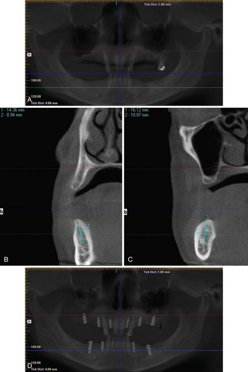
Treatment

The planned treatment was discussed in detail with the patient and the restorative dentist. A surgical guide prepared by the restorative dentist was used, indicating the ideal position for the implants in the planned restorative schema. Underlying bone dictates where implants can be placed; if bone is not present where implants need to be placed, various grafting or distraction techniques (which are beyond the scope of the current discussion) can be performed. In the current patient, it was predetermined that no such additional surgery was indicated. Chlorhexidine mouth rinse was used before surgery. No preoperative prophylactic antibiotics were prescribed for this patient, because there is no literature support for this practice in low- and moderate-risk dental implant patients.

Under intravenous sedation and local anesthesia, implant surgery was performed according to the particular system protocol. The wound was sutured with nonresorbable sutures, and a gauze sponge was placed to obtain hemostasis. The patient was recovered and discharged with detailed instructions for home care, analgesics, and a return appointment in 1 week for suture removal. Postoperative CBCT images (see Figure 6-1, D) revealed satisfactory placement of implants well above the mandibular canal, with adequate spacing for a three-unit fixed partial denture. A final restoration was placed 4 months later, and a satisfactory cosmetic and functional outcome resulted, with contours and emergence similar to those of the replaced natural dentition.

Although research indicates equally satisfactory results, during the first 16 weeks of healing after implant placement, for implants placed under three different loading regimens (loaded immediately, early [6 weeks], or using conventional/delayed timing [12 weeks]), the decision was made to follow the conventional/delayed plan in this patient.

Complications

The direct anchorage of dental implants to the host surrounding bone (osseointegration) is a good indication of clinical success, and although a high success rate is seen in implantology, endosseous implants do fail. Early implant failure is associated with a lack of primary stability (possibly aggravated by early loading protocols), surgical trauma, and infection. Infection noted early is much more problematic than infection later in the course of treatment because of the disturbance in primary bone healing associated with the former. Occlusal overburden and peri-implantitis are the most important factors associated with delayed or late implant failure. The primary limiting factor for lower premolar and molar implants is the height of bone above the mandibular canal. If it is determined that an implant has been placed in contact with the mandibular canal, causing postoperative hypoesthesia or anesthesia, the implant should be backed off of this structure as soon as possible or removed and replaced with a shorter implant (Figure 6-2). CBCT, unlike a panoramic film, can differentiate superimposition from direct contact with the nerve. In the presence of symptoms (e.g., paresthesia) and a CBCT scan showing no direct contact with the nerve, removal of the implant is unlikely to resolve the paresthesia, and interval neurosensory testing determines whether microneurosurgical exploration is indicated.
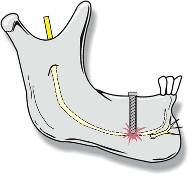
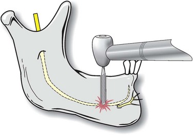
Perforation with the implant drill and direct injury to the mandibular nerve are possible (Figure 6-3). Hemorrhage, salivary gland involvement, painful impingement of implants on the lingual surface of the mandible, and associated misalignment are all complications associated with lingually directed surgical misadventures. Such problems can be minimized by careful attention to the anatomic features of the lower posterior jaw as depicted on the CBCT. Frequently a slight buccal inclination of posterior mandibular implants is necessary to avoid lingual perforation, and this fact is best determined and discussed preoperatively with the restorative dentist to minimize misunderstandings. Should infection occur in the early postoperative period, conservative management, including antibiotics, debridement, incision and drainage, and irrigation, is indicated, with implant removal as a last resort. Occasionally, inflammation and the development of a fistula over an implant site occur, and this frequently indicates that the cover screw has loosened. In this case the implant site should be exposed, the screw removed, the site debrided and irrigated, and a new, sterile cover screw placed and tightened.

Other complications with implant surgery include aspiration or swallowing of instruments or implant components. This can be prevented by paying close attention to positioning, using a well-placed gauze curtain, and attaching a 12- to 16-inch floss cord to such instruments as screwdrivers so retrieval is possible should the instrument be dropped.

Discussion

Implant surgeons can choose from a wide range of implant designs, materials, and surfaces, although a detailed analysis of these variables is beyond the scope of this discussion. Currently, a textured-surface titanium alloy implant is indicated, because such surfaces have been shown to be better receptors for fibrin strands forming initial attachments of implants to bone in the microgap. A recently reported, long-term outcome study of titanium implants with a sand-blasted and acid-etched (SLA) surface in a large cohort of partially edentulous patients showed a 10-year implant survival rate of 98.8% and a restoration success rate of 97%. In addition, the prevalence of peri-implantitis in this large cohort of orally healthy patients was low (1.8% during the 10-year period).

Several techniques that can increase inadequate bone volume above the inferior alveolar canal and in the buccal and lingual dimensions have been documented, including onlay block or particulate grafting, guided tissue regeneration techniques, distraction osteogenesis, and nerve repositioning. When any technique is used other than what is considered standard implant surgery (as described in this discussion), there are tradeoffs, with additional potential complications associated with each of these techniques. Discussion of the particulars of these techniques is beyond the scope of the present text.

Bibliography

Ahmad, N, Saad, N. Effects of antibiotics on dental implants: a review. J Clin Med Res. 2012; 4:1–6.

Barewal, RM, Stanford, C, Weesner, TC. A randomized controlled clinical trial comparing the effects of three loading protocols on dental implant stability. Int J Oral Maxillofac Implants. 2012; 27:945–956.

Buser, D, Janner, SF, Wittneben, JG, et al. Ten-year survival and success rates of 511 titanium implants with a sand-blasted and acid-etched surface: a retrospective study in 303 partially edentulous patients. Clin Implant Dent Relat Res. 2012; 14(6):839–851.

Lambert, PM, Morris, HF, Ochi, S. The influence of 0. 12% chlorhexidine digluconate rinses on the incidence of infectious complications and implant success. J Oral Maxillofac Surg. 1997; 55(12 Suppl 5):25–30.

Liddelow, G, Klineberg, I. Patient-related risk factors for implant therapy: a critique of pertinent literature. Aust Dent J. 2011; 56:417–426.

Marchand, F, Raskin, A, Dionnes-Hornes, A, et al. Dental implants and diabetes: conditions for success. Diabetes Metab. 2012; 38:14–19.

Misch, CE. Density of bone: effect on treatment plans, surgical approach, healing and progressive bone loading. Int J Oral Implantol. 1990; 6:23–31.

Renouard, F, Rangert, B. Risk factors in implant dentistry: simplified clinical analysis for predictable treatment, ed 2. Hanover Park, Ill: Quintessence; 2008.

Sakka, S, Baroudi, K, Nassani, MZ. Factors associated with early and late failure of dental implants. J Investig Clin Dent. 2012; 3(4):258–261.

Posterior Maxillary Implant Supported Fixed Prostheses

PMHX/PDHX/Medications/Allergies/SH/FH

The patient has a significant past medical history. He had an ST-elevated myocardial infarction (STEMI) 3 years earlier and had three-vessel coronary bypass 2 years ago for angina that interfered with his ability to work. He also has hypertension and hyperlipidemia. He denies any chest pain or exercise intolerance since his surgery and works as a structural engineer on outdoor jobs, where he is regularly engaged in physical exertion. For his hypertension he takes benazepril (Lotensin), an angiotensin-converting enzyme inhibitor (ACE inhibitor); for his hyperlipidemia he takes simvastatin (Zocor), a 3-hydroxy-3-methylglutaryl coenzyme ( HMG-CoA ) reductase inhibitor. Both of these medications can be given preoperatively in their usual dosages. In addition, he takes clopidogrel (Plavix), along with an 81-mg aspirin (low dose) for prophylaxis against thromboembolic events.

Clopidogrel reduces thrombosis by blocking adenosine diphosphate receptors on platelet cell membranes, thereby preventing platelet adhesion and aggregation. Aspirin irreversibly inhibits platelet-dependent cyclooxygenase (COX), which decreases platelet aggregation for the life of the platelet. Aspirin is a much more potent inhibitor of COX-1 than COX-2, so higher doses also inhibit endothelial cell synthesis of prostacyclin, a vasodilator that serves a protective role against ischemia (higher aspirin doses may be deleterious). It is suggested that clopidogrel be discontinued 5 days before elective surgery. Low-dose aspirin should have no clinically significant effect on bleeding and can be continued. The patient has no known drug allergies and no additional medical problems. He is a nonsmoker with excellent oral hygiene.

Examination

The quality and quantity of bone must be adequate for successful implant placement.

Clinical examination reveals that the posterior right maxilla has a rounded form and is free of soft tissue abnormalities. In the bicuspid region, the width of the ridge is about 7 mm, with a minimal facial undercut; in the molar region, 8 mm of width is noted. Because of the buccal undercut, perforation may occur at implant placement; however, if this happens, the defect can be augmented or repaired with a xenograft and collagen membrane. The mesiodistal dimension is a free end, so adequate room is present for three implants (two 4-mm-diameter anterior implants and one 5-mm-diameter posterior implant); however, the decision was made to place only two implants, spaced to allow for a three-unit bridge, as an economy measure. The interarch space is adequate, because the opposing dentition is absent (5 mm or more is needed for placement of the restorative stack). The patient opens 52 mm (limited opening can make implant placement difficult).

Imaging

A cone-beam computed tomography (CBCT) panoramic image (Figure 6-4, A) reveals that there is adequate bone for placement of implants in both areas selected, without the necessity of a sinus graft. The bone quality is judged to be type II. Bone quality is determined by radiographic appearance and also by the tactile feeling on drilling (see Box 6-1). If CBCT is not available, an implant tracing template at 25% larger than normal size (due to a 25% magnification factor on a panoramic radiograph) can be used with a panoramic radiographic image for surgical planning. CBCT helps to accurately determine the position of the floor of the maxillary sinus and the condition of the sinus membrane, which aids in determining the necessity and feasibility of sinus grafting. Coronal CBCT images in the area planned for the placement of the more anterior implant in this case (2 mm posterior to tooth #11) demonstrates that the buccolingual dimension is 7.55 mm, and a vertical height of 18.8 mm is available (Figure 6-4, B). In the area proposed for the placement of the more posterior implant, 8.16 mm is noted as the buccolingual dimension, and 12.65 mm of bone is present from the crest of the ridge to the floor of the right maxillary sinus (Figure 6-4, C). It is relatively uncommon to have adequate bone below the maxillary sinus for placement of implants of satisfactory length, as is seen in this case. Various strategies can be used when adequate volume is absent. Bone graft augmentation by open and closed technique using various graft materials (e.g., onlay grafting with particulate and/or block grafts of various natures), sinus membrane elevation followed by implant placement without grafting, and short implants are all methodologies used to overcome inadequate bone stock below the sinus floor in posterior maxillary constructs. Immediate, rather than delayed, implant placement in grafted sinuses is determined by the presence of at least 5 mm of bone, which allows for initial stability of immediately placed implants. In the case of two-stage sinus grafts, the primary graft placement is followed 4 months later by secondary implant surgery, with or without immediate loading as determined by initial stability at placement.
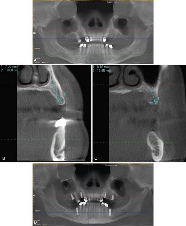
Labs

No routine laboratory studies are indicated for implant placement unless dictated by the medical history.

In the current patient, because hyperlipidemia is a risk factor for atherosclerosis and associated cardiovascular disease, in addition to an increased risk of stroke, it would be pertinent to evaluate the most recent total body cholesterol level, LDL, HDL, and triglyceride levels. The patient’s total cholesterol was 167 mg/dl (a value below 200 mg/dl is associated with a relatively low risk of myocardial infarction unless other risks factors are present). The LDL was 70 mg/dl (normal range, 70 to 130 mg/dl, although lower values are better), the HDL was 64 mg/dl (normal range, 40 to 60 mg/dl, higher values are better), and triglycerides were 58 mg/dl (normal range, 10 to 150 mg/dl, lower values are better).

Treatment

The planned surgery was discussed in detail with the restorative dentist, who showed the patient on a diagnostic wax-up what the final restoration will look like and has provided a surgical guide indicating the desired position and inclination for the implants. The patient had been off of clopidogrel for 6 days on presentation. He was given 2 gm of amoxicillin by mouth 1 hour before surgery, and chlorhexidine oral rinse was used immediately before surgery. Under intravenous sedation and local anesthesia, the implant surgery was performed in accordance with the manufacturer’s protocol. After the osteotomies were performed, the sites were probed with a blunt instrument, and the sinus floors were found to be intact at both sites. The implants were placed, and wound closure was obtained with 4-0 polyglactin (Vicryl) sutures. The patient recovered and was discharged with instructions for home care, analgesics, and a return appointment in 1 week for suture removal. Postoperative CBCT panoramic imaging revealed satisfactory implant placement (Figure 6-4, D).

Although there is research indicating good results with immediate placement of healing caps and even immediate loading, fewer problems can be expected from implants submerged and unloaded for 4 months in the mandible and 6 months in the maxilla and in grafts (two-stage placement).

Complications

Success rates for fixed partial dentures on implants in the posterior maxilla have been reported to be about 95% at 5 years and about 93% at 10 years, and the quality of bone appears to have little influence on the success rate. Patients should be informed of the risks associated with the surgical placement of implants in the posterior maxilla, including sinus penetration, buccal perforation, infection, and failure to integrate, even though survival data suggest an adequate success rate for this application of dental implants. The most common implants lost in the posterior maxilla are shorter fixtures; wide fixtures show the lowest failure rates. Other complications associated with implants used to treat partial posterior maxillary edentulism are fractures of the occlusal surface of restorations and loose anchorage components.

Discussion

Implant surgeons can choose from a wide range of implant designs, materials and surfaces. Although a detailed analysis of the array of implant variables is beyond the scope of this discussion, a textured-surface, titanium alloy implant is currently indicated, because it is known to improve initial attachment of implants to bone in the microgap between the bone and the implant. Immediate loading, one-stage placement, and the traditional two-stage implant surgery all have their proponents. In the posterior maxilla, two-stage placement seems ideal, because no cosmetic considerations come into play. There are several techniques that can increase inadequate bone volume for implant placement below the maxillary sinus, decrease buccal undercuts, or treat buccal perforations. Onlay grafting of particulate or block bone or bone substitute, guided tissue regeneration, distraction, and sinus grafting are well documented. However, when any technique is used other than standard implant surgery, there are tradeoffs, with potential complications specific to the technique used.

Bibliography

Bahat, O. Brånemark system implants in the posterior maxilla: clinical study of 660 implants followed for 5 to 12 years. Int J Oral Maxillofac Implants. 2000; 15:646–653.

Benavides, E, Rios, HF, Ganz, SD, et al. Use of cone beam computed tomography in implant dentistry: the International Congress of Oral Implantologists consensus report. Implant Dent. 2012; 21:78–86.

Cheng-Ching, E, Samaniego, EA, Reddy Naravetla, B, et al. Update on pharmacology of antiplatelets, anticoagulants, and thrombolytics. Neurology. 2012; 79(13 Suppl 1):S68–S76.

Cho-Lee, GY, Naval-Gias, L, Castrejon-Castrejon, S, et al. A 12-year retrospective analytic study of the implant survival rate in 177 consecutive maxillary sinus augmentation procedures. Int J Oral Maxillofac Implants. 2010; 25:1019–1027.

Dent, CD, Olsen, JW, Farish, SE, et al. The influence of preoperative antibiotics on success of endosseous implants up to and including stage II surgery: a study of 2,641 implants. J Oral Maxillofac Surg. 1997; 55(Suppl 5):19–24.

Felice, P, Soardi, E, Pellegrino, G, et al. Treatment of the atrophic edentulous maxilla: short implants versus bone augmentation for placing longer implants: five-month post-loading results of a pilot randomised controlled trial. Eur J Oral Implantol. 2011; 4:191–202.

Kaufman, E. Maxillary sinus elevation surgery: an overview. J Esthet Restor Dent. 2003; 15:272–282.

Lambert, PM, Morris, HF, Ochi, S. The influence of 0. 12% chlorhexidine digluconate rinses on the incidence of infectious complications and implant success. J Oral Maxillofac Surg. 1997; 55(12 Suppl 5):25–30.

Liddelow, G, Klineberg, I. Patient-related risk factors for implant therapy: a critique of pertinent literature. Aust Dent J. 2011; 56:417–426.

Renouard, F, Rangert, B. Risk factors in implant dentistry: simplified clinical analysis for predictable treatment, ed 2. Hanover Park, Ill: Quintessence; 2008.

Sinus Grafting for Implants

HPI

The patient lost most of his posterior teeth at a younger age due to decay and failed endodontics. Early loss of posterior maxillary teeth is associated with increased pneumatization of the maxillary sinus, and frequently inadequate bone for satisfactory implants does not exist below such a sinus. The patient has found himself at a point in life where he can seek optimal care, and his case has been followed by a periodontist, who believes that the patient’s periodontal condition is stable. All remaining teeth are sound except for tooth #15, which shows extensive restoration. The referring dentist has informed the patient that he may require a “sinus lift” procedure. The patient wants to know the details of this surgery, and he wants treatment planned for implant-based restorations, because he is unable to wear his existing removable partial dentures comfortably.

PMHX/PDHX/Medications/Allertgies/SH/FH

The medical history is significant for paroxysmal supraventricular tachycardia (PSVT), for which he takes metoprolol (Lopressor), and for chronic maxillary sinusitis. He is a one-half pack per day smoker with a 10 pack-year history.

Cigarette smoking (nicotine) increases platelet adhesiveness, raises the risk of microvascular occlusion, and causes tissue ischemia. Tobacco smoking causes catecholamine release and associated vasoconstriction, resulting in decreased tissue perfusion. Smoking is additionally believed to suppress the immune responses by affecting the function of neutrophils. A perioperative smoking cessation program has been shown to reduce respiratory and wound-healing complications. Good health care mandates smoking cessation in any patient, and the possibility of an increased risk of failure of osseointegration in smokers should be discussed with the patient and included in the consent as a shared liability.

The risk of PSVT is increased with alcohol or caffeine use and with smoking. It can be associated with bothersome palpitations, anxiety, chest tightness, and shortness of breath. This patient takes metoprolol (Lopressor), a β-blocker, to prevent PSVT episodes, and he reports very infrequent episodes in the recent past. PSVT is typically a benign disease but can be associated with profound hypotension and require emergency management; therefore, it is an anesthesia consideration.

A history of acute or chronic sinusitis may be problematic for an implant surgery with an associated sinus graft procedure planned. Prolonged inflammation and/or infection creates an inappropriate environment for the procedure. Maxillary sinusitis results from a secondary bacterial infection of an obstructed sinus. The mucosal edema, increased mucous production, bacterial accumulation, and inflammatory debris associated with sinusitis create an unfavorable environment for surgery and subsequent healing. Infections of the maxillary sinus after sinus grafting surgery occur in a small percentage of cases and are usually managed conservatively, with preservation of uninfected graft and subsequent implant success. The two most common bacteria involved in acute maxillary sinusitis are Haemophilus influenzae and Streptococcus pneumoniae. Staphylococcus aureus, α-hemolytic streptococci, and Bacteroides and Pseudomonas spp. are most frequently found in chronic bacterial sinusitis. Any form of sinus infection should be treated with decongestants and antibiotics, and some infections require functional endoscopic sinus surgery (FESS) before performance of a sinus grafting procedure can be contemplated. A broad-spectrum antibiotic, such as amoxicillin with clavulanic acid (Augmentin), is often the initial antibiotic used in the management of infections caused by nasal or sinus flora.

Examination

The patient is a well-developed, well-nourished man in no acute distress. There are no signs or symptoms of sinus infection currently.

The patient is missing all of his lower posterior teeth (teeth #22 through #27 remain). In the maxilla he is missing teeth #1 through #5, #7, #10, #12 through #14, and #16. Tooth #15 is present but is considered nonrestorable and slated for removal. No lesions or bony irregularities are noted in the oral cavity, and the remaining teeth, except for tooth #15, are in satisfactory condition. In the maxilla the ridge form in the posterior is rounded without undercuts and is of adequate width clinically for implant placement. The maximal incisal opening is 48 mm (limited opening may prevent ideal implant placement and angulation, especially of posterior implants). There is adequate interarch space for the restorative stack because the patient is edentulous in the mandibular arch in the posterior, and full mouth, implant-based rehabilitation is planned.

Imaging

A panoramic cone-beam computed tomography (CBCT) image showed that there was inadequate bone beneath the maxillary sinus bilaterally (especially on the right) in the posterior maxilla to allow for adequate implant placement (Figure 6-5, A). A coronal CBCT cut (Figure 6-5, B) determined that an implant of 5 mm in diameter could be placed in the most posterior right maxilla site; however, only 3.6 mm of bone is present below the sinus membrane. Consequently, a sinus graft must be completed to place an implant of adequate length to support a fixed partial denture on two implants in this area of relatively high occlusal load. On the left side at the site of the more posterior implant, 9.7 mm of bone in the buccolingual dimension is seen, and 10 mm of bone is noted below the sinus floor in the area of the most posterior implant placement site (Figure 6-5, C). An 8-mm implant could be placed in this site; however, because this implant will be the posterior stop in a high occlusal force area, it was determined that a longer implant with a sinus graft was indicated here. It should be noted that on both the right and the left, the more anterior implant site showed adequate bone for fixtures of 4 mm in diameter and 13 mm or greater in length and no need for a sinus graft (Figure 6-5, D and E). Implants will be placed at the time of grafting, because adequate bone for initial stability (greater than 4 mm) is present in all sites planned for fixtures.
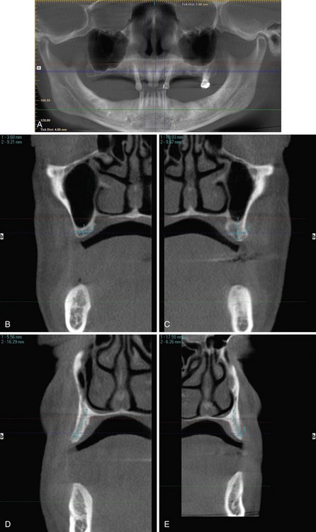
Treatment

Treatment with dental implants is initiated with a thorough physical examination, appropriate radiology (CBCT), and a coordinated treatment plan devised in cooperation with the restorative dentist. Numerous modalities for the management of the atrophic posterior maxilla in different clinical circumstances are available to oral and maxillofacial surgeons; these techniques include maxillary sinus augmentation, zygomatic implants, LeFort I downfracture osteotomy, distraction osteogenesis, onlay cortical or cancellous grafting associated with various containment protocols, and guided tissue regeneration with various membrane technologies. Maxillary sinus floor augmentation has become the most popular strategy among surgeons due to its predictability, low morbidity, and technical simplicity. Various methods can be used to augment the excessively pneumatized maxillary sinus to accommodate an implant of at least 10 mm in length. Sinus membrane elevation followed by implant placement without grafting has its advocates also. A lateral wall antrostomy, or window (open technique), is the most common technique used to expose the sinus floor. Alternatively, the Summer osteotome technique (closed technique) can be used for selected cases when less than 4 mm of sinus floor elevation is needed. The grafting material or materials are selected based on the surgeon’s preference. If a decision is made to use autogenous bone, the harvest technique planned must be explained to the patient so that informed consent can be obtained (the tibial plateau bone harvesting technique is discussed later). The decision on simultaneous or staged augmentation and implant placement is made based on the quality and quantity of host bone at the surgical site.

There are four primary types of grafting material available for sinus augmentation:

These materials can be used alone or in combination (composite graft) for sinus augmentation. Autogenous bone (cancellous marrow or cortical shavings) is a popular and predictable material for sinus grafting. Donor sites for bone harvest include intraoral sites (maxillary tuberosity, zygomatic buttress, mandibular ramus, posterior body or symphysis) and extraoral sites (the tibial plateau and anterior iliac crest are the most commonly used). Donor site selection is based on the clinical situation and the amount and type of bone needed. Intraoral sites must be considered a limited source of cancellous marrow but are a good source of surface-derived autogenous cortical bone (cortical shavings). Extraoral sites can provide sufficient autogenous cancellous marrow for large, bilateral augmentations. Some surgeons prefer to construct a composite graft by mixing autogenous bone with allogenic, alloplastic, or xenogenic graft materials, especially when inadequate autogenous bone is available.

Another alternative modality for maxillary sinus floor augmentation is the use of recombinant human bone morphogenetic protein 2 (rhBMP-2), which has been shown to induce de novo bone formation. rhBMP-2 in combination with a collagen sponge (Infuse; Medtronic, Inc. Minneapolis, Minnesota) is placed on the sinus floor in a fashion similar to bone graft material; it acts as an osteoinductive factor that stimulates undifferentiated mesenchymal cells to transform into osteoprogenitor cells and produce bone. De novo bone formation for sinus augmentation and placement of functional implants has been shown to be predictable and comparable to that seen with autogenous bone grafting; however, recent reports of increased adverse events with this modality have been published.

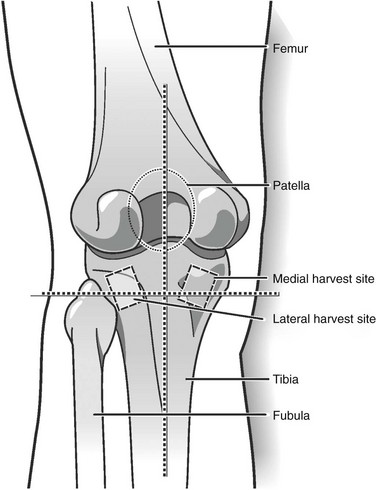
The technique of tibial bone harvest can be briefly discussed by pointing out that cancellous bone marrow of the tibial plateau can be approached by a medial or lateral route. Because the medial approach seems now to be the preferred method, this technique is discussed. The patient is placed in the supine position in the operating room or in the recumbent position in a dental chair if the procedure is performed in a clinic setting. A broad-spectrum antibiotic (generally a cephalosporin) is given as prophylaxis, and the surgical site is prepped and draped appropriately. The tibial tuberosity is located, and lines perpendicular and parallel to its long axis, intersecting at the center of the tuberosity, are scribed. A point 15 mm medial to the vertical line and 15 mm superior to the horizontal line is marked; this is the center of the incision. A 1- to 1.5-cm oblique incision is made over this point to the underlying bone (Figure 6-6). The periosteum is reflected, and a 1-cm circular osteotomy is prepared. The thin cortical window is removed, and cancellous bone is harvested with a curette. The upper boundary is 1 cm above the window to avoid the articular surface of the tibial plateau. Lateral and medial harvesting is done until the cortical bone is reached. Lower harvesting can proceed as far as the curette will reach. The wound is closed in layers, with attention paid to not tightly closing the periosteum. A pressure wrap is placed to complete the procedure. About 15 cc of noncompressed bone can be obtained in such a harvest. The strength of the tibia is unaffected by the surgery.

In the current case, because the bone quantity needed was minimal, the decision was made to use a human bone allograft in a putty combination (RegenerOss; Biomet 3-I, LLC, Palm Beach Gardens, Florida). For this type of surgery, the maxillary sinus wall (just below the zygomatic buttress) is exposed with a crestal incision and vertical releases as needed. An antrostomy, or window to access the maxillary sinus, is created with a carbide or diamond burr (a piezosurgery unit may be used if preferred). The bony window can be completely removed or infractured, as preferred, and the sinus membrane is carefully elevated off of the antral floor and walls and positioned medially and superiorly. Occasionally septae can be encountered that make dissection of the membrane more difficult and increase the risk of membrane perforation. If sinus membrane perforations occur, they can be enfolded (if small) or repaired using a collagen membrane (if larger). After reflection of the sinus membrane, the implant osteotomies are completed, the graft is placed, and the implants are inserted. The antrostomy osteotomy is covered with a slow-resorbing collagen membrane, and closure is obtained with resorbable sutures.

For the current patient, the immediate postoperative CBCT is shown in Figure 6-7, A. Subsequently, when the implants were uncovered and the healing caps placed (about 4 months after surgery), the sinus grafts demonstrated good consolidation (Figure 6-7, B). A CBCT cut from the area of the most posterior fixture on the right (Figure 6-7, C) further demonstrated graft remodeling and consolidation.
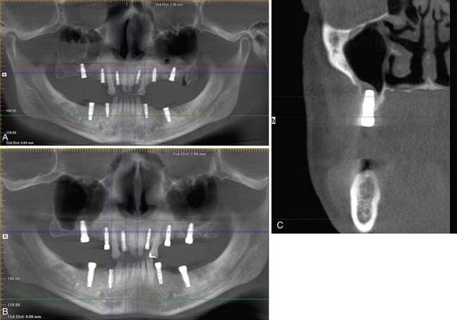
Complications

Complications of sinus grafting can be divided into intraoperative and postoperative types. Significant intraoperative complications are unusual; however, considerable bleeding can occasionally be encountered. Sinus membrane perforation occurs in more than 20% of cases but is not considered a risk factor for implant survival generally. Infection is the most consistent and serious postoperative complication, although most series report an occurrence rate of less than 5%. Infections are managed by incision and drainage, along with antibiotic therapy; many respond to this conservative management and heal uneventfully. However, infection is the major cause of implant fixture loss in most series reported. If tibial harvesting is performed, the complication rate is similarly low, and most problems associated with this procedure resolve with conservative management.

Discussion

Implant cases involving sinus grafting show a cumulative implant survival rate of more than 93% up to 5 years. Prosthesis survival rates are reported to be even higher. The patient’s age, gender, health status, and smoking habits; the implant’s size, shape, and surface; the residual ridge height; the timing of implant placement with respect to grafting; the graft material; and the occurrence of surgical complications have been evaluated in analyses in an attempt to identify significant risk factors for implant failure in sinus grafting. Smoking more than 15 cigarettes per day and a residual ridge height of less than 4 mm are reported to be associated with significant reductions in implant survival rates. Smoking habits and residual ridge height should be carefully evaluated prior to sinus elevation procedures.

Bibliography

Balaji, SM. Tobacco smoking and surgical healing of oral tissues: a review. Indian J Dent Res. 2008; 19:344–348.

Herford, AS, King, BJ, Audia, F, et al. Medial approach for tibial bone graft: anatomic study and clinical technique. J Oral Maxillofac Surg. 2003; 61:358–3563.

Jensen OT, ed. The sinus bone graft, ed 2, Chicago: Quintessence, 2006.

Jensen, T, Schou, S, Svendsen, PA, et al. Volumetric changes of the graft after maxillary sinus floor augmentation with Bio-Oss and autogenous bone in different ratios: a radiographic study in minipigs. Clin Oral Implants Res. 2012; 23:902–910.

Liddelow, G, Klineberg, I. Patient-related risk factors for implant therapy: a critique of pertinent literature. Aust Dent J. 2011; 56:417–426.

Lin, IC, Gonzalez, AM, Chang, HJ, et al. A 5-year follow-up of 80 implants in 44 patients placed immediately after the lateral trap-door window procedure to accomplish maxillary sinus elevation without bone grafting. Int J Oral Maxillofac Implants. 2011; 26:1079–1086.

Padmanabhan, TV, Gupta, RK. Comparison of crestal bone loss and implant stability among the implants placed with conventional procedure and using osteotome technique: a clinical study. J Oral Implantol. 2010; 36:475–483.

Renouard, F, Rangert, B. Risk factors in implant dentistry: simplified clinical analysis for predictable treatment, ed 2. Hanover Park, Ill: Quintessence; 2008.

Rodriguez-Argueta, OF, Figueiredo, R, Valmaseda-Castellon, E, et al. Postoperative complications in smoking patients treated with implants: a retrospective study. J Oral Maxillofac Surg. 2011; 69:2152–2157.

Singh, D, Teo, SG, Poh, KK. Regular narrow complex tachycardia. Singapore Med J. 2011; 52:146–149.

Testori, T, Weinstein, RL, Taschieri, S, et al. Risk factor analysis following maxillary sinus augmentation: a retrospective multicenter study. Int J Oral Maxillofac Implants. 2012; 27:1170–1176.

Triplett, RG, Nevins, M, Marx, RE, et al. Pivotal, randomized, parallel evaluation of recombinant human bone morphogenetic protein-2/absorbable collagen sponge and autogenous bone graft for maxillary sinus floor augmentation. J Oral Maxillofac Surg. 2009; 67:1947–1960.

Urban, IA, Nagursky, H, Church, C, et al. Incidence, diagnosis, and treatment of sinus graft infection after sinus floor elevation: a clinical study. Int J Oral Maxillofac Implants. 2012; 27:449–457.

Vittayakittipong, P, Nurit, W, Kirirat, P. Proximal tibial bone graft: the volume of cancellous bone and strength of decancellated tibias by the medial approach. Int J Oral Maxillofac Surg. 2012; 41:531–536.

Wallace, SS, Tarnow, DP, Froum, SJ, et al. Maxillary sinus elevation by lateral window approach: evolution of technology and technique. J Evid Based Dent Pract. 2012; 12(3 Suppl):161–171.

Wong, J, Lam, DP, Abrishami, A, et al. Short-term preoperative smoking cessation and postoperative complications: a systematic review and meta-analysis. Can J Anaesth. 2012; 59:268–279.

Woo, EJ. Adverse events reported after the use of recombinant human bone morphogenetic protein 2. J Oral Maxillofac Surg. 2012; 70:765–767.

Zygomatic Implants

CC

Only gold members can continue reading. Log In or Register to continue

Stay updated, free dental videos. Join our Telegram channel

Jan 12, 2015 | Posted by in Oral and Maxillofacial Surgery | Comments Off on 6: Dental Implant Surgery

VIDEdental - Online dental courses

Get VIDEdental app for watching clinical videos