14: Syndromes of the Head and Neck

Syndromes of the Head and Neck

A multitude of anomalies and syndromes occur in the head and neck, most of which are beyond the scope of this book. The chapters in this section cover some of the most common anomalies or syndromes associated with the craniomaxillofacial region (Table 14-1). Congenital anomalies include nonsyndromic craniosynostosis, hemifacial microsomia, and cleft lip and palate; congenital syndromes include Apert and Crouzon. Obstructive sleep apnea syndrome, although not congenital in nature, is included in this section because its pathogenesis and manifestation are based on anatomic anomalies of the head and neck. Oral and maxillofacial surgeons are uniquely trained to play an integral role in the surgical management of these patients, syndromic or not.

The intent of these chapters is to familiarize the readers with the pathogenesis, presentation, and management of such anomalies. The chapters are structured so that key features of each syndrome or anomaly are emphasized. The reconstructive strategies and rationale for treatment are discussed. Because of the complexity of the craniofacial deformities involved in the growing child, the reconstructive efforts are generally staged. There is no consensus on the best timing of each stage; however, the general guidelines are presented. Various surgical strategies can be used, depending on the surgeon’s preference and the clinical situation. These are presented in the various sections, along with the rationale for treatment.

Cleft Lip and Palate

PMHX/PDHX/Medications/Allergies/SH/FH

Except for the CLP, the child has no other medical problems. She was born with Apgar scores of 8 and 9, at 1 and 5 minutes, respectively. There are no facial or systemic anomalies characteristic of any known syndromes (see Discussion later in this section), including any associated cardiac, respiratory, ophthalmologic, or musculoskeletal abnormalities. There is no family history of CLP or CP.

Pedigree analysis has demonstrated an increased incidence when a family member has CLP. In addition, if a couple has a child with CLP, the risk of having an affected second child is significantly increased (fortyfold).

Infants born with CLP are at increased risk for associated congenital syndromes, particularly congenital cardiac disease. As such, obtaining a screening echocardiogram should be considered to rule out congenital valvular heart disease or transposition of the great vessels. In addition, the presence of CLP interferes with appropriate feeding, which in turn can lead to failure to thrive in the infant. The use of special cleft nipples is recommended, in addition to consultation with a maxillofacial prosthodontist for obturator fabrication.

Examination

General. Female infant is in the 25th percentile for weight and height (likely due to difficulty with feeding).

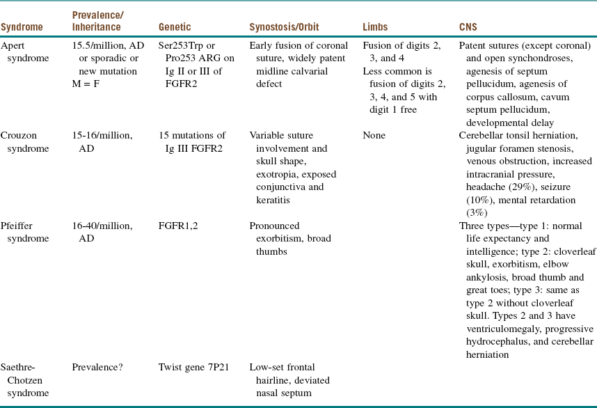
Maxillofacial. The cleft lip (CL) is complete, penetrating the entire thickness of the lip, alveolus, nasal tip cartilages, and floor of the nose (Figure 14-1). The cleft is unilateral, right of midline (left side prevalence), and continuous with the palate (CLP is most commonly expressed unilaterally, with a 2 : 1 predilection for the left side).

Intraoral. The cleft continues through the hard palate and soft palate (structures anterior to the incisive foramen form the primary palate and posterior to the incisive foramen form the secondary palate). Throughout the cleft, the nasal cavity, nasal conchae, and posterior pharyngeal wall are readily visible. The nasal mucosa appears inflamed and ulcerated (due to irritation of the fragile tissue from feeding). Bidigital palpation identifies solid supportive bone along the palatal shelves bordering the cleft site (it is important to palpate the hard palate of any infant to detect the presence of a notch in the posterior border of the hard palate, suggesting the presence of a submucous cleft).

Assessment

Female infant with an isolated, complete, nonsyndromic CLP.

Although there are several classification systems for facial clefting, including one by Tessier, they are not routinely used at most centers.

CL is a unilateral or bilateral gap in the upper lip and jaw that forms during the third through seventh weeks of embryonic development. It develops from failure of fusion of the medial nasal process and the maxillary process. CLs are described as either complete or incomplete. A complete CL is a cleft of the entire lip and alveolar arch or premaxilla; an incomplete CL involves only the lip and often spares the soft tissue of the associated alar base. A CP is a gap in the hard and/or soft palate that forms during the fifth through twelfth weeks of development. CP forms as a result of failure of attachment and alignment of the levator veli, tensor veli palatini, uvular, palatopharyngeus, and palatoglossus muscles. The primary palate is formed by the lip, alveolar arch, and palate anterior to the incisive foramen (known as the premaxilla). The secondary palate is formed by the soft and hard palates posterior to the incisive foramen.

Treatment

There is no general consensus regarding the timing and techniques used for CLP surgery. Individual craniofacial centers and craniofacial surgeons follow various protocols according to their own experience, rationale, and preferences. The functional needs, esthetic concerns, and ongoing growth of affected individuals all create specific concerns that complicate the treatment process. Presurgical dentofacial orthopedics is increasingly used to optimize primary CL repair.

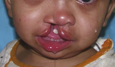
Table 14-2 outlines the sequence of management of patients with CLP. CL repair is usually addressed at 10 to 14 weeks of age. One advantage of waiting until this age is that it allows time for a thorough medical evaluation to determine whether the infant has any congenital defects. The surgical procedure is generally easier to perform when the child is slightly larger, because anatomic landmarks are more prominent and well defined. In addition, it has historically been accepted that the safest anesthesia time period for infants is based on the “rule of tens”—surgery can be performed when the child is at least 10 weeks of age, weighs at least 10 pounds, and has a minimum hemoglobin value of 10 mg/dl (however, there is no current scientific rationale to support this rule). With modern intraoperative pediatric monitoring techniques, general anesthesia can be performed safely at an earlier age as needed, although there is no documented benefit to performing lip repair before 3 months of age. In addition, excessive scarring and inferior esthetic results have been found to occur when surgery is performed earlier than age 3 months.

Table 14-2

Sequence of Management of the Cleft Palate

image

Modified from Kaban LB, Troulis MJ (eds): Pediatric oral and maxillofacial surgery, St Louis, 2004, Saunders.

CP repair is usually performed between 9 and 18 months of age. It is intended to coincide with the progression of natural speech development and growth. In deciding upon the timing of repair, the surgeon must consider the delicate balance between facial growth restriction after early surgery and early speech development, which requires an intact palate. Most children require an intact palate to produce certain sounds by 18 months of age. If developmental delay is present and speech is not anticipated to develop until later, CP repair can be delayed. Coincident with CP repair is placement of myringotomy tubes, because CLP patients have a higher incidence of middle ear disease.

There is very little evidence to support palate repair before 9 months of age. Surgical repairs before this time are associated with a higher incidence of maxillary hypoplasia later in life and show no improvement in speech. After initial CP repair, 20% of the children develop inadequate closure of the velopharyngeal mechanism (velopharyngeal insufficiency). This is usually diagnosed at 3 to 5 years of age, when a detailed speech examination can be performed. Surgery is performed to correct the anatomic defect, with the goal of improving closure between the oral and nasal cavities and reducing nasal air escape during the production of certain sounds.

Approximately 75% of patients with any type of cleft present with clefting of the maxilla and alveolus. Bone graft reconstruction of the alveolus is performed during the mixed dentition period, before eruption of the permanent canine and/or permanent lateral incisor. The timing of this procedure is based on dental development and not chronologic age. Reconstruction of the alveolus before the mixed dentition stage has been associated with a high degree of maxillary growth restriction, requiring orthognathic correction later in life. Autogenous bone grafted from the iliac crest has provided the best results for reconstruction of alveolar cleft defects. Although globally there are multiple recommendations with regard to the timing of the bone graft, most commonly in the United States the alveolar grafting is completed when the associated canine root is two thirds formed; that is, typically at 9 to 11 years of age. Prior to grafting, orthodontic expansion of the maxilla is indicated to maximize the amount of graft placed in the alveolus.

Orthognathic reconstruction of maxillary and mandibular discrepancies is generally performed from 14 to 18 years of age based on individual growth characteristics. This is performed in conjunction with orthodontics before and after surgery. Orthognathic surgery before this time frame is performed only for severe cases of dysmorphology.

Lip and nasal revision is best done once the majority of growth is complete, which generally occurs after 5 years of age; it is usually performed only for severe deformities. When orthognathic reconstruction is planned, rhinoplasty is best performed after orthognathic surgery, because maxillary advancement improves many characteristics of nasal form.

Treatment techniques for CL. Unilateral CL repair, as previously mentioned, is usually performed after 10 weeks of age. The technique most commonly used is the Millard rotation-advancement technique. The basic intent of the repair is to create a three-layer closure of skin, muscle, and mucosa that approximates the normal tissue and excises hypoplastic tissue at the cleft margins. The orbicularis oris muscle is adapted to form a continuous sphincter. The incision lines of the Millard technique also fall within the natural contours of the lip and nose on closure, which helps promote a natural form of symmetry. The C-flap can be used to lengthen the columella or create a nasal sill.

The Randall-Tennison technique is a Z-plasty technique used by some surgeons for unilateral CL repair. This technique does not achieve the same semblance of symmetry obtained with the Millard technique.

Primary nasal reconstruction may be performed at the time of lip repair to reposition the displaced lower lateral cartilages and alar tissues. Various techniques have been advocated, each with considerable variation. The repair essentially involves releasing the alar base, augmenting the area with allogenic subdermal grafts, or proceeding with open rhinoplasty with minimal dissection to avoid scar formation.

Bilateral lip repair is a very challenging technical procedure, primarily due to the lack of quality tissue present and the manner of separation of the tissues caused by the clefting. The typically shortened columella and rotation of the premaxillary segment make achieving acceptable aesthetic results difficult. Variations to surgical approaches range from aggressive lengthening of the columella with preservation of hypoplastic tissue to conservative primary nasal reconstruction as performed with McComb’s unilateral CL technique. McComb’s technique involves release and repositioning of the lower lateral cartilages and alar base on both sides without aggressive degloving of the entire nasal complex. Aggressive corrective techniques often produce initial results that are very good. Long-term results, however, are not so favorable due to the progression of natural growth processes. Excessive angulations and lengthened structures provide a less-than-optimal esthetic effect. Revision of these deformities is usually very difficult, and sometimes impossible. In general, if hypoplastic tissue is excised and incisions within the medial nasal base and columella are avoided, long-term esthetic results are excellent.

Treatment techniques for CP. Successful CP repair during infancy depends on two objectives. The first involves water-tight closure of the entire oronasal communication involving the hard and soft palate. The second involves anatomic repair of the musculature within the soft palate, which is critical for the creation of normal speech. The soft palate functions in coupling and decoupling of the oral and nasal cavities in the production of speech. The tensor and levator palatini and uvularis muscles, which usually join at the midline, forming a continuous sling, are separated and insert along the posterior edge of the hard palate. Surgical treatment of a CP is concerned with closing the palatal defect and releasing the abnormal muscle insertions. The timing for CP repair is correlated with the development of speech, which usually occurs around 18 months for a normally developing child. The velum, or soft palate, must be closed before the development of speech. If repair occurs after this time, compensatory speech articulations may result. The timing of surgery must also be balanced with the known biologic consequences of performing surgery during infancy, specifically during the growth phase, which could result in maxillary growth restriction. When repair of the palate is performed between 9 and 18 months of age, the incidence of maxillary restriction is approximately 25%. If repair is carried out earlier than 9 months of age, the incidence of severe growth restriction requiring future orthognathic surgery is greater. In addition, CP repair before 9 months of age is not associated with any increased benefit in terms of speech development. Performing CP repair between 9 and 18 months of age seems to best address the functional concerns of speech development and the potential negative impact surgery has on growth.

An approach used to address the speech issues with growth-related concerns involves staging the closure of the secondary palate with two procedures. This involves repair of the soft palate early in life, followed by closure of the hard palate later during infancy. The intent of this approach is to accomplish timely repair of the soft palate, which is critical for speech, while delaying hard palate repair until further growth has occurred. This technique offers the advantage of less growth restriction, easier repair of larger clefts, and less chance for fistula formation.

The basic premise of CP repair involves mobilization of multilayered flaps to close the defect created due to the failed fusion of the palatal shelves. The nasal mucosa is first closed, followed by reconstruction of the levator and tensor palatini muscles. The abnormal insertion of the levator and tensor palatini muscles on the hard palate must be removed and reconstructed to join in the midline. The musculature making up the velopharyngeal mechanisms are also reconstructed to allow the soft palate to close the space between the nasopharynx and oropharynx in order to create certain speech sounds. Closure of the oral mucosa completes the repair.

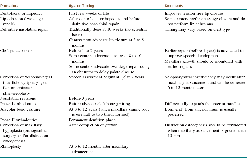
Many techniques have been devised for CP repair. The Bardach technique involves creation of two large, full-thickness flaps on each palatal shelf, which are layered and brought to the midline for closure (Figure 14-2, A and B). This technique allows for preservation of the palatal neurovascular bundle, which is contained within the pedicle of each flap. The Von Langenbeck technique is similar to the Bardach technique, but it preserves an anterior pedicle for increased blood supply to the flaps. It also involves elevation of large mucoperiosteal flaps from the palate with midline approximation of the cleft margins. Long lateral releasing incisions are made at the border of the palatal and alveolar bone to allow mobilization. The levator muscles are detached from their abnormal insertion along the hard palate. The Furlow double-opposing Z-plasty technique involves two Z-plasties, one on the oral mucosa and one in the reverse orientation on the nasal mucosa (Figure 14-2, C). The levator muscle on one side is included in the posteriorly based oral mucosa Z-plasty, whereas the levator muscle from the opposite side is included within the posteriorly based nasal mucosal Z-plasty flap. This procedure produces palatal lengthening and reorients and provides overlap of the malpositioned levator muscles. The Furlow Z-plasty has been reported to be associated with a higher rate of fistula formation at the junction of the soft and hard palates.

The Wardill-Kilmer-Veau technique is a VY advancement of the mucoperiosteum of the hard palate and is intended to lengthen the palate in the anteroposterior plane at the time of palatoplasty. Bone is left exposed in the area where the flaps were advanced. These areas granulate and epithelialize within 2 to 3 weeks but form excessive scat tissue that may contribute to maxillary growth disturbances. The vomer flap is used to achieve closure of the hard palate. A wide, superiorly based flap of nasal mucosa is elevated from the vomer and attached to the palatal shelf to close the defect. The vomer flap eliminates the need to elevate large mucoperiosteal flaps from the hard palate, thus avoiding possible maxillary growth disturbances.

For very wide clefts, a pharyngeal flap may be used. This technique allows the central portion of the cleft to be filled with posterior pharyngeal wall tissue, making the closure of the nasal and palatal mucosa easier. Patients with Pierre Robin sequence (malformation) or Treacher Collins syndrome have exceptionally wide clefts that are difficult to close without tension. The pharyngeal flap seems to address the concerns for CP repair with these patients. Pharyngeal flaps, however, pose an increased risk of bleeding, snoring, obstructive sleep apnea, and hyponasality.

Figure 14-3 shows the 1-year postoperative view of the patient seen in Figure 14-1.

image

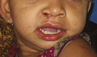
Figure 14-3 Postoperative photograph of patient in Figure 14-1 after lip and palate repair at 1 year of age. (Courtesy of Dr. Shahid Aziz.)

Complications

The complications associated with cleft repair are essentially related to the technique used for treatment. The overall goals include nasal lining closure, adequate exposure, and release of soft tissue attachment along the bony borders of the cleft from the alveolar crest to the pyriform rim and closure of the oral mucosa with well-vascularized tissue that contains attached mucosa at the alveolar crest. Failure to address these concerns during treatment ultimately leads to complications that include wound infection with fistula formation, mucosal dehiscence, hypertrophic scar formation, and hemorrhage.

It is also important to note that many patients with CLP have coexisting systemic abnormalities that may negatively affect the outcome of the treatment provided. Patients who present with systemic abnormalities in general are expected to have a higher incidence of complications compared with healthy patients. With surgical correction of CLP, Lees and Pigott observed a high incidence of intraoperative and postoperative complications related to the respiratory system.

The most significant complication or unfavorable result of palate closure is the development of velopharyngeal insufficiency. This is manifested as a resonance problem, with creation of hypernasal or hyponasal speech. Velopharyngeal insufficiency can be secondary to maxillary orthognathic (advancement) surgery or the use of pharyngeal flaps. The resonance occurs from the modification of oronasal portals that are either too large or too narrow. Correction of this problem involves reconstructive surgery with revision or creation of a new pharyngeal flap, accompanied by aggressive speech therapy.

A common complication of lip repair is the “whistle” deformity, which occurs due to vertical retraction of the scar or from inadequate advancement and rotation of the skin flap. Various lip-lengthening procedures can be performed secondarily, such as the V-Y advancement, which corrects the deformity and creates a normal lip seal.

In cases of complete bilateral CLP, collapse of the alveolar segments posterior to the premaxilla is a common occurrence when orthodontic or palatal retention devices are not used. Correction of segment collapse is very complex, involving multiple surgeries over an extended period.

Discussion

CL and/or CP malformations are the most common congenital abnormalities in the facial region. Worldwide, the incidence of CL is approximately 1 : 700 live births. The incidence of CP is approximately 1 : 2,000 live births. CLP patients routinely have impaired facial growth, dental anomalies, speech disorders, poor hearing, psychological difficulties, and poor social relationships. Due to the multiple factors associated with CLP, specialty multidisciplinary teams are involved in the overall care of these patients. The involvement of the team starts during the immediate neonatal period and continues through completion of growth and adolescence. The multidisciplinary team is composed of a craniofacial surgeon, an oral and maxillofacial surgeon, a pediatrician, an otolaryngologist, a pediatric dentist, an orthodontist, an audiologist, a speech and language therapist, a geneticist, a psychologist, and a social worker.

CP deficiencies commonly go undetected during infancy, only to be identified later during childhood when the resultant anomaly becomes very apparent with the emergence of speech, feeding, and growth complications. It is therefore very important to accurately assess the palatal anatomy of any infant before such deficiencies create significant problems. During the initial inspection and examination of the palatal anatomy of infants, the presence of a submucous cleft is quite frequently missed. With submucous clefts, on visual inspection the palate appears intact; however, the overlying oral and nasal mucous membranes are expanded against the cleft area, giving the illusion of an intact palate. Digital palpation identifies a notch or discontinuity along the posterior aspect of the bony hard palate. The submucous cleft represents a deficiency in the musculature of the palate due to failed midline fusion of the palatal muscles, namely, the levator veli palatini, tensor veli palatini, uvulus, palatoglossus, and palatopharyngeus muscles. A bluish midline streak is often present over the soft palate, which indicates the splitting of the muscle layers.

A number of concerns are associated with CLP.

• Feeding. Approximately 25% of CLP infants have early feeding difficulties, with poor weight gain for the first 2 to 3 months. Feeding sessions are prolonged due in part to ulceration of the nasal mucosa. Some infants also have increased metabolic needs due to congenital heart disease or airway obstruction. Initial poor weight gain usually resolves after cleft closure, and any deficiency in growth is corrected by 6 months of age. Height and weight progressions are routinely monitored.

• Speech and language development. Even after the palate has been repaired, children are still at risk for subsequent speech disorders. It is reported that 25% of children with CLP develop normal speech after primary surgery, whereas the remaining 75% require many surgical interventions throughout childhood and adolescence. Speech problems arise from velopalatal insufficiency, dental and occlusal problems, oronasal fistulas, and hearing problems. Approximately 15% to 20% of patients who have CP repair within the first 12 to 15 months of life have velopharyngeal insufficiency. As mentioned earlier, surgical intervention must be coordinated with the development of speech. Speech and language therapy must also be provided during this time. The monitoring of speech continues into adolescence and adulthood in conjunction with active orthodontic and surgical management.

• Hearing. Patients with CP are at increased risk of middle ear effusions and subsequent infections. The attachment of the levator veli palatini muscle around the eustachian tube is abnormal and leads to poor aeration and drainage of the middle ear. Regular assessment by the ear, nose, and throat (ENT) surgeon and audiologist is recommended to ensure that poor hearing is not a contributing factor to compromised speech.

• General dental welfare. Children with CLP are at great risk for developing malocclusion. When the cleft involves the alveolar process, odontogenic structures within this region are routinely absent or malformed. Orthodontic intervention is generally initiated during the preschool years. Active occlusal manipulation and correction should not be instituted until the permanent dentition has erupted.

• Genetics. There are three types of genetic risk groups for CLP: the syndromic group, identified by physical examination; the familial group, identified by history; and isolated defects, identified by exclusion of the first two groups. As mentioned earlier, the incidence of CL is approximately 1 : 700 live births, and the incidence of CP is approximately 1 : 2,000 live births. With one parent and one child affected, the chance of a second child having a cleft is 10%. When both parents are without clefts and two children have clefts, the chance of a third child having a cleft is 19%. When one parent has a cleft and two offspring are normal, the chance of the third child being born with a cleft is 2.5%.

• Environment. Epidemiologic studies have demonstrated a relationship between maternal exposure to environmental factors or teratogens during pregnancy and the development of CLP. These factors or teratogens include alcohol consumption, cigarette smoking, folic acid deficiency, corticosteroids, benzodiazepines, and anticonvulsants.

Bibliography

Bryne, PJ, Sands, NR. Secondary bone grafting of residual alveolar and palatal clefts. J Oral Surg. 1972; 30:87–92.

Carici, F. Recent developments in orofacial cleft genetics. J Craniofac Surg. 2003; 142(2):130–143.

Chung, K, Kowalski, C, Kim, HM, et al. Maternal cigarette smoking during pregnancy and the risk of having a child with cleft lip/palate. Plast Reconstr Surg. 2000; 105(2):485–491.

Copeland, M. The effect of very early palatal repair on speech. Br J Surg. 1990; 43:676.

Costello, BJ, Ruiz, RL, Turvey, T. Surgical management of velopharyngeal insufficiency in the cleft patient. Oral Maxillofac Surg Clin North Am. 2002; 14:539–551.

Costello, BJ, Shand, J, Ruiz, RL. Craniofacial and orthognathic surgery in the growing patient. Select Readings Oral Maxillofac Surg. 2003; 11(5):1–20.

Dorf, DS, Curtin, JW. Early cleft repair and speech outcome: a ten year experience. In: Bardach J, Morris HL, eds. Multidisciplinary management of cleft lip and palate. Philadelphia: Saunders; 1990:341–348.

Glenny, AM, Hooper, L, Shaw, WC, et al. Feeding interventions for growth and development in infants with cleft lip, cleft palate or cleft lip and palate. Cochrane Database Syst Rev. 2, 2005.

Habel, A, Sell, D, Mars, M. Management of cleft lip and palate. Arch Dis Child. 1996; 74(4):360–366.

Lees, VC, Pigott, RW. Early postoperative complications in primary cleft lip and palate surgery: how soon may we discharge patients from hospital? Br J Plast Surg. 1992; 45:232–234.

Marsh, JL. Craniofacial surgery: the experiment on the experiment of nature. Cleft Palate Craniofac J. 1996; 33:1.

McComb, H. Primary correction of unilateral cleft lip nasal deformity: a ten year review. Plast Reconstr Surg. 1985; 75:791–799.

Murray, JC. Gene/environment causes of cleft lip and/or palate. Clin Genet. 2002; 61(4):248–256.

Padwa, BL, Sonis, A, Bagheri, SC, et al. Children with repaired bilateral cleft lip/palate: effect of age at premaxillary osteotomy on facial growth. Plast Reconstr Surg. 1999; 105:1261.

Posnick, JC. Cleft orthognathic surgery: the unilateral cleft lip and palate deformity. In: Posnick JC, ed. Craniofacial and maxillofacial surgery in children and young adults. Philadelphia: Saunders; 2000:860–907.

Posnick, JC. The staging of cleft lip and palate reconstruction: infancy adolescence. In: Posnick JC, ed. Craniofacial and maxillofacial surgery in children and young adults. Philadelphia: Saunders; 2000:785–826.

Ruiz, RL, Costello, BJ, Turvey, T. Surgical correction of midface deficiency in cleft lip and palate malformation. Oral Maxillofac Surg Clin North Am. 2002; 14(4):491–507.

Spritz, RA. The genetics and epigenetics of orofacial clefts. Curr Opin Pediatr. 2001; 13(6):556–600.

Takato, T, Yonohara, Y, Mori, Y. Early correction of the nose in unilateral cleft lip patients using an open method: a ten year review. J Oral Maxillofac Surg. 1995; 53A:28–33.

Nonsyndromic Craniosynostosis

HPI

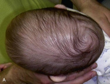
The mother of this 7-month-old, otherwise healthy male (craniosynostosis has a male predilection) has been concerned about the abnormal shape of his head, which was noticed immediately after birth. (Craniosynostosis is the premature fusion of the cranial sutures during intrauterine life. The deformity is often noticeable early [Figure 14-4].) The pediatrician has been closely observing this skull deformity for changes and resolution. It was initially assumed to be deformational plagiocephaly (skull deformity caused by vaginal delivery or early fetal decent into the pelvis) and later was thought to be secondary to a positional plagiocephaly, an acquired skull deformity caused by a repetitive head position during sleep (nonsynostotic posterior plagiocephaly has increased since the American Academy of Pediatrics issued a recommendation that infants be placed on their back during sleep to reduce the risk of sudden infant death syndrome [SIDS]). Despite conservative management, the child continued to exhibit the cranial deformity, which appeared to slightly worsen over time. He is otherwise in good health, and the mother denies any behavioral abnormalities. He is referred for craniofacial evaluation (the rate of detectable cranial abnormalities secondary to craniosynostosis has been reported to be as high as 1 : 1,700 to 1 : 1,900 births).

Examination

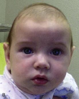
General. The patient is a well-developed and well-nourished, pleasant child in no apparent distress.

Maxillofacial. Examination of the skull reveals a mild dysmorphology (the exact dysmorphology varies greatly and depends on which portion or portions of the sagittal suture are involved) in which the cranial vault is narrow in the bitemporal and biparietal dimensions and abnormally elongated in the anteroposterior dimension (this is called scaphocephaly, meaning “long and narrow”). Frontal and occipital bossing is apparent (described as a “keel-like” appearance).

There is no midfacial or mandibular hypoplasia or asymmetry and no orbital dystopia (a relative discrepancy in globe position in the vertical and/or horizontal planes) or exophthalmos (anterior position of the globe relative to the orbital rims).

The fundoscopic examination is normal, with no evidence of papilledema (edema of the optic disc, which is indicative of elevated intracranial pressure).

Intraoral. The results of the examination are within normal limits (nonsyndromic craniosynostosis is not associated with an increased incidence of cleft lip/palate [CLP]).

Extremities. There are no deformities (nonsyndromic craniosynostosis does not have any associated abnormalities of the extremities).

Imaging

Plain film complete skull series comprise the initial diagnostic radiographs of choice (the clinical diagnosis of craniosynostosis must be confirmed radiographically). In the current patient, the radiographs showed the absence of the entire sagittal suture. (Sagittal suture synostosis can involve the entire suture, the anterior portion only, or the posterior portion only. If the sutures appear patent on a radiographic study of diagnostic quality, craniosynostosis can be ruled out.)

Craniofacial axial and coronal (or reformatted) cut CT scans and three-dimensional reconstructions provide more detailed morphologic information, which is very useful during surgical planning (CT scans are also indicated when plain films are nondiagnostic). In the current patient, CT scans showed a scaphocephalic skull deformity, which is consistent with synostosis of the sagittal suture. CT scans of the head showed no masses (the possibility of an intracranial mass should be included in the differential diagnosis of cranial vault abnormalities) and no hydrocephalus (this is usually not encountered in single-suture craniosynostosis but may occur independently; hydrocephalus is seen in approximately 10% of cases in which multiple sutures are involved).

Treatment

There are two primary goals in the surgical management of nonsyndromic craniosynostosis: (1) release of the fused suture or sutures to allow unrestricted growth of the brain and (2) reconstruction of all dysmorphic skeletal components to correct the anatomic form. The surgical team should be composed of a pediatric craniofacial surgeon and a pediatric neurosurgeon for optimal results (“strip craniectomy,” previously performed by neurosurgeons working independently, did not address the dysmorphology of the craniofacial skeleton and resulted in residual deformities). Modern craniofacial management includes a formal craniotomy performed by a neurosurgeon and simultaneous skeletal reconstruction by the craniofacial surgeon. Reconstruction and reshaping include the removal, dismantling, and reassembly of all dysmorphic skeletal components into an anatomically desirable shape. The extent of the surgery depends on the suture or sutures involved and the resultant skeletal deformity.

Although craniosynostosis is surgically addressed during the first year of life, the exact timing of craniosynostosis repair is controversial. Some surgeons prefer early surgical correction, when the child is 3 to 6 months of age. In theory, early release of the suture or sutures allows the expanding brain to naturally reshape the cranial vault, minimizing the later-staged reconstructive efforts. Other surgeons prefer delaying the surgical correction until 9 to 11 months of age, permitting more growth of the cranial vault before reconstruction. The more stable cranial skeleton may result in fewer postsurgical deformities. Also, increased bony calcification allows for easier rigid fixation of the bony segments.

Surgical correction of nonsyndromic sagittal suture craniosynostosis involves a biparietal craniotomy for release of the fused suture and reshaping of the posterior and anterior cranial vault. The abnormal cranial components are dismantled and osteotomized into strips for reshaping of the cranial vault. The objectives are to increase the bitemporal and biparietal width and to decrease the anteroposterior length of the cranial vault (reduce frontal and occipital bossing). The bony segments are placed in the correct anatomic position and secured with rigid miniplates with monocortical screws. When the surgery is performed by age 2, most bony gaps, including full-thickness defects, completely fill with bone because of the osteogenic potential of the periosteum and dura mater. Complete healing of these defects is less predictable when surgery is performed between 2 and 4 years of age. After age 4, these defects may not heal without immediate reconstruction (with bone grafting or other alloplastic material).

If the entire sagittal suture is involved, reconstruction can be done in a single-stage procedure (associated with increased difficulty, surgical time, blood loss, and morbidity) or in a two-stage operation. The staged reconstruction involves addressing and reshaping the posterior two thirds of the cranial vault; this corrects the bitemporal and biparietal width and improves the anteroposterior cranial dimension. It does not correct the frontal bossing, which must be addressed during the second surgical phase, 6 to 12 weeks later. Others in the past have advocated strip craniectomy (at least 3 cm wide) from the anterior fontanelle to just beyond the lambdoidal suture as adequate treatment when the child is between 3 months and 1 year of age. However, this technique (along with its endoscopically assisted variation) has been criticized for its less-than-ideal cosmetic results.

When only the posterior portion of the sagittal suture is fused, surgical reshaping of the posterior two thirds of the cranial vault can be accomplished via a postauricular coronal scalp incision, with the patient in a prone position. Formal biparietal and occipital craniotomy is performed by the neurosurgeon. The bone flaps are removed, osteotomized, placed in the correct anatomic position, and secured with bone plates using monocortical screws. If only the anterior portion of the sagittal suture is involved, the resulting deformity is primarily frontal bossing. A coronal flap is elevated, and a bifrontal craniotomy is performed with the patient supine. The anterior cranial vault is reshaped and fixated as previously described.

Complications

Despite the very low complication rates associated with craniofacial surgery, both intraoperative and postoperative complications can occur. Massive blood loss and postoperative infection are the most common and most feared complications (Box 14-1). There is a high likelihood that homologous blood transfusion will be required during cranial vault reshaping. This is in part due to the low effective blood volume in infants and children. Acute normovolemic hemodilution (ANH) is a technique commonly performed intraoperatively to reduce the need for transfusion. Additional techniques include intraoperative blood salvage. Maintaining a “safe” hematocrit level, between 28% and 35%, has been recommended. However, the incidence of transfusion has been estimated to be as high as 90%.

Stay updated, free dental videos. Join our Telegram channel

Jan 12, 2015 | Posted by in Oral and Maxillofacial Surgery | Comments Off on 14: Syndromes of the Head and Neck

VIDEdental - Online dental courses

Get VIDEdental app for watching clinical videos