5: Dentoalveolar Surgery

Dentoalveolar Surgery

Dentoalveolar surgery is the surgical procedure that oral and maxillofacial surgeons perform most often. These procedures are associated with the dentate segment of the maxilla or mandible, termed the alveolar ridge. They include a variety of procedures, including simple tooth extractions, alveoplasty (recontouring of the alveolar bone), removal of tori, exposure of impacted teeth for orthodontic treatment, and extraction of impacted third molars. The origins and the current practice of oral and maxillofacial surgery are heavily based on dentoalveolar surgery. Such procedures account for more than 50% of the practice of oral and maxillofacial surgeons worldwide. Recently, placement of dental implants was added to the rehabilitative and reconstructive options for the maxillofacial region, replacing the more traditional preprosthetic procedures.

Since its establishment in 1918, the American Association of Oral and Maxillofacial Surgeons (AAOMS) has gone through extensive change, reflecting progressive changes in the specialty. Initially called the American Society of Exodontists, it was originally formed by a group of oral surgeons in Chicago after the National Dental Association meeting. As it has grown, the association has gone through several name changes, each reflecting the expansion of the specialty. Despite the wide scope of training of graduating oral and maxillofacial surgeons, as evidenced by the sections in this book, dentoalveolar surgery remains the foundation of our specialty.

In this chapter we present teaching cases representing some of the important aspects of this branch of oral and maxillofacial surgery. Three cases focus on complications of dentoalveolar surgery (dry socket, lingual nerve injury, and displacement of a tooth fragment during surgery), and two discuss the current issues in the treatment of impacted canines and third molars.

Third Molar Odontectomy

Examination

General. The patient is a well-developed and well-nourished man in no apparent distress (higher levels of anxiety may require a deeper level of sedation/anesthesia).

Maxillofacial. There is no soft tissue abnormality or lymphadenopathy (LAD). The patient has a good range of mandibular motion with an maximal interincisal opening (MIO) of 45 mm. Examination of the TMJ reveals no abnormalities (clicks or pain upon palpation). The muscles of mastication are nontender to palpation (important to detect preexisting symptoms of TMD).

Intraoral. Oral soft tissue is free of lesions, and there is no evidence of acute infection. The mandibular third molars are partially erupted, with approximately 20% of the crown visible in the oral cavity with insufficient room for functional eruption. The overlying operculum appears slightly inflamed, with evidence of food debris and periodontal pockets of greater than 6 mm on the distal of the left and right mandibular second molars. The right and left maxillary third molars are partially erupted. Oral hygiene is fair. An examination of the oropharynx is without tonsillar hypertrophy, and the patient has a Mallampati score of 1.

Indications for the removal of third molars include tooth malposition, periodontal conditions (e.g., probing lengths greater than 4 mm at distal of second molars or third molars), pericoronitis, symptoms of pain, evidence of infection or caries, orthodontic considerations, lack of space, associated pathology, and inability to maintain oral hygiene, especially when the third molars are incompletely erupted. Although some controversy exists as to the timing of or necessity for removal of asymptomatic third molars, there is evidence that younger patients (under 25 years old) have a decreased risk of complications and improved recovery after surgery. The decision to remove asymptomatic third molars should be driven by evidence-based decision making; the predominant guiding principle should be the patient’s preference. The clinical findings should be the main driving factors in treatment decisions.

Imaging

A panoramic radiograph is the minimum imaging modality necessary for the evaluation and treatment of impacted third molars. Computed tomography (CT) scans are not necessary for the routine evaluation, but they may be used in select cases of suspected maxillofacial pathology or for accurate determination of the inferior alveolar nerve anatomy. Partial odontectomy (coronectomy) may at times be an alternative treatment in patients requiring removal of a third molar that is in close proximity to the inferior alveolar nerve; however, this procedure does not eliminate the risk of inferior alveolar nerve injury and possible future infectious complications due to retention of root fragments.

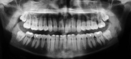
In the current patient, the panoramic radiograph reveals a partial lack of space to accommodate the eruption of the mildly mesioangularly impacted mandibular molars with 75% root development (Figure 5-1). The roots are not fused and do not extend below the level of the neurovascular bundle. The outlines of mandibular canals are easily discerned on the radiograph. There is no diversion of the inferior alveolar canal, darkening of the third molar root, or interruption of the cortical white line (risk factors associated with inferior alveolar nerve injury) (Box 5-1). The maxillary third molars are vertically positioned with partial bony impaction. The maxillary sinuses and the remainder of the radiograph are within normal limits.

Treatment

Two major professional organizations have made contradictory recommendations on the prophylactic removal of impacted third molars. The researchers for AAOMS Third Molar Clinical Trials published several scientific articles that linked third molars to future health problems in adults. In light of these findings, in 2005 the AAOMS suggested that removal of the third molars during young adulthood may be the most prudent option. In contrast, the National Health Service (NHS) of Great Britain and an associated agency, the National Institute of Clinical Excellence (NICE), published a series of guidelines recommending that “the practice of prophylactic removal of pathology-free impacted third molars should be discontinued in the NHS.” These guidelines, made public in 2000, did acknowledge the ongoing AAOMS Third Molar Clinical Trials. In 2012 Renton and colleagues published an article chronicling the United Kingdom’s experience with retention of third molars. They concluded that “admissions for M3

surgery activity under the NHS have decreased from the mid-1990s and into the 2000s, in association with professional and policy guidelines.” They found that the average age for third molar surgery had risen, and the indications for the surgery were “increasingly associated with other pathologic features such as dental caries or pericoronitis, in line with NICE guidelines.”

Although in some regions of the world, socioeconomic and available resources play a major role in the determination of guidelines for third molar extractions, current scientific evidence remains unchanged. The cumulative financial costs of treating the health complications of retained third molars in the older population should be considered. Although there is cost associated with the procedure to remove third molars, there is also the cost of monitoring retained third molars. Subsequent removal at an older age may also be associated with the cost of lost income in recuperation time, in addition to the greater risks of removal at an older age.

It is clear that the extraction of third molars poses some risks to the patient. However, the determination of extraction versus nonextraction of asymptomatic third molars must compare the cost and risks of surgical extraction with the lifetime health and cost benefits of preventing and eliminating any pathologic processes associated with retention of the third molars.

The effectiveness, safety, and relatively minimal cost of extraction of third molars using outpatient, office-based anesthesia, along with the currently available scientific evidence linking asymptomatic third molars to multiple health hazards, generally support the extraction of asymptomatic third molars in young adults; however, as mentioned, the patient’s preference and an informed decision arrived at by the surgeon and patient are the most important deciding factors.

The current patient was seen in the clinic for extraction of his teeth under intravenous sedation. Monitors (pulse oximetry, capnography, blood pressure, and three-lead electrocardiography) were placed, and oxygen was delivered via a nasal mask at 4 L/min, followed by nitrous oxide. Midazolam and fentanyl were slowly titrated until a comfortable state of conscious sedation was achieved. A local anesthetic with epinephrine was injected, and adequate time was allowed for the local anesthetic block. A bite block was place for TMJ stabilization. An oral screen with loosely packed, moist gauze was placed to protect the airway from accidental aspiration. A full mucoperiosteal flap was elevated using a buccal envelope incision with a distal hockey-stick extension for the mandibular third molars. Special consideration was given to preventing trauma to the lingual tissue.

A buccal trough was made using a high-speed instrument (impaction drill and burr with irrigation), and the teeth were elevated and extracted. Careful attention must be given to avoiding violation of the lingual cortex (although at times, disrupting the lingual cortex is unavoidable). The neurovascular bundle was not visualized, and there was no excessive hemorrhage from the socket (visualization of the neurovascular bundle and excessive hemorrhage from the socket are associated with an increased risk of inferior alveolar nerve injury). The wound was irrigated with normal saline, and the flaps were closed with chromic suture, with careful attention paid to suturing only the superficial lingual mucosa and thus preventing lingual nerve injury.

The upper third molars were removed through an envelope mucoperiosteal flap. Care was taken to avoid the roots of the maxillary second molars (a possible complication). There was no evidence of an oral antral communication. The tooth follicles were removed, and the sites were irrigated. Gauze was placed between the teeth to promote hemostasis, and the patient was monitored in the recovery room until he was fully awake and alert.

Complications

As mentioned, third molar extraction is the surgical procedure that oral and maxillofacial surgeons perform most often. A well-planned surgical approach, with the goal of prevention, is the best way to minimize complications. Yet, despite our best efforts, complications are expected, and it is best to counsel patients preoperatively for potential risks. Clinicians need to be aware of the risk factors associated with an increased risk of complications for this commonly performed procedure.

Pogrel concluded, “The age of 25 years appears in many studies to be a critical time after which complications increase more rapidly.” No studies indicate that complications decrease as age increases. In fact, the older a patient is, the more likely it is that the recovery from complications will be prolonged, less predictable, and less complete.

Sensory nerve injury is well documented. Injury to the inferior alveolar nerve can lead to a range of symptoms along its distribution (anesthesia, hypoesthesia, dysesthesia, or paresthesia). A review of the literature demonstrates an incidence of nerve injury between 0.4% and 5%. In one large study with 367,170 patients, the incidence of nerve injury was 0.4% (22% of whom had symptoms lasting longer than 12 months). The risk of nerve injury is greater with increasing patient age, degree of root development, degree of impaction, and the radiographic relationship of the roots to the inferior alveolar canal. The incidence of injury to the inferior alveolar nerve is slightly higher than that for the lingual nerve, but the inferior alveolar nerve has a higher incidence of spontaneous recovery (due to its position in the bony canal, which allows a greater possibility that the nerve endings will reapproximate); however, older patients are more likely to have incomplete recovery. Injury to the long buccal nerve is also possible, but it is less of a concern, causing minimal to no subjective disability. Patients with severe inferior alveolar nerve or lingual nerve injury should be referred to a microneurosurgeon for prompt evaluation and potential surgical intervention (decompression, neurolysis, or neurorrhaphy). Complications from local anesthesia also have been reported, probably due to direct needle trauma to the inferior alveolar nerve. The reported incidence ranges from 1 in 400,000 to 1 in 750,000 patients.

Not unlike with any other procedure, infections are commonly associated with third molar removal, both preoperatively and postoperatively. This appears to be more common after removal of partial and full bony impactions. Infections can occur as early as several days after the procedure, or they may present late (within several weeks). They can be localized to the area of the third molar or occasionally can spread to adjacent fascial spaces to cause life-threatening conditions. Most infections are easily managed with local measures and the use of antibiotics. The incidence of postoperative infection is approximately 3%. Increasing evidence supports the use of antibiotic prophylaxis, which has been shown to decrease the risk of postoperative infection. However, the decision on whether to prescribe antibiotics is multifactorial.

Localized osteitis (dry socket) is a well-known complication of tooth extractions and is discussed in detail elsewhere (see Alveolar Osteitis [Dry Socket] later in this chapter). Other complications associated with third molar surgery include periodontal complications, maxillary sinus involvement (oral antral communications, displacement of a fragment into the sinus), displacement of a tooth into adjacent fascial spaces, breaking of instruments, aspiration or swallowing of foreign objects, TMJ pain, maxillary tuberosity fractures, root fracture, injury to adjacent teeth, hemorrhage/hematoma, wound dehiscence, mandible fracture, and soft tissue emphysema.

Discussion

Indications for the removal of third molars are variable and influenced by many factors. Insufficient room for adequate eruption of the teeth can create difficulty with maintenance of oral hygiene in these areas, affecting the adjacent soft tissues and teeth. The increased difficulty and risks of third molar removal with increasing age, inadequate oral hygiene, and tooth position, in addition to periodontal health and orthodontic considerations, should be taken into account. Erupted or partially erupted third molars have been shown to have a negative impact on periodontal health. In a study by Dodson, attachment levels and probing depths improved after third molar removal. Pogrel reported that a periodontal condition may persist or may be created on the distal aspect of the second molar after third molar removal, especially in some older patients. Dodson has suggested that in this subgroup of patients, immediate reconstruction may be beneficial in the long term. However, the relationship between third molars and periodontal disease pathogenesis requires further study. There is no clear consensus on the ability of mandibular third molars to cause crowding of the anterior teeth. Although some investigators have shown a statistical association of third molars and late anterior crowding, this association is not strong. The majority of the literature does not support this hypothesis.

Offenbacher and colleagues published a study on periodontal disease and the risk of preterm delivery. The study involved 1,020 pregnant women who received antepartum and postpartum periodontal examinations. The findings clearly demonstrated that maternal periodontal disease increases the relative risk of preterm or spontaneous preterm birth. The mothers with third molar periodontal pathology had elevated serum markers of systemic inflammation (C-reactive protein, isoprostanes). Periodontal disease was also a predictor of more severe adverse pregnancy outcomes.

For extraction of third molars, there is a wide range of choices of anesthetic and surgical techniques related to the surgeon’s training and experience. As the common dictum proclaims, “There is more than one way to do it.” Many different surgical flaps and instruments have been developed over the years. A variation of the buccal hockey-stick incision appears to be the most commonly used and has the lowest incidence of permanent neurosensory injury. Similarly, the choice of anesthesia can vary from local anesthesia, to intravenous sedation using a variety of medications, to general anesthesia with endotracheal intubation. This choice is influenced by many factors, including the patient’s preference, available resources, surgeon’s training, and practice patterns in the region. Various regimens of perioperative care are also followed. Common practices include the use of a long-acting local anesthetic (e.g., 0.5% Marcaine), corticosteroids, and nonsteroidal antiinflammatory drugs (NSAIDs) to improve postoperative pain management.

The anatomy of the inferior alveolar nerve is variable, but the canal is usually located inferior and buccal to the impacted mandibular third molars. In the largest cadaveric study of lingual nerve anatomy, by Behnia and associates, 669 nerves from 430 fresh cadavers were examined. In 94 cases (14%), the nerve was above the lingual crest, and in one case the nerve was in the retromolar pad region. In the remaining 574 cases (86%), the mean horizontal and vertical distances of the nerve to the lingual plate and the lingual crest were 2.1 mm and 3 mm, respectively. In 149 cases (22%), the nerve was in direct contact with the lingual plate of the alveolar process. The unpredictable anatomy of the lingual nerve in relation to the mandibular third molar increases this nerve’s susceptibility to injury.

Bibliography

Al-Belasy, FA. The relationship of “Shisha” (water pipe) smoking to postextraction dry socket. J Oral Maxillofac Surg. 2004; 62:10.

Alling, CC. Dysesthesia of the lingual and inferior alveolar nerves following third molar surgery. J Oral Maxillofac Surg. 1986; 44:454.

Alling, C, Helfrick, J, Alling, R. Impacted teeth. Philadelphia: Saunders; 1993.

American Association of Oral and Maxillofacial Surgeons: Research study links wisdom teeth to health problems in young adults, September 20, 2005 (news release).

Bagheri, SC, Khan, HA. Extraction versus non-extraction management of third molars. Oral Maxillofac Surg Clin North Am. 2007; 19(1):15–21.

Behnia, H, Kheradvar, A, Shahrokhi, M. An anatomic study of the lingual nerve in the third molar region. J Oral Maxillofac Surg. 2000; 58(6):649–651.

Blaeser, BF, August, MA, Donoff, RB, et al. Panoramic radiographic risk factors for inferior alveolar nerve injury after third molar extraction. J Oral Maxillofac Surg. 2003; 61(4):417–421.

Blakey, GH, Jacks, MT, Offenbacher, S, et al. Progression of periodontal disease in the second/third molar region in subjects with asymptomatic third molars. J Oral Maxillofac Surg. 2006; 64(2):189–193.

Blakey, GH, Marciani, RD, Haug, RH, et al. Periodontal pathology associated with asymptomatic third molars. J Oral Maxillofac Surg. 2002; 60(11):1227–1233.

Cilasun, U, et al. Coronectomy in patients with high risk of inferior alveolar nerve injury diagnosed by computed tomography. J Oral Maxillofac Surg. 2011; 69:1557–1561.

Dodson, TB. Management of mandibular third molar extraction sites to prevent periodontal defects. J Oral Maxillofac Surg. 2004; 62(10):1213–1224.

Dodson, TB. Management of asymptomatic wisdom teeth: an evidence-based approach. In: Bagheri SB, Bell RB, Khan HA, eds. Current therapy in oral and maxillofacial surgery. Philadelphia: Elsevier/Saunders; 2011:122–126.

Dodson, TB. Surveillance as a management strategy for retained third molars: Is it desirable? J Oral Maxillofac Surg. 2012; 70(9):S20–S24.

Elter, JR, Cuomo, CJ, Offenbacher, S, et al. Third molars associated with periodontal pathology in the Third National Health and Nutrition Examination Survey. J Oral Maxillofac Surg. 2004; 62(4):440–445.

Hall, HD, Bildman, BS, Hand, CD. Prevention of dry socket with local application of tetracycline. J Oral Surg. 1971; 29:35.

Herpy, AK, Goupil, MT. A monitoring and evaluation study of third molar surgery complications at a major medical center. Military Medicine. 1991; 156:1.

Kiesselbach, JE, Chamberlain, JG. Clinical and anatomic observations on the relationship of the lingual nerve to the mandibular third molar. J Oral Surg. 1984; 42:565.

Koumaras, GM. What costs are associated with the management of third molars? J Oral Maxillofac Surg. 2012; 70(9):S8–S10.

Long, H, Zhou, Y, Liao, L, et al. Coronectomy vs total removal for third molar extraction: a systematic review. J Dent Res. 2012; 91(7):659–665.

Nakamori, K, Fujiwara, K, et al. Clinical assessment of the relationship between the third molar and the inferior alveolar canal using panoramic images and computed tomography. J Oral Maxillofac Surg. 2008; 66:2308–2313.

Nakayama, K, Nonoyama, M, Takaki, Y, et al. Assessment of the relationship between impacted mandibular third molars and inferior alveolar nerve with dental 3-dimensional computed tomography. J Oral Maxillofac Surg. 2009; 67:2587–2591.

National Institute for Clinical Excellence. Guidance on the extraction of wisdom teeth. www. nice. org. uk/pdf/wisdomteethguidance. pdf, 2013.

Offenbacher, S, Boggess, KA, Murtha, AP, et al. Progressive periodontal disease and risk of very preterm delivery. Obstet Gynecol. 2006; 107(1):29–36.

Offenbacher, S, Beck, JD, Moss, KL, et al. What are the local and systemic implications of third molar retention? J Oral Maxillofac Surg. 2012; 70(9):S58–S65.

Osborn, TP, Frederickson, G, Small, IA, et al. A prospective study of complications related to mandibular third molar surgery. J Oral Maxillofac Surg. 1985; 43:767–771.

Patel, V, Gleeson, CF, Kwok, J, et al. Coronectomy practice. Paper 2. Complications and long term management. Br J Oral Maxillofac Surg. 2012; 50(8):739–744.

Piecuch, JF. What strategies are helpful in the operative management of third molars? J Oral Maxillofac Surg. 2012; 70(9):S25–S32.

Pogrel, MA. Complications of third molar surgery. In: Kaban LB, Pogrel MA, Perrott DH, eds. Complications of oral and maxillofacial surgery. Philadelphia: Saunders, 1997.

Pogrel, MA. What are the risks of operative intervention? J Oral Maxillofac Surg. 2012; 70(9):S33–S36.

Pogrel, MA. What is the effect of timing of removal on the incidence and severity of complications? J Oral Maxillofac Surg. 2012; 70(9):S37–S40.

Renton, T, et al. What has been the United Kingdom’s experience with retention of third molars? J Oral Maxillofac Surg. 2012; 70(9):S48–S57.

Rood, JP, Shehab, BA. The radiological predictors of inferior alveolar nerve injury during third molar surgery. Br J Oral Maxillofac Surg. 1990; 28:20.

Sweet, JB, Butler, DP. Predisposing and operative factors: effect on the incidence of localized osteitis in mandibular third molar surgery. J Oral Surg. 1978; 46:206.

White, RP, Jr., Madianos, PN, Offenbacher, S, et al. Microbial complexes detected in the second/third molar region in patients with asymptomatic third molars. J Oral Maxillofac Surg. 2002; 60(11):1234–1240.

Alveolar Osteitis (Dry Socket)

Treatment

The primary goal of treatment is relief of pain through this phase of delayed healing. Medicaments such as eugenol and lidocaine jelly have been advocated for packing the socket, although dry sockets resolve with symptomatic therapy alone. If the clinician chooses to use packs, they can be changed every day or every other day for approximately 3 to 6 days until the pain has resolved. It is imperative to avoid using eugenol-based packs if the alveolar nerve is suspected to be in close proximity, as mentioned, because of eugenol’s neurotoxic properties. It is important to evaluate the preoperative panoramic radiograph and the operative note for evidence of close proximity to or exposure of the inferior alveolar ne/>

Stay updated, free dental videos. Join our Telegram channel

Jan 12, 2015 | Posted by in Oral and Maxillofacial Surgery | Comments Off on 5: Dentoalveolar Surgery

VIDEdental - Online dental courses

Get VIDEdental app for watching clinical videos