5: Cardiac Arrhythmias

Chapter 5

Cardiac Arrhythmias

Cardiac arrhythmia, which refers to any variation in the normal heartbeat, includes disturbances in rhythm, rate, or the conduction pattern of the heart. Cardiac arrhythmias are present in a significant percentage of the population, many of whom will seek dental treatment. Most arrhythmias are of little clinical concern, for either the patient or the dentist; however, some can produce symptoms, and a few may be life-threatening. Potentially fatal arrhythmias can be precipitated by strong emotion such as anxiety or anger,< ?xml:namespace prefix = "mbp" />1,2 and by various drugs,3 both of which are factors likely to be encountered in the dental setting. Therefore, patients with significant arrhythmias must be identified before undergoing dental treatment.

General Description

Incidence and Prevalence

Cardiac arrhythmias are relatively common in the general population; and their prevalence increases with age. They occur more frequently in elderly persons, people with a long history of smoking, patients with underlying ischemic heart disease, and patients taking certain drugs or have various systemic diseases.4 In the United States, arrhythmias are present in 12.6% of people older than 65 years of age,5 with a rate of 13.6 per 100,000 reported for the general population.6 Arrhythmias directly account for more than 36,000 deaths annually and constitute the underlying or contributing cause in almost 460,000 cases.7 The most common type of persistent arrhythmia is atrial fibrillation (AF), which affects approximately 2.6 million people.7

Little and associates8,9 found the prevalence of cardiac arrhythmias in a large population of more than 10,000 general dentistry patients to be 17.2%, and more than 4% of those were serious, potentially life-threatening cardiac arrhythmias. In two similar studies performed in health care settings, a prevalence of 15% for arrhythmias, with 1.7% to 4% considered as potentially serious, has been reported.10,11 To manage their arrhythmias, more than 500,000 people in North America have implanted pacemakers.12

Etiology

Cardiac contractions are controlled by a complex system of specialized excitatory and conductive neuronal circuitry (Figure 5-1). The normal pattern of sequential depolarization involves the structures of the heart in the following order: (1) sinoatrial (SA) node, (2) atrioventricular (AV) node, (3) bundle of His, (4) right and left bundle branches, and finally (5) subendocardial Purkinje network.13 The electrocardiogram (ECG) is a recording of this electrical activity. The primary anatomic pacemaker for the heart is the SA node, a crescent-shaped structure 9 to 15 mm long that is located at the junction of the superior vena cava and the right atrium. The SA node regulates the functions of the atria and is responsible for production of the P wave (atrial depolarization) on the ECG (Figure 5-2). The ends of the sinus nodal fibers connect with atrial muscle fibers. The generated action potential travels along the muscle fibers (internodal pathways) and eventually arrives at and excites the AV node, which serves as a gate that regulates the entry of atrial impulses into the ventricles. It also slows the conduction rate of impulses generated within the SA node. From the AV node, impulses travel along the AV bundle (His bundle) within the ventricular septum, which divides into right and left bundle branches. The bundle branches then terminate in the small Purkinje fibers, which course throughout the ventricles and become continuous with cardiac muscle fibers. Simultaneous depolarization of the ventricles produces the QRS complex on ECG. The T wave is formed by repolarization of the ventricles. Repolarization of the atria occurs at about the same time as depolarization of the ventricles and thus is usually obscured by the QRS wave.13

image

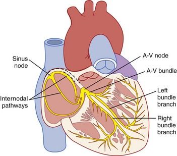
FIGURE 5-1 The electrical conduction system of the heart.

(From Hall JE: Guyton and Hall textbook of medical physiology, ed 12, Philadelphia, 2011, Saunders.)

image image

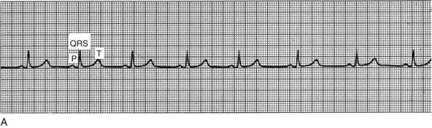
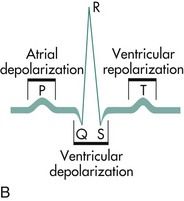
FIGURE 5-2 A, Electrocardiographic tracing of the cardiac cycle. B, Normal electrocardiographic deflections. The normal electrocardiogram consists of a P wave, representing atrial depolarization; a QRS complex, representing ventricular depolarization; and a T wave, representing rapid repolarization of the ventricles.

(A, From Goldberger AL, Goldberger E: Clinical electrocardiography: a simplified approach, ed 4, St. Louis, 1990, Mosby. B, From Pagana KD, Pagana TJ: Mosby’s manual of diagnostic and laboratory tests, ed 4, St. Louis, 2010, Mosby.)

Normal cardiac function depends on cellular automaticity (impulse formation), conductivity, excitability, and contractility. Disorders in automaticity and conductivity constitute the underlying cause of the vast majority of cardiac arrhythmias. Under normal conditions, the SA node is responsible for impulse formation, resulting in a sinus rhythm with a normal rate of 60 to 100 beats per minute.14 However, other cells or groups of cells also are capable of generating impulses (ectopic pacemakers), and under certain conditions, these may emerge outside of the normal conduction system. After a normal impulse is generated (depolarization), cells of the SA node need time for recovery and repolarization and are said to be refractory; during this time, they cannot conduct an impulse. Disturbances causing complete refractoriness result in a block, and those inducing partial refractoriness result in delay of conductivity.

Disorders of conductivity (block or delay) paradoxically may lead to rapid cardiac rhythm through the mechanisms of reentry. Reentry arrhythmias occur when accessory or ectopic pacemakers reexcite previously depolarized fibers before they would become depolarized in the normal sequential impulse pathway, typically producing tachyarrhythmias. The type of arrhythmia may suggest the nature of its cause. For example, paroxysmal atrial tachycardia with block suggests digitalis toxicity.14 However, many cardiac arrhythmias are not specific for a given cause. In such cases, a careful search is undertaken to identify the cause of the arrhythmia. The most common causes include primary cardiovascular disorders, pulmonary disorders (e.g., embolism, hypoxia), autonomic disorders, systemic disorders (e.g., thyroid disease), drug-related adverse effects, and electrolyte imbalances.3,15 Cardiac arrhythmias also are associated with many systemic diseases (Table 5-1) and various drugs or other substances including foods3,14,16,17 (Table 5-2).

TABLE 5-1 Cardiac Arrhythmias Associated with Various Systemic Diseases

Arrhythmia Associated Systemic Conditions
Sinus bradycardia Infectious diseases, hypothermia, myxedema, obstructive jaundice, increased intracranial pressure, myocardial infarction
Atrial extrasystoles Congestive heart failure, coronary insufficiency, myocardial infarction
Sinoatrial block Rheumatic heart disease, myocardial infarction, acute infection
Sinus tachycardia Febrile illness, infection, anemia, hyperthyroidism
Atrial tachycardia Obstructive lung disease, pneumonia, myocardial infarction
Atrial flutter Ischemic heart disease, mitral stenosis, myocardial infarction, open heart surgery
Atrial fibrillation Myocardial infarction, mitral stenosis, ischemic heart disease, thyrotoxicosis, hypertension
Atrioventricular block Rheumatic heart disease, ischemic heart disease, myocardial infarction, hyperthyroidism, Hodgkin disease, myeloma, open heart surgery
Ventricular extrasystole Ischemic heart disease, congestive heart failure, mitral valve prolapse
Ventricular tachycardia Mitral valve prolapse, myocardial infarction, coronary atherosclerotic heart disease
Ventricular fibrillation Blunt cardiac trauma, mitral valve prolapse, anaphylaxis, cardiac surgery, rheumatic heart disease, cardiomyopathy, coronary atherosclerotic heart disease

TABLE 5-2 Drugs/Foods that Can Induce Cardiac Arrhythmias

Cardiac Arrhythmia Precipitating Drugs/Food Substance
Bradycardia

Digitalis

Morphine

Beta blockers

Calcium channel blockers

Tachycardia

Atropine

Epinephrine

Nicotine

Ephedrine

Caffeine

Premature atrial beats

Alcohol

Nicotine

Tricyclic antidepressants

Caffeine

Ventricular extrasystoles

Digitalis

Alcohol

Epinephrine

Amphetamines

Ventricular tachycardia

Digitalis

Quinidine

Procainamide

Potassium

Sympathetic amines

Pathophysiology and Complications

The outcome of an arrhythmia often depends on the nature of the arrhythmia and the physical condition of the patient. For example, a young healthy person with paroxysmal atrial tachycardia may have minimal symptoms, whereas an elderly patient who has heart disease with the same arrhythmia is at risk for developing shock, congestive heart failure, or myocardial ischemia. Furthermore, evidence suggests that patients with certain types of cardiac arrhythmias (e.g., AF) are susceptible to ischemic events within the dental office.18

Arrhythmias are classified by site of origin (Box 5-1). Any arrhythmia that arises above the bifurcation of the His bundle into right and left bundle branches is classified as supraventricular.19 Supraventricular cardiac arrhythmias may be broadly categorized into tachyarrhythmias and bradyarrhythmias. Brief descriptions of some of the more common arrhythmias likely to be encountered in dental patients are provided.

image

Box 5-1 Classification of Common Cardiac Arrhythmias

Supraventricular Arrhythmias

Sinus nodal disturbances

    Sinus arrhythmia
    Sinus tachycardia
    Sinus bradycardia

Disturbances of atrial rhythm

    Premature atrial complexes
    Atrial flutter
    Atrial fibrillation
    Atrial tachycardias

Tachycardias involving the atrioventricular (AV) junction

    Preexcitation syndrome (Wolff-Parkinson-White)

Heart block

    AV block/complete AV block

Ventricular Arrhythmias

Premature ventricular complexes

Ventricular tachycardia

Ventricular fibrillation

Disorders of Repolarization

Long QT syndrome

image

Supraventricular Arrhythmias

Sinus Nodal Disturbances

Sinus arrhythmia. Sinus arrhythmia is characterized by phasic variation in sinus cycle length.14 In the respiratory type, heart rate increases with inhalation and decreases with exhalation. It is seen predominantly in the young and reflects variations in parasympathetic and sympathetic signals to the heart and is considered a normal event. Nonrespiratory sinus arrhythmia is unrelated to respiratory effort and is seen in digitalis intoxication.

Sinus tachycardia. Tachycardia in an adult is defined as a heart rate greater than 100 beats per minute, with otherwise normal findings on the ECG.14 The rate usually is between 100 and 180 beats per minute. This condition most often is a physiologic response to exercise, anxiety, stress, or emotion. Pathophysiologic causes include fever, hypotension, hypoxia, infection, anemia, hyperthyroidism, and heart failure. Drugs that may cause sinus tachycardia include atropine, epinephrine, alcohol, nicotine, and caffeine.

Sinus bradycardia. Bradycardia is defined as a heart rate less than 60 beats per minute, with an otherwise normal ECG tracing.14 It often coexists with a sinus arrhythmia. It is relatively common among well-conditioned athletes and healthy young adults and decreases in prevalence with advancing age. Pathophysiologic causes of bradycardia include intracranial tumor, increased intracranial pressure, myxedema, hypothermia, and gram-negative sepsis. Bradycardia may occur during vomiting and vasovagal syncope and as the result of carotid sinus stimulation. Drugs that may cause bradycardia include lithium, amiodarone, beta blockers, clonidine, and calcium channel blockers.

Disturbances of Atrial Rhythm

Premature atrial complexes. Impulses arising from ectopic foci anywhere in the atrium may result in premature atrial beats. Premature atrial complexes, or contractions, occur frequently in otherwise healthy people but often occur during infection, inflammation, or myocardial ischemia.14 They may be provoked by smoking, lack of sleep, excessive caffeine, or alcohol.13 They are common in conditions associated with dysfunction of the atria such as congestive heart failure.

Atrial flutter. Atrial flutter is characterized by a rapid, regular atrial rate of 250 to 350 beats per minute. It is rare in healthy persons and most often occurs in association with septal defects, pulmonary emboli, mitral or tricuspid valve stenosis or regurgitation, or chronic ventricular failure.14 It ialso may be noted in patients with hyperthyroidism, alcoholism, or pericarditis.

Atrial fibrillation. AF is the most common sustained arrhythmia in adults.19 It is characterized by rapid, disorganized, and ineffective atrial contractions that occur at a rate of 350 to 600 beats per minute. The ventricular response is highly irregular. The atria do not contract effectively, thereby promoting the formation of intraarterial clots, along with consequent embolism and stroke. Thus, patients with AF who are at risk for stroke (e.g., history of previous stroke, systemic emboli, valvular heart disease, hypertension, diabetes, coronary heart disease, heart failure) should be placed on a regimen of warfarin for antithrombotic therapy, with a target international normalized ratio (INR)20 of 2.0 to 3.0.14,21 Patients who cannot take warfarin, as well as those who do not have risk factors for stroke, may be managed with dabigatran or aspirin therapy. AF is associated with a history of congestive heart failure, valvular heart disease and stroke, left atrial enlargement, abnormal mitral or aortic valve function, or treated systemic hypertension, as well as with advanced age.12,22 It may occur intermittently or may be chronic. Symptoms are variable and depend on underlying cardiac status, ventricular rate, and loss of atrial contraction. Treatment consists of medication or cardioversion.

Atrial tachycardias. Any tachycardia arising above the AV junction for which the ECG shows a P wave configuration different from that for sinus rhythm is called atrial tachycardia.19 Atrial tachycardia is characterized by an atrial rate between 150 and 200 beats per minute14 and may result from enhanced normal automaticity, abnormal automaticity, triggered activity, or reentry. It commonly is seen in patients with coronary artery disease, myocardial infarction (MI), cor pulmonale (right ventricular hypertrophy and pulmonary hypertension), or digitalis intoxication.

Tachycardias Involving the AV Junction

Preexcitation syndrome (e.g., Wolff-Parkinson-White syndrome). The atria and ventricles are electrically insulated from each other by fibrous tissue that forms the anatomic AV junction. Normally, impulses are transmitted from atria to ventricles across this electrical bridge; however, in some persons, additional electrical bridges connect the atria and ventricles, bypassing the normal pathways and forming the basis for preexcitation syndromes such as Wolff-Parkinson-White syndrome.19 The basic defect in this disorder involves premature activation (preexcitation) of the ventricles by way of an accessory AV pathway that allows the normal SA-AV pathway to be bypassed. This accessory pathway allows rapid conduction and short refractoriness, with impulses passed rapidly between atria and ventricles, and it provides a route for reentrant (backflow) tachyarrhythmias. Resultant paroxysmal tachycardia is characterized by a normal QRS complex, a regular rhythm, and ventricular rates of 150 to 250 beats per minute, along with sudden onset and termination.14 Wolff-Parkinson-White syndrome is found in all age groups but is more prevalent among men and decreases with age. For most patients with recurrent tachycardia, the prognosis is good, but sudden death occurs rarely, at a frequency of 0.1%.14

Heart Block

AV block. Heart block is a disturbance of impulse conduction that may be permanent or transient, depending on the underlying anatomic or functional impairment. Conduction impairment in heart block is classified by severity, with the various forms divided into three categories.14 During first-degree heart block, conduction time is prolonged, but all impulses are conducted. Second-degree heart block occurs in two forms: Mobitz type I (Wenckebach) and type II. Type I heart block is characterized by progressive lengthening of conduction time until an impulse is not conducted. Type II heart block denotes occasional or repetitive sudden block of conduction of an impulse without previous lengthening of conduction time. When no impulses are conducted, complete or third-degree block is present. AV block occurs when the atrial impulse is conducted with delay or is not conducted at all to the ventricles at a time when the AV junction is not physiologically refractory.14 Conduction delay may occur at the AV node, within the His-Purkinje system (bundle branches), or at both sites. AV block may be first-degree or second-degree block, or it may be complete. AV block may be caused by a multitude of conditions such as surgery, electrolyte disturbance, myoendocarditis, tumor, myxedema, rheumatoid nodules, Chagas’ disease,* calcific aortic stenosis, polymyositis, and amyloidosis. In children, the most common cause is congenital. Drugs (e.g., digitalis, propranolol, potassium, quinidine) also may cause AV heart block. Symptoms increase in severity with increasing degree of block.

Ventricular Arrhythmias

Premature ventricular complexes. Premature ventricular complexes (PVCs) (or contractions) are very common arrhythmias that are characterized by the premature occurrence of an abnormally shaped QRS complex (ventricular contraction), followed by a pause. PVCs may occur alone, as bigeminy (every other beat is a PVC), as trigeminy (every third beat is a PVC), or with higher periodicity. The combination of two consecutive PVCs is called a couplet; three or more in a row at a rate of 100 beats per minute are referred to as ventricular tachycardia.23 PVCs may be provoked by a variety of medications, by electrolyte imbalance, by tension states, and by excessive use of tobacco, caffeine, and alcohol. In patients without structural heart disease, PVCs have no prognostic significance and no impact on longevity or limitation of activity.18 The prevalence of PVCs increases with age; they are associated with male gender and are related to low serum potassium concentration. Among patients with previous MI or valvular heart disease, however, frequent PVCs are associated with an increased risk of death.12

Ventricular tachycardia. The occurrence of three or more ectopic ventricular beats (PVCs) at a rate of 100 or more per minute is defined as ventricular tachycardia (VT). VT may be sustained or episodic. Sustained VT that persists for 30 seconds or longer may require termination because of hemodynamic instability. VT can quickly degenerate into ventricular fibrillation. A variant of VT called torsades de pointes is characterized by QRS complexes of changing amplitude that appear to twist around the isoelectrical line; this rhythm occurs at rates of 200 to 250 beats per minute.24 VT almost always occurs in patients with heart disease, most commonly ischemic heart disease and cardiomyopathy.14 Certain drugs such as digitalis, sympathetic amines (epinephrine), potassium, quinidine, and procainamide may induce VT.25

Ventricular flutter and fibrillation. Ventricular flutter and ventricular fibrillation (VF) are lethal arrhythmias characterized by chaotic, disorganized electrical activity that results in failure of sequential cardiac contraction and inability to maintain cardiac output.23 The distinction between flutter and fibrillation can be difficult and is of academic interest only; therefore, the two can be discussed together. If these disorders are not rapidly treated within 3 to 5 minutes, death will ensue. VF occurs most commonly as a sequela of ischemic heart disease.

Disorders of Repolarization

Long QT syndrome. Long QT syndrome is a disorder of the conduction system in which the recharging of the heart during repolarization (i.e., the QT interval) is delayed. It is caused by a genetic mutation in myocardial ion channels and by certain drugs, or may be the result of a stroke. The condition can lead to fast, chaotic heartbeats, which can trigger unexplained syncope, a seizure, or sudden death.24,26

Clinical Presentation

Signs and Symptoms

Arrhythmias may be symptomatic or asymptomatic; however, symptoms alone cannot be relied on to determine the seriousness of an arrhythmia. Some arrhythmias such as PVCs may be highly symptomatic, yet are not associated with an adverse outcome, whereas some patients with atrial fibrillation have no symptoms at all but may be at significant risk for stroke.27 The symptoms most commonly associated with cardiac arrhythmias include palpitations, lightheadedness, feeling faint, syncope, and those related to congestive heart failure (e.g., shortness of breath, orthopne/>

Only gold members can continue reading. Log In or Register to continue

Stay updated, free dental videos. Join our Telegram channel

Jan 4, 2015 | Posted by in General Dentistry | Comments Off on 5: Cardiac Arrhythmias

VIDEdental - Online dental courses

Get VIDEdental app for watching clinical videos