16: Thyroid Diseases

Chapter 16

Thyroid Diseases

Thyroid disease in a patient who presents for dental treatment is a cause for concern on several fronts. Undiagnosed or poorly controlled disorders of the thyroid can be expected to compromise outcomes with otherwise perfectly appropriate dental management plans. Detection of early signs and symptoms of such disorders during the dentist’s initial assessment, however, can lead to referral of the patient for medical evaluation and treatment. In some instances, such intervention may be lifesaving, whereas in others, quality of life can be improved and complications of certain thyroid disorders avoided, particularly in the context of delivery of dental care.

The focus of this chapter is on disorders involving hyperfunction of the gland (hyperthyroidism or thyrotoxicosis), hypofunction of the gland (hypothyroidism or myxedema or cretinism), thyroiditis, and the detection of lesions that may be cancerous (< ?xml:namespace prefix = "mbp" />Table 16-1). In an average dental practice of 2000 patients, an estimated 20 to 150 patients will have a thyroid disorder.

TABLE 16-1 Etiology of Thyroid Conditions

Thyroid Condition Causes
Hyperthyroidism

Primary thyroid hyperfunction

    Grave’s disease
    Toxic multinodular goiter
    Toxic adenoma

Secondary thyroid hyperfunction

    Pituitary adenoma—TSH secretion
    Inappropriate TSH secretion (pituitary)
    Trophoblastic hCG secretion

Without thyroid hyperfunction

    Hormonal leakage—subacute thyroiditis
    Thyroid hormone use (factitia)
    Bovine thyroid in ground beef
    Metastatic thyroid cancer
    Iatrogenic (overdosage of thyroid hormone)
Hypothyroidism (cretinism, myxedema)

Primary atrophic hypothyroidism

    Insufficient amount of thyroid tissue
       Destruction of tissue by autoimmune process
         Hashimoto’s thyroiditis (atrophic and goitrous)
         Graves’ disease—end-stage
       Destruction of tissue by iatrogenic procedures

         131I therapy
         Surgical thyroidectomy
         External radiation to thyroid gland
       Destruction of tissue by infiltrative process

         Amyloidosis, lymphoma, scleroderma
 

Defects of thyroid hormone biosynthesis

    Congenital enzyme defects
    Congenital mutations in TSH receptor
    Iodine deficiency of excess
    Drug-induced: thionamides, lithium, others

Agenesis or dysplasia

Secondary hypothyroidism

    Pituitary
       Panhypopituitarism (neoplasm, irradiation, surgery)
       Isolated TSH deficiency
    Hypothalamic
       Congenital
       Infection
       Infiltration (sarcoidosis, granulomas)

Transient hypothyroidism

    Silent and subacute thyroiditis
    Thyroxine withdrawal

Generalized resistance to thyroid hormone

Thyroiditis

Acute suppurative

Subacute painful

Subacute painless

Hashimoto’s

Chronic fibrosing (Riedel’s)

Thyroid neoplasms

Adenomas

Carcinomas

Others

hCG, Human chorionic gonadotropin; TSH, Thyroid-stimulating hormone.

Thyroid Gland

Location

The thyroid gland, which is located in the anterior portion of the neck just below and bilateral to the thyroid cartilage, develops from the thyroglossal duct and portions of the ultimobranchial body1,2 (Figure 16-1). It consists of two lateral lobes connected by an isthmus. A superior portion of glandular tissue, or a pyramidal lobe, can be identified. Thyroid tissue may be found anywhere along the path of the thyroglossal duct, from its origin (midline posterior portion of the tongue) to its termination (thyroid gland, in the neck).1,2 In rare cases, the entire thyroid lies within the anterior mediastinal compartment; in most people, however, remnants of the duct atrophy and disappear.1,2 The thyroglossal duct passes through the region of the developing hyoid bone, and remnants of the duct may become enclosed or surrounded by bone.2,3 Ectopic thyroid tissue may secrete thyroid hormones or may become cystic (Figure 16-2) or neoplastic.2,4 In a few people, the only functional thyroid tissue is found in these ectopic locations.2

image

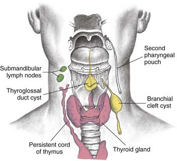
FIGURE 16-1 Thyroglossal duct cyst and branchial cleft cyst development.

(From Seidel HM, et al: Mosby’s guide to physical examination, ed 7, St. Louis, 2011, Mosby.)

image

FIGURE 16-2 Thyroglossal duct cyst.

The parathyroid glands develop from the third and fourth pharyngeal pouches and become embedded within the thyroid gland.5 Neural crest cells from the ultimobranchial body give rise to thyroid medullary C cells, which produce calcitonin, a calcium-lowering hormone.2,6 These C cells are found throughout the thyroid gland.2,6

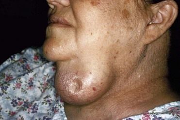
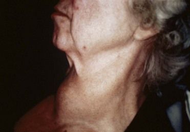
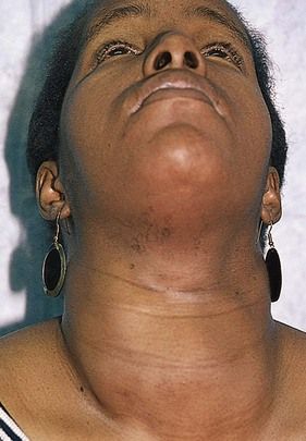
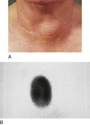
Enlargement and Nodules of the Thyroid Gland

Generalized enlargement of the thyroid gland, referred to as a goiter, may be diffuse (Figure 16-3) or nodular (Figure 16-4), and the goiter may be functional or nonfunctional.1,7,8 On a functional basis, thyroid enlargement can be divided into three types: primary goiter (simple goiter and thyroid cancer), thyrostimulatory secondary goiters (Graves’ disease and congenital hereditary goiter), and thyroinvasive secondary goiters (Hashimoto’s thyroiditis, subacute painful thyroiditis, Riedel’s thyroiditis, and metastatic tumors to the thyroid). Simple goiter accounts for about 75% of all thyroid swellings.7 Most of these goiters are nonfunctional and thus do not cause hyperthyroidism. The goiter of Graves’ disease is associated with hyperthyroidism.1,7 Hashimoto’s thyroiditis leads to hypothyroidism and thyroid enlargement.7 By contrast, patients with enlargement due to subacute thyroiditis experience a transient period of hyperthyroidism.7 Nodules found in the thyroid may be hyperplastic nodules, adenomas, or carcinomas. Hyperplastic nodules and adenomas can be functional (Figure 16-5) or nonfunctional. Most carcinomas are nonfunctional.1,7,9 Thyroid cancer most often manifests as a single nodule but can arise as multiple lesions or, in rare cases, can occur within a benign goiter.1,7,9

image

FIGURE 16-3 Diffuse enlargement of the thyroid gland due to Graves’ disease (goiter).

image

FIGURE 16-4 Multinodular goiter.

(From Swartz MH: Textbook of physical diagnosis: history and examination, ed 6, Philadelphia, 2010, Saunders.)

image

FIGURE 16-5 A, Toxic adenoma of the thyroid gland causing hyperthyroidism. B, Toxic adenoma in the right thyroid demonstrated with the use of Tc-pertechnetate scanning.

(From Forbes CD, Jackson WF: Color atlas and text of clinical medicine, ed 3, Edinburgh, 2003, Mosby.)

Function of the Thyroid Gland

The thyroid gland secretes three hormones: thyroxine (T4), triiodothyronine (T3), and calcitonin.8,10,11 Thyroxine and triiodothyronine collectively, they are termed thyoid hormone. Thyroid hormone influences the growth and maturation of tissues, cell respiration, and total energy expenditure. This hormone is involved in the turnover of essentially all substances, vitamins, and hormones.8,10,11

Most thyroid actions (metabolic and developmental) are mediated through activity of nuclear receptors that are tissue site–specific.8 Thyroid receptors work by altering gene expression in response to changes in thyroid hormone concentrations (mostly T3). This alteration in gene transcription profile is believed to account for most of the observed physiologic effects of thyroid hormones, although there are also actions of thyroid hormones that do not involve transcription.11 Thyroid hormone increases oxygen consumption, thermogenesis, and expression of the low-density lipoprotein (LDL) receptor, resulting in accelerated LDL cholesterol degradation. In myocardium, T3 increases myocyte contractility and relaxation by altering myosin heavy chain and sarcoplasmic reticulum adenosine triphosphatase (ATPase). In the cardiac conducting system, T3 increases the heart rate by altering sinoatrial node depolarization and repolarization. Other physiologic effects of thyroid hormone include increased mental alertness, ventilatory drive, gastrointestinal motility, and bone turnover. During fetal development, thyroid hormone plays a critical role in brain development and skeletal maturation.1

Calcitonin is involved, along with parathyroid hormone and vitamin D, in regulating serum calcium and phosphorus levels and skeletal remodeling. (This hormone and its actions are considered further in Chapter 12.)8,10,11

Epidemiology

Incidence and Prevalence

Graves’ disease occurs in up to 2% of women and 0.2% of men. Graves’ disease is rare before adolescence; the usual age at presentation is between 20 and 50 years, although it does occur in elderly persons.7,12 Congenital hypothyroidism is present in about 1 in 4000 newborns. Most cases (80% to 85%) are due to thyroid gland dysgenesis, and developmental abnormalities are twice as common in girls. The annual incidence rate of autoimmune hypothyroidism is 4 cases per 1000 women and 1 per 1000 men. Prevalence increases with age, and mean age at diagnosis is 60 years. Subclinical hypothyroidism is diagnosed in 6% to 8% of women (10% in women over 60 years of age) and 3% of men.13

Subacute painful thyroiditis accounts for 5% of all medical consultations regarding thyroid disorders, and is three times more common in women than men. Subacute painless thyroiditis occurs in patients with underlying autoimmune thyroid disease and is reported in up to 5% of women 3 to 6 months after pregnancy. In these circumstances it is called postpartum thyroiditis. Riedel’s thyroiditis is a rare form of chronic thyroiditis that typically occurs in middle-aged women. Acute suppurative thyroiditis is rare.1,7,12,13

Thyroid nodules can be found in about 5% of the adult population in the United States.1,7,9 The frequency of cancer in solitary thyroid nodules has been reported to be about 1% to 5%.1,7,9 During the past 10 years or so, the incidence of thyroid cancer has increased at a rate of about 5% per year.9,14 In 2007, 434,000 people (96,000 men and 338,000 women) were living with thyroid cancer.15 For 2010, the National Cancer Institute estimated a total of 44,670 new cases of thyroid cancer, with about 1690 deaths.16

Pathophysiology and Etiology

Blood levels of T4 and T3 are controlled through a servofeedback mechanism mediated by the hypothalamic-pituitary-thyroid axis (Figure 16-6). Increased or decreased metabolic demand appears to be the main modifier of the system. Drugs, illness, thyroid disease, and pituitary disorders may affect control of this balance.7,8,11,17 Studies also show that age has some effect on the system.

image

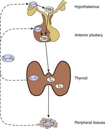
FIGURE 16-6 Hypothalamic-Pituitary-Thyroid Axis.

Solid lines correspond to stimulatory effects, and dotted lines depict inhibitory effects. Conversion of T4 to T3 in the pituitary and the hypothalamus is mediated by 5′-deiodinase type II. This event also is important throughout the central nervous system, thyroid, and muscle. 5′-Deiodinase type I (propylthiouracil-sensitive) plays a major role in liver, kidney, and thyroid function. TRH, Thyrotropin-releasing hormone; TSH, Thyroid-stimulating hormone.

(Redrawn from DeGroot LJ, Jameson JL: Endocrinology, ed 5, vol 2, Philadelphia, 2006, Saunders.)

Under normal conditions, thyrotropin-releasing hormone (TRH) is released by the hypothalamus in response to external stimuli (e.g., stress, illness, metabolic demand, low levels of T3 and, to a lesser extent, T4). TRH stimulates the pituitary to release thyroid-stimulating hormone (TSH), which causes the thyroid gland to secrete T4 and T3. T4 and T3 also have a direct influence on the pituitary. High levels turn off the release of TSH, and low levels turn it on. In the blood, T4 and T3 are almost entirely bound to plasma proteins.7,8,11,17

Binding plasma proteins consist of thyroxine-binding globulin (TBG), transthyretin, and thyroid-binding albumin (TBA). Small amounts of T3 and T4 are bound to high-density lipoproteins.1 The most important thyroid hormone–binding serum protein is TBG, which binds about 70% of T4 and 75% to 80% of T3.1 Only 0.02% to 0.03% of free thyroxine (FT4) and about 0.3% of free triidothyroxine (FT3) is found in plasma.1,7

Low T4 and T3 plasma levels often are found in ill and medicated older persons. Protein abnormalities can affect total T4 and T3 levels. Illness can reduce the conversion of T4 to T3. Drugs and illness also can affect free levels of T4 and T3. The main age-related change seen in much older individuals is a fall in T3 due to the reduced peripheral conversion of T4 to T3.18

Antibodies to various structures within the thyroid are associated with autoimmune diseases of the thyroid. Graves’ disease and Hashimoto’s thyroiditis have such an association. Three autoantibodies are most often involved in autoimmune thyroid disease: TSH receptor antibodies (TSHRAb), thyroid peroxidase antibodies (TPoAb), and thyroglobulin antibodies (TgAb).19 TSHRAb are not found in the general population but are present in 80% to 95% of patients with Graves’ disease and in 10% to 20% of those with autoimmune thyroiditis. Most TSHRAb in Graves’ disease are stimulating antibodies, which stimulate the release of thyroid hormone. However, blocking antibodies to the TSH receptor (TSHR-blocking Ab) also are found, which block the release of thyroid hormone. The ratio of these TSH receptor antibodies determines the clinical status of the patient and the functional status of the thyroid gland.12,19

TgAb are found in about 10% to 20% of the general population. These antibodies are present in 50% to 70% of patients with Graves’ disease and in 80% to 90% of those with autoimmune thyroiditis.7,12,19 TPoAb are found in 8% to 27% of the general population. About 50% to 80% of patients with Graves’ disease have these antibodies. TPoAb are found in 90% to 100% of patients with autoimmune thyroiditis.7,12,19

Laboratory Tests

Direct tests of thyroid function involve the administration of radioactive iodine. Measurement of thyroid radioactive iodine uptake (RAIU) is the most common of these tests. 131I has been used for this test, but 123I is preferred because it exposes the patient to a lower radiation dose. RAIU, which is measured 24 hours after administration of the isotope, varies inversely with plasma iodide concentration and directly with the functional status of the thyroid. In the United States, normal 24-hour RAIU is 10% to 30%. RAIU discriminates poorly between normal and hypothyroid states. Values above the normal range usually indicate thyroid hyperfunction.1,7,8,20

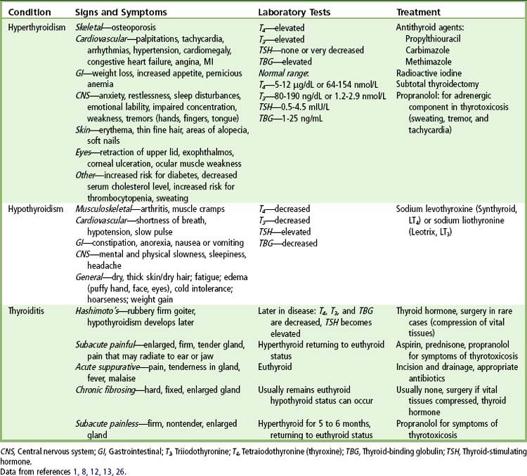
Several tests are available that measure thyroid hormone concentration and binding in blood. Highly specific and sensitive radioimmunoassays are used most often to measure serum T4 and T3 concentrations and rarely to measure reverse T3 (rT3) concentration. The normal range for T4 is 64 to 154 nmol/L (5 to 12 µg/dL). The normal range for T3 is 1.2 to 2.9 nmol/L (80 to 190 ng/dL).20 Elevated levels usually indicate hyperthyroidism, and lower levels usually indicate hypothyroidism. Free hormone levels usually correlate better with the metabolic state than do total hormone levels.8,20

Measurement of basal serum TSH concentration is useful in the diagnosis of hyperthyroidism and hypothyroidism. Very sensitive methods, such as immunoradiometric or chemiluminescent techniques, are now available to measure serum TSH. The normal range for TSH is 0.5 to 4.5 mIU/L (µIU/mL). In cases of hyperthyroidism, the TSH level is almost always low or nondetectable. Higher levels indicate hypothyroidism8,20 (Table 16-2).

TABLE 16-2 Laboratory Tests

Test Normal Range Interpretation
Radioactive iodine uptake (RIU) 5-30% Elevated:  hyperthyroidism
Decreased:  hypothyroidism
Thyroid-stimulating hormone (TSH) 0.5-4.5 mIU/L Elevated:  hypothyroidism
Suppressed:  hyperthyroidism
Total serum T4 (TT4) 5-12 µg/dL
64-154 nmol/L
High: hyperthyroidism
Low:  hypothyroidism
Free T4 (FT4) 1.0-3.0 ng/dL
13-39 pmol/L
Increased:  hyperthyroidism
Decreased:  hypothyroidism
Total serum T3 (TT3) 1.2-2.9 nmol/L
80-190 ng/dL
High: hyperthyroidism
Low:  hypothyroidism
Free T3 (FT3) 0.25-0.65 ng/dL
3.8-10 nmol/L
Increased:  hyperthyroidism
Decreased:  hypothyroidism

Other tests used in selected cases include the TSH stimulation test, the T3 suppression test, and radioassay techniques for measuring TSHRAb, TSHR-blocking Ab, TPoAb, and TgAb.8,20 A thyroid scan commonly is used to localize thyroid nodules and to locate functional ectopic thyroid tissue. 123I or 99Tc (technetium) is injected, and a scanner localizes areas of radioactive concentration. This technique allows for the identification of nodules 1 cm or larger. When a pinhole thyroid scan is used, 2- to 3-mm lesions may be detected.7,2022

Ultrasonography may be used to detect thyroid lesions. Nodules 1 to 2 mm in size can be identified. This technique also is used to distinguish solid from cystic lesions, to measure the gland, and to guide needles for aspiration of cysts or for biopsy of thyroid masses. Computed tomography (CT) and magnetic resonance imaging (MRI) are helpful mainly in the postoperative management of patients with thyroid cancer. These forms of imaging also are used for the preoperative evaluation of larger lesions of the thyroid (greater than 3 cm in diameter) that extend beyond the gland into adjacent tissues.7,2022

Thyrotoxicosis (Hyperthyroidism)

Etiology, Pathophysiology, and Complications

The term thyrotoxicosis refers to an excess of T4 and T3 in the bloodstream. This excess may be the result of production by ectopic thyroid tissue, multinodular goiter, or thyroid adenoma or may be associated with subacute thyroiditis (painful and painless), ingestion of thyroid hormone (thyrotoxicosis factitia) or of foodstuffs containing thyroid hormone, or pituitary disease involving the anterior portion of the gland (see Table 16-1). In this section, the signs and symptoms, laboratory tests, treatment, and dental considerations for the patient with Graves’ disease are considered in detail; this disease serves as a model for other conditions that can result in similar clinical manifestations. Of note, multinodular goiter, ectopic thyroid tissue, and neoplastic causes of hyperthyroidism are rare compared with toxic goiter.1,8,12

Graves’ disease is an autoimmune disease in which thyroid-stimulating immunoglobulins bind to and activate thyrotrophic receptors, causing the gland to grow and stimulating the thyroid follicles to increase the synthesis of thyroid hormone.1,8,12 The chief risk factors for Graves’ disease are genetic mutations (i.e., in susceptibility genes for CD40, cytotoxic T lymphocyte antigen [CTLA-4], thyroglobulin, TSH receptor, and PTPN2212) and female gender, in part because of modulation of the autoimmune response by estrogen. This disorder is much more common in women (with a male-to-female ratio of 10 : 1) and may manifest during puberty or pregnancy, or at menopause (see Figure 16-3). Genetic predisposition along with emotional stress such as severe fright or separation from loved ones has been reported to be associated with its onset. The disease may occur in a cyclic pattern and may then “burn itself out” or continue in an active state.1,8,12

Clinical Presentation

Signs and Symptoms

Direct and indirect effects of excessive thyroid hormones contribute to the clinical picture in Graves’ disease. The most common symptoms and signs are nervousness, fatigue, rapid heartbeat or palpitations, heat intolerance, and weight loss (Table 16-3). These manifestations are reported in more than 50% of all diagnosed patients. With increasing age, weight loss and decreased appetite become more common, and irritability and heat intolerance are less common. Atrial fibrillation is rare in patients younger than 50 years of age but occurs in approximately 20% of older patients. The patient’s skin is warm and moist and the complexion rosy; the patient may blush readily. Palmar erythema may be present, profuse sweating is common, and excessive melanin pigmentation of the skin is evident in many patients; however, pigmentation of the oral mucosa has not been reported. In addition, the patient’s hair becomes fine and friable, and the nails soften.1,8,12

TABLE 16-3 Clinical Findings and Treatment of Thyroid Disorders

image

Graves’ ophthalmopathy, which is identified in approximately 50% of patients, is characterized by edema and inflammation of the extraocular muscles, as well as an increase in orbital connective tissue and fat. Ophthalmopathy is an organ-specific autoimmune process that is strongly linked to Graves’ hyperthyroidism. Although hyperthyroidism may be successfully treated, ophthalmopathy often produces the greatest long-term disability for patients with this disease. Figures 16-7 and 16-8 demonstrate the changes associated with ophthalmopathy (eyelid retraction, proptosis, periorbital edema, chemosis, and bilateral exophthalmos). This disease may progress to visual loss through exposure keratopathy or compressive optic neuropathy.23,24

image

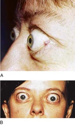
FIGURE 16-7 Eyelid changes in Graves’ disease. A, Lid retraction is a common eye sign in Graves’ disease. It is recognized when the sclera is visible between the lower margin of the upper lid and the cornea. B, Proptosis in Graves’ disease results from enlargement of muscles and fat within the orbit as a result of mucopolysaccharide infiltration.

(A, From Goldman L, Ausiello D: Cecil textbook of medicine, ed 23, Philadelphia, 2008, Saunders. B, From Seidel H: Mosby’s guide to physical examination, 4th ed 4, St. Louis, 1999, Mosby.)

image

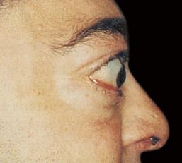
FIGURE 16-8 Exophthalmos of Graves’ disease can be unilateral or bilateral. The forward protrusion of the globe results from an increase in volume of the orbital contents.

(From Stein HA, Stein RM, Freeman MI: The ophthalmic assistant: a text for allied and associated ophthalmic personnel, ed 8, Philadelphia, 2006, Mosby.)

Most thyrotoxic patients show eye signs not related to the ophthalmopathy of Graves’ disease as well. These signs (i.e., stare with widened palpebral fissures, infrequent blinking, lid lag, jerky movements of the lids, and failure to wrinkle the brow on upward gaze) result from sympathetic overstimulation and usually clear when thyrotoxicosis is corrected.23,24

Another complication, which is found in about 1% to 2% of patients with Graves’ disease, is dermopathy (Figure 16-9). In focal areas of the skin, hyaluronic acid and chondroitin sulfate concentrations in the dermis are increased. This may occur as the result of lymphokine activation of fibroblasts. Accumulation causes compression of the dermal lymphatics and nonpitting edema. Early lesions contain a lymphocytic infiltrate. Nodular and plaque formation may occur in chronic lesions. These lesions are most common over the anterolateral aspects of the shin. Patients with dermopathy almost always develop severe ophthalmopathy.1,12,23,24

image

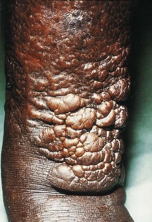
FIGURE 16-9 Infiltrative dermopathy seen in Graves’ disease. Hyperpigmented, nonpitting induration of the skin of the legs usually is found in the pretibial area (pretibial myxedema). Lesions are firm, and clear edges can be seen.

(From Melmed S, et al: Williams textbook of endocrinology, ed 12, Philadelphia, 2011, Saunders. Courtesy Dr. Andrew Werner, New York, New York.)

Thyroid acropachy is another rare manifestation of Graves’ disease. This feature is associated with presence of TgAbs (Figure 16-10). It is characterized by clubbing and soft tissue swelling of the last phalanx of the fingers and toes. The overlying skin often is discolored and thickened. Subperiosteal new bone formation occurs, along with glycosaminoglycan deposits in the skin. The patho/>

Only gold members can continue reading. Log In or Register to continue

Stay updated, free dental videos. Join our Telegram channel

Jan 4, 2015 | Posted by in General Dentistry | Comments Off on 16: Thyroid Diseases

VIDEdental - Online dental courses

Get VIDEdental app for watching clinical videos