Chapter 5
Additional Diagnostic Procedures
Diagnostic Imaging
Radiographic examination appropriate to the proposed treatment should be carried out in order to establish the presence or absence of pathology, the assessment of the hard tissues and as an aid to planning treatment.36,37
Implant dentistry requires specific information from diagnostic imaging additional to that required for other branches of dentistry involving advanced restorative treatment. These are listed below.
- • Bone volume.
- • Bone height. This is usually measured from the crest of a ridge to the opposing border of the jaw or relevant anatomical structure. It includes alveolar and basal bone. Selection of the implant length is based on this.
- • Bone width. This refers to the bucco-lingual width. Selection of the diameter of implant is based on this.
- • Length of the edentulous ridge. This refers to the distance between two adjacent teeth and of their roots in a bound gap or the length along the crest of an edentulous ridge. This determines the number of implants and the distance between the implants and adjacent teeth.
- • Bone quality. This refers to the density and thickness of the cortical portion as well as the trabecular density in the cancellous portion. This will influence the success of treatment or the method by which osteotomy preparation may be carried out. It may be used for the assessment of bone for immediate loading.
- • Ridge orientation. This is the inclination of the edentulous ridge to the adjacent teeth and the proposed restoration. It will influence the angulation at which the implant is placed.
- • Spatial relationship of bone to the proposed restoration. This provides information relating tooth position to the bony ridge in horizontal (bucco-lingual and mesiodistal) and vertical (corono-apical) dimensions. It, therefore, relates the proposed tooth position to the proposed implant position.
- • Relationship to anatomical structures. Implant dentistry requires the precise identification and location of anatomical structures in order to prevent damage during hard and soft tissue surgery.
The diagnostic imaging techniques currently available are outlined with some of the benefits specific to implant dentistry.
Dental Panoramic Tomography (Orthopantomography)
A good diagnostic dental panoramic tomograph (DPT), otherwise known as orthopantomograph, provides the clinician with a good overview of the mouth.
The presence of teeth, any pathology and related anatomical structures, as well as bone heights, all on one image makes it an invaluable treatment planning aid. Judicious measurement of the magnification of each section will provide the clinician with a reasonably accurate assessment of bone height available for implant placement. It is indicated for patients requiring implant treatment as a screening and planning aid.
Periapical Radiography
Periapical radiographs provide details of the region being investigated. The structure, periodontal and endodontic status of the teeth can be established. The level of bone in the edentulous ridge and its level of attachment to the adjacent teeth can be measured accurately, and residual roots and pathology within the ridge can be seen in detail.
Lateral Cephalography
Lateral cephalography provides excellent information regarding the facial profile (using soft tissue filters; Figs 5-1 and 5-2), the relation of the jaws and some indication about the width of bone in the midline (Fig 5-3).
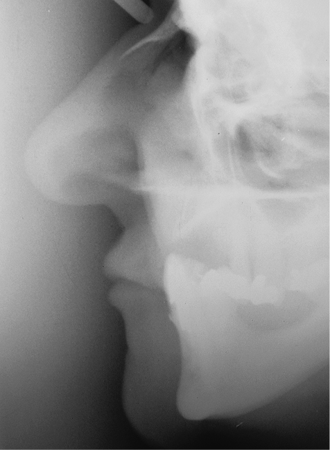
Fig 5-1 Lateral cephalograph using soft tissue filter prior to treatment. The soft tissue contours can be seen with the patient’s mandible in an overclosed position due to an absence of the maxillary denture.
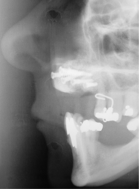
Fig 5-2 Lateral cephalograph after augmentation. Same patient as Fig 5-1. The onlay graft can be seen with its effect on lip support.
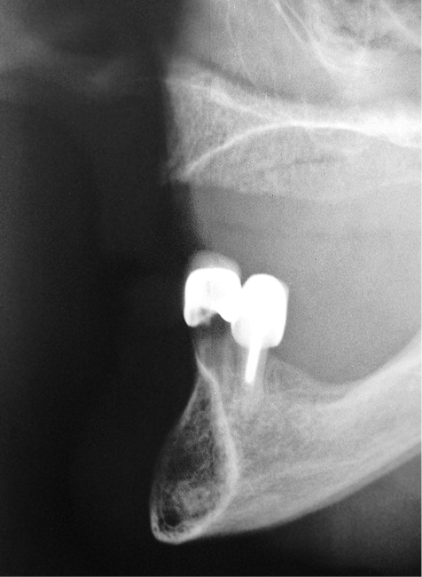
Fig 5-3 The lateral cephalograph may be used to provide an indication of bone availability in the midline, both in the maxilla and the mandible. It will also provide information about the intermaxillary relationship.
Conventional Tomography
Conventional tomography provide cross-sectional information about the area being investigated. However, it should be used with caution, as the magnification and mesiodistal positioning may lack accuracy,38 and multiple exposures increase the radiation dose significantly. They do not have a place anymore in imaging in implant dentistry.
Computed Tomography
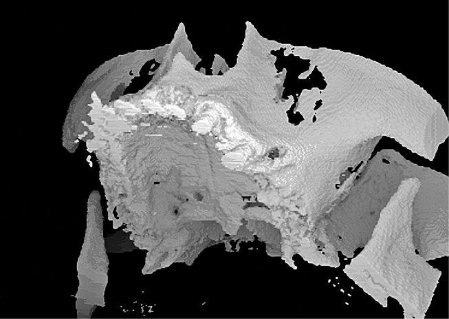
Computed tomography (CT) scans provides the implant clinician with a substantial amount of valuable information (Figs 5-4–5-8). The CT scan (and cone beam CT) is the only investigation that ideally provides the information that the implant surgeon requires for the planning of treatment. This includes bone volume and density, ridge orientation and spatial relationship to the proposed prosthesis, opposing jaw and anatomical structures. It is, therefore, the ideal investigation from the point of view of data provision. Accurate measurements can be made, which is particularly important when working within the posterior mandible.
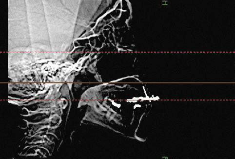
Fig 5-4 CT Scan in scout view (lateral view of the skull). This is used to determine the area to be scanned (between red lines).
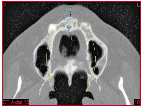
Fig 5-5 CT scan in axial section through the maxilla passing through the canine teeth and depicting the maxillary sinuses. The mandibular rami and the pterygoid plates as well as the surrounding soft tissues are visible. The greater density of the muscles is discernible. The yellow line indicates the position of the panoramic section (Fig 5-7) and the blue lines the position of the cross-sectional reformations of the alveolar ridge (Fig 5-6).
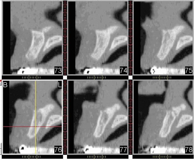
Fig 5-6 Cross-section of the maxilla in the midline showing the ridge width, density and inclination. The naso-palatine foramen and canal can be seen. The buccal aspect is on the left of each section and the palatal on the right. The radiopacity at the bottom of each image is due to the metal framework of the provisional fixed partial denture.
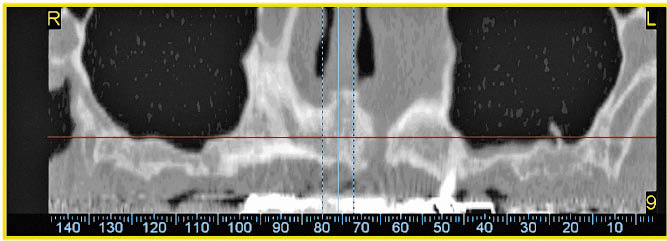
Fig 5-7 A panoramic section through the maxilla depicting the structures within that section: one root can be seen, on the left-hand side, the nasal cavities, and both maxillary sinuses with a small septum in the left sinus. A bony deficiency in the region of the left lateral incisor can also be noted.
Stay updated, free dental videos. Join our Telegram channel
VIDEdental - Online dental courses


