43
Enamel Disorders
Chronological Disturbances
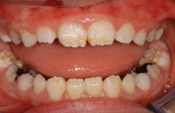
Teeth represent a fossil record of in utero, perinatal and childhood illness. Developmental defects of enamel can be acquired or inherited, localised to single teeth, generalised affecting the entire dentition or chronological affecting all teeth in different patterns dependent on the stage of formation of those teeth at the time of the insult. Tooth development begins at 3–4 months in utero and continues until the root formation of the third molars is complete. Any severe systemic upset may affect ameloblast function resulting in death of these cells, abnormalities in the secretion of enamel proteins, interrupted calcification or maturation of the enamel. This will result in the characteristic patterns of chronological hypoplasia affecting teeth at different stages of development (Fig. 43.1). In utero disturbances will cause defects in the primary dentition but the permanent teeth should be unaffected. Perinatal illness will affect the first permanent molars but the incisors should be unaffected as they do not begin to calcify until 3–4 months after birth.
Figure 43.1 Chronological hypoplasia associated with a febrile illness in the first 12 months of life. This has led to damage to the ameloblasts secreting enamel at this time, affecting the teeth at different levels depending on their developmental stage.
Hypoplasia
Hypoplasia is a disorder in the quantity of the enamel. Hypoplastic enamel may also be hypomineralised but there are some teeth that are purely hypoplastic with no change in the quality of the enamel. This occurs during the secretory phase (apposition and calcification stages) of tooth development. Generally, composite resin will bond well to enamel that is deficient in quantity but poorly to hypomineralised enamel. Breakdown may occur very rapidly following tooth eruption and treatment of these teeth should not be delayed.
Hypomineralisation (Opacities)
Hypomineralisation is a defect in the quality of the enamel. It occurs principally during the maturation phase of tooth development and results in incomplete enamel crystal growth or a cessation in prism formation. There may be an incorporation of pigments leading to discolouration (Table 43.1) or it may be simply an opacity that indicates air voids within the prism. Hypomineralised posterior teeth are extremely susceptible to caries and are often extremely sensitive.
Table 43.1 Discolouration of teeth.
| Green | Localised extrinsic | Chromogenic bacteria |
| Black/grey | Localised extrinsic | Ferrous sulphate |
| Localised intrinsic | Amalgam | |
Stay updated, free dental videos. Join our Telegram channel
VIDEdental - Online dental courses


