41: Immunotherapy

CHAPTER 41 Immunotherapy

Immunopharmacology is the study of the interaction between drugs and the immune system. Immunotherapy is the application of clinical strategies to modulate the activities of certain components of the immune system in order to improve immune function and to prevent or treat disease. This chapter reviews the immune system with regard to the pathways used toward adaptive, specific immunity that are or can be targeted for immunotherapy and discusses immunotherapeutic strategies that are of clinical importance today or show promise for the future. The pharmacologic manipulation of innate immune mechanisms involved in inflammation is covered in Chapters 21 and 35.

OVERVIEW OF SPECIFIC IMMUNITY

Components of the Immune System

Research in immunology has progressed rapidly over the last three decades. Spanning this period, major technologic feats (e.g., the development of hybridomas for the production of monoclonal antibodies) and major strides in our understanding of the immune system (including elucidation of cytokines and intracellular signaling) have resulted in significant advances in immunotherapeutics. Today immunopharmacologists use these new insights to pinpoint therapeutic targets among (1) a constellation of cytokines and other factors that influence cellular growth, differentiation, and function and (2) myriad receptors responsive to these mediators, specific antigens, or ligands found on other cells.

Cells

The immune system is composed of two major arms, innate immunity and adaptive immunity. These two components of the immune system differ in the types of effectors, their specificity for antigens, speed of action, and induction of memory. Innate immune cells are the primary initiators of the immune response and support functional activation of adaptive immune effectors. They differ from the adaptive immune effectors by their lack of antigen specificity and their fast acting capabilities. Unlike the adaptive immune effectors, the innate immune effectors do not generate memory. The main effectors of innate immune system are granulocytes, macrophages, and natural killer (NK) cells, whereas T and B lymphocytes are the main effectors of adaptive immunity.

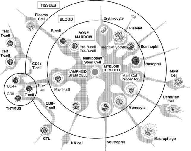
All the effectors of the immune system are derived from the bone marrow. Pluripotent hematopoietic stem cells in bone marrow give rise to either myeloid progenitor cells or lymphoid progenitor cells (Figure 41-1). Myeloid progenitor cells are the precursors of red blood cells, platelets, granulocytes (polymorphonuclear leukocytes [PMNs]: neutrophils, eosinophils, and basophils), monocyte-macrophages, dendritic cells (DCs), and mast cells. Lymphoid progenitor cells give rise to the T, B, and NK cells. Lymphocytes are generated and mature in the bone marrow and thymus, which are considered to be the primary or central lymphoid organs. From there, they travel to reside in secondary or peripheral lymphoid organs, such as lymph nodes, spleen, mucosa-associated lymphoid tissues (MALT), gut-associated lymphoid tissues (GALT), and bronchus-associated lymphoid tissues (BALT).

The various cell types exit the bone marrow at different levels of maturation, circulate through the bloodstream, and may take up residence in specific tissues. Certain cells involved in the body’s defense system provide rapid responses, whereas others support slower, adaptive responses. Neutrophils and NK cells exit the bone marrow in a relatively mature state and require very little time to become activated. In contrast, monocytes, dendritic cells, and most lymphoid cells leave the bone marrow in a relatively immature state and complete their maturation at some other tissue site, where they can be activated to respond to local cues.

Lymphocytes circulate between blood and lymphatics continuously. Lymphocytes that have not encountered antigen are called naïve lymphocytes; those that have encountered antigen and have become mature are effector lymphocytes. Activation of antigen-presenting cells (APCs)—monocytes, B cells and dendritic cells—is the necessary first step for the induction of adaptive immunity. In adaptive immunity, lymphocytes activated by antigens give rise to clones of antigen-specific cells, which are then selected either positively or negatively. Clonal selection is the central principle of adaptive immunity. The four basic postulates of clonal selection are that (1) each lymphocyte bears a single type of receptor with a unique specificity to self and nonself; (2) lymphocytes expressing receptors for specificity for self-antigens are deleted at an early stage and therefore are absent from the pool of mature lymphocytes; (3) interaction between a foreign antigen and the receptor capable of binding to it leads to lymphocyte activation; and (4) the differentiated effector cells derived from an activated lymphocyte will bear receptors of identical specificity to those of the parental cells. In adaptive immunity, unique antigen receptors are generated by gene rearrangement, and signals received through antigen receptors determine the development and survival of lymphocytes. Binding of antigen activates lymphocytes, resulting in the generation of effector cells and the establishment of immunologic memory.

T cells exit the bone marrow as CD3-CD4-CD8–null cells according to the cluster of differentiation (CD) classification of leukocyte antigens before entering the thymus. In young individuals, the thymus contains large numbers of developing T-cell precursors embedded in a network of epithelia known as the thymic stroma, which provides a supportive environment for the developing T cells. T-cell precursors proliferate extensively in the thymus, but most eventually are eliminated there. The thymus of a young adult mouse contains 1-2 × 10⁸ thymocytes. Although 50 million new cells are generated each day, only 2 million of these cells survive to leave the thymus. The developmental pathway of T cells in the thymus is the following. Early on, CD3-CD4-CD8-null cells develop into CD3+pTαCD4+CD8+ (triple–positive) thymocytes. About 95% of triple-positive thymocytes will be eliminated by apoptosis, and the remaining 5% will exit thymus as either CD3+CD4+ or CD3+CD8+ double-positive T cells.

Once T cells have completed their developmental stages in the thymus, they enter the bloodstream and then are carried to the secondary lymphoid tissues, such as the lymph nodes or spleen. If they encounter their specific antigen, they will become activated, proliferate, and differentiate to armed effector T cells. Otherwise, they will continue to travel back and forth from the blood to the secondary lymphoid tissues. Meanwhile, these naïve T cells will receive survival signals from self-peptide-self-MHC complexes.

Immature dendritic cells also travel from the tissues to secondary lymphoid tissues where they mature to activate T cells. The first encounter of a naïve T cell with the antigen on a mature dendritic cell is called priming, to distinguish it from the response of an armed effector T cell to antigen.

The sampling of different peptide-MHC complexes by naïve T cells on dendritic cells is important for the specific encounter of antigen-specific T cells, since only 0.0001% to 0.01% of T cells are specific for any particular antigen. Once they encounter the antigen they cease migration, proliferate, and differentiate to effector T cells. This process may take several days. At the end of that time, they leave the lymphoid tissues and re-enter the bloodstream to migrate to the site of the infection.

CD4+ T cells differentiate further outside the thymus into four distinct subsets. T helper (TH)1 cells primarily secrete interleukin (IL)-2 and interferon (IFN)-γ and activate monocytes and B cells, whereas TH2 cells primarily secrete IL-4, IL-5, IL-6, and IL-10 and activate B cells. TH17 cells secrete IL-17 and activate neutrophil responses to extracellular bacteria; T regulatory (Treg) cells function to suppress T cell responses.

TH1 cells upon activation produce IL-2 and IL-2 receptors, both of which are important in the induction of cell proliferation. The IL-2 receptor is composed of three different chains; α, β, and γ chains. Resting T cells express the β and γ chains, which together form the intermediate affinity IL-2 receptor and can respond to high concentrations of IL-2. The high affinity IL-2 receptor is formed when all three chains associate; it responds to low concentrations of IL-2 in activated T cells.

CD8+ T cells differentiate into cytotoxic cells. CD8+ cytotoxic T cells kill virally infected cells or tumor-target cells via two different mechanisms, namely apoptosis and necrosis. Apoptosis is distinguished from necrosis by the intact plasma membrane at the initial stages of programming for cell death, nuclear membrane blebbing, and DNA fragmentation.

Receptors and other cell-surface proteins

Cells involved in specific immunity express various surface glycoproteins to help coordinate their functions and interactions. These glycoproteins include adhesion molecules, cytokine receptors, and receptors that bind and respond to specific antigens and to co-receptors and costimulatory receptors expressed on other cells.

Clonally distributed antigen-specific receptors

As mentioned above, T cells and B cells possess receptors that specifically recognize the antigen and are distributed in a clonal manner. These receptors, both members of the immunoglobulin superfamily, are the T cell receptor (TCR) mentioned previously and the B-cell antigen receptor (BCR). Secreted forms of the BCR constitute the immunoglobulins found in plasma, extracellular fluid, and secretions. The BCR and TCR recognize short oligomeric sequences of a molecule and exhibit primary sequence specificity. In addition, the BCR (but not the TCR), which is designed to react with unprocessed antigen, may recognize secondary, tertiary, and quaternary structural features. The diversity of the TCR/BCR repertoire is generated by four main mechanisms: (1) somatic recombination, in which variable regions of the receptor chains, which are encoded in several pieces called gene segments, are assembled in the developing lymphocytes by somatic DNA recombination (a process known as gene rearrangement); (2) pairing of heavy and light chains of the BCR, or α and β chains of the TCR; (3) junctional diversity; and (4) somatic hypermutation. In both BCRs and TCRs the diversity is significantly increased by the addition and subtraction of nucleotides at the junction between the gene segments. Because the total number of nucleotides added by junctional diversity is random, the added nucleotides may disrupt the reading frame of the coding sequences, causing frameshifts that will lead to a nonfunctional protein. This outcome is called nonproductive rearrangement. Since two out of three rearrangements are nonproductive, many B cell progenitors never succeed in producing BCRs and never mature (into plasma cells and memory cells). Junctional diversity is achieved only at the expense of considerable cellular waste.

Rearranged genes in B cells (but not T cells) are further diversified by somatic hypermutation. Somatic hypermutation is the process of introducing point mutations into the variable region of the rearranged heavy and light chain genes at a very high rate. Somatic hypermutation occurs only when the B cells respond to the antigen along with the signals from activated T cells. Mutations that are deleterious and cannot bind antigen will remove the B cells (negative selection). Those that are positive will select for B cells with an even more improved antigen binding capacity (affinity maturation) causing the clonal expansion of these B cells.

Nonclonally distributed antigen-binding receptors

Most cells possess receptors that bind and present antigen specifically, but are not clonally distributed. These receptors are members of the immunoglobulin superfamily and include two classes of proteins encoded within the major histocompatibility gene complex (MHC) named MHC I and MHC II. T cells recognize self- and non–self-antigens as peptide fragments bound to MHC antigens. The MHC I and MHC II molecules recognize peptides derived from proteins that have been processed internally. MHC I proteins deliver peptides originating from cytosolic compartments, whereas MHC II proteins deliver peptides from vesicular compartments. The MHC I and II molecules do not discriminate the entire primary structure of the peptide, but instead recognize two (or more) “anchor” positions in the peptide separated by a short sequence of amino acids of virtually any composition or sequence. Peptide fragments presented through MHC I activate CD8+ T cells to kill; peptide fragments presented by MHC II activate the function of CD4+ T cells to aid B cells and macrophages to become activated.

Polygenic and polymorphic properties of the MHC make it difficult for pathogens to evade immune responses. Polygenic refers to the fact that the MHC contains several different MHC I and II genes; polymorphic indicates there are multiple variants of each gene.

The MHC is located on chromosome 6 in humans. There are three class I genes in humans named HLA-A, HLA-B, and HLA-C (classical MHC I genes). There are also three pairs of class II genes named HLA-DR, HLA-DP, and HLA-DQ. The HLA-DR cluster contains an extra β chain gene, where its product can pair with the DRα chain, giving rise to four types of MHC II proteins. Expression of MHC II genes is induced by IFN-γ via the production of a transcriptional activator known as MHC class II transactivator (CIITA). The absence of CIITA causes severe immunodeficiency.

MHC restriction refers to the recognition of the antigen by the T cells in the context of self- MHC. Thus a non–self-MHC I presenting the same peptide would not be recognized and would not activate T cells. Non–self-MHC molecules are recognized by 1% to10% of T cells, an event termed alloreactivity. In alloreactivity, recognition of either the peptide antigen (peptide-dominant binding) or the foreign MHC molecule irrespective of the peptide with which it is complexed (MHC-dominant binding) leads to T-cell activation.

Other genes that map to the MHC locus include components of the complement cascade, such as C2, C4, and factor B, and cytokines including tumor necrosis factor (TNF)-α. These genes are referred to as MHC class III genes. In addition to highly polymorphic MHC I and II genes, there are many genes encoding MHC I–like molecules that show little polymorphism termed MHC class Ib. Some MHC Ib genes (e.g., the MIC gene family) are induced during cellular stress and regulate NK function.

Most individuals are heterozygous at each MHC locus. MHC polymorphism affects T-cell recognition of antigen by influencing both the peptide binding and the contact between the TCR and MHC molecule.

Mediators

Numerous water-soluble proteins affect or create specific immune reactions. Two principal groups of interest in immunotherapy are the cytokines and humoral antibodies.

Cytokines

Cytokines are produced by a wide variety of cells. They play crucial roles in stimulating the production of blood cells of all types and in regulating the differentiation, activation, and suppression of cells involved in specific immunity. Some cytokines are referred to as interleukins; cytokines also include interferons, and colony-stimulating factors. Cytokines have local and distant effects and activate cells in an autocrine (causing a self-response) and paracrine (affecting other cells) fashion.

An important feature of cytokine action is that multiple cytokines often work in concert to foster a particular change in cellular activity. The proliferation and differentiation of effector T cells important in cell-mediated immunity (CMI) depend on the interplay of TH1 cytokines such as IL-2, IL-12, and IFN-γ. Activation of B cells for humoral immunity is based on the release of several interleukins (IL-4, IL-5, IL-10, and IL-13) by TH2 cells. TH1 cytokines are important in stimulating B-cell differentiation leading to the production of immunoglobulins IgG1, IgG2, and IgG3. TH2 cytokines stimulate IgE and IgG4 production. The actions of selected cytokines are summarized in Table 41-1.

TABLE 41-1 Selected Cytokines and Their Functional Relationships

CYTOKINE SECRETED BY FUNCTIONS
Lymphoid Hematopoiesis    
IL-7 SC

T-Cell–Stimulating Factors     IL-1 (α and β) APC, B, Ep, En

IFN-α B, Ma

IFN-β Fb, Ep Same as for IFN-α IFN-γ T

IL-2 T T-cell proliferation and formation of cytotoxic T lymphocytes IL-10 T, B

B-Cell–Stimulating Factors     IL-4 T IL-5 T IL-6 T, M, Fb IL-13 CD4+ T Tumor Killers and Cell-Mediated Immunity TNF-α Ma TNF-β T Death of tumor cells (lymphotoxin) IL-12 Ma Myeloid Factors     IL-3 L, My Multiple granulocytic cell type CSF IL-8 Ma, En IL-9 T IL-11 SC Megakaryocyte growth and differentiation GM-CSF L, My Granulocyte/monocyte CSF M-CSF L, My Monocyte CSF G-CSF L, My Erythropoietin RC Erythroid growth and differentiation

This summary table is not intended to provide a complete listing of all the biologic functions of the cytokines, but rather to point out the relationships among them.

Ag, Antigen; APC, antigen-presenting cell; B, B cell; CMI, cell-mediated immunity; CSF, colony-stimulating factor; En, endothelial cell; Ep, epithelial cell; Fb, fibroblast; G, granulocyte; L, lymphoid cell; M, monocyte; Ma, macrophage; My, myeloid cell; OAF, osteoclast-activating factor; RC, renal cortex; SC, stromal cell; T, T cell; TNF, tumor necrosis factor.

Humoral antibodies

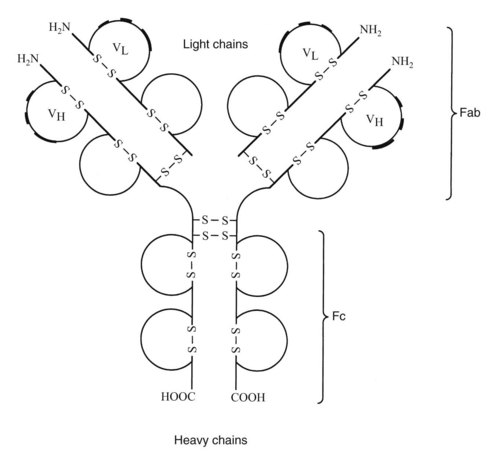
Antibodies synthesized and released by plasma cells directly mediate humoral immunity. As shown in Figure 41-2, the basic immunoglobulin structure consists of two heavy chains and two light chains covalently linked by interchain disulfide bonds. Both chains consist of two or more domains, each defined by a single intrachain disulfide bond. The heavy chain is composed of three or four constant domains and one variable domain. The light chain incorporates one constant and one variable domain. The relatively flexible hinge regions found in certain immunoglobulins are believed to be remnants of primordial constant domains. Terminal sequences on the amino end of each chain make up the variable regions of the molecule. Within each variable region are hypervariable sequences that are responsible for specific antigen binding. There are two types of light chains, λ and κ. The ratio of λ and κ chains differs in various species, being 1 : 20 in mice and 1 : 2 in humans. Sometimes the ratio of λ to κ is used to identify multiple myeloma.

The class of an antibody is determined by its heavy chain. There are five heavy chains or isotypes: IgM, IgG, IgD, IgA, and IgE. IgG is the most abundant subtype and has several subclasses: IgG1, IgG2 (IgG2a and IgG2b), IgG3; and IgG4. The amino terminal region in the variable domain (v domain) of the heavy and light chains (VH and VL) binds to antigen, whereas constant domain (C domain) of the heavy and light chains (CH, CL) makes up the constant regions.

These domains form the Fc region of the molecule (Fc is the “crystallizable fragment” of a polyclonal immunoglobulin). The Fc region dictates the specific binding of each isotype to different Fc receptors on phagocytes, mast cells, and other cells involved in inflammatory reactions. The Fc region also dictates complement activation by IgG and IgM.

There are three discrete regions in the VH and VL domains of the antibodies named HV1 (hypervariable 1), HV2, and HV3 (HV3 is the most variable site of each domain). These are separated by structural framework regions (FRs), namely FR1, FR2, FR3, and FR4. The six hypervariable regions in each arm of the antibody that form the antigen binding site when brought together are complementary to the antigen, thus they are called complementarity determining regions or CDRs. There are three CDRs, CDR1, CDR2 and CDR3. Antigen molecules contact antibody over a broad area of its surface and binds noncovalently through electrostatic forces, hydrogen bonds, van der Waals forces, and hydrophobic interactions.

Transmembrane and secreted forms of Igs are generated through alternative splicing of heavy chain domains. There are two polyadenylated sites that dictate the generation of transmembrane or secreted forms of Igs. All B cells initially express IgM in its transmembrane form, but upon contact with antigen they generate the secreted forms and undergo isotype switching to generate other antibodies.

Sequence differences between Ig heavy chains cause the various isotypes to differ in the number and location of their disulfide bonds, the number of attached oligosaccharide moieties, the number of C domains, and the length of the hinge region.

IgM, which is co-expressed with IgD on the surface of cells, predominates in neonates and during initial, or primary, immune responses to antigenic challenges. IgG, IgA, and IgE are important in responses to antigens on secondary exposure. Because of an extended half-life (approximately 21 days), IgG constitutes approximately 75% of circulating immunoglobulins. IgG is the principal opsonic antibody important for phagocytosis; it plays a central role in immune responses against submucosal antigens. Fc receptors for IgG are found on neutrophils, monocytes, and dendritic cells.

On a daily basis, approximately three times more IgA is produced than all other immunoglobulins combined. IgA has two subclasses, IgA1 and IgA2. The IgA2 isotype is found in most mucous secretions and constitutes about 40% of total salivary IgA. It is important in mucosal immunity and caries immunology. Both IgA and IgM are bound by the polymeric immunoglobulin receptor (pIgR), which is not within the Fc receptor family. Found on the basolateral surface of ductal epithelial and intestinal epithelial cells, the pIgR enables the two immunoglobulins to be transported by transcytosis across the epithelium and into secretions. IgA is important in specific immunity against supramucosal antigens and in anti-inflammatory reactions below the mucosa. Although circulatory IgA production (mostly IgA1) is equal to that of IgG, it has a shorter half-life (approximately 7 days) than IgG and a lower plasma concentration.

IgE is evolutionarily related to IgG in that both their ancestries can be traced back to IgY, the predominate inflammatory immunoglobulin found in nonmammalian vertebrates. IgE is bound with extremely high affinity by Fc receptors found on mast cells and basophils; it promotes immediate inflammation (which is important in initiating acute and chronic inflammation). IgE is also bound by low-affinity receptors present on eosinophils and enables these cells to exert anthelmintic and antiparasitic effects. IgE production is tightly controlled.

The role of IgD is less clear; it is not a secondary response antibody because it can be co-expressed with IgM on a single B cell. IgD and IgM mRNA is produced as a single transcript. Post-transcriptional modifications dictate whether IgM or IgD will be translated. IgD is believed to prevent the induction of B-cell tolerance, which can occur in B cells expressing IgM alone. Fc receptors for IgD have been found on T cells.

Each plasma cell produces a unique antibody because of the clonal distribution of the BCR. Initially, the differentiation of B cells to plasma cells results in the production of IgM antibodies. If antigen has also been presented to CD4+ T cells by B cells, the T cells can guide B-cell differentiation along the memory pathway as opposed to the plasma cell differentiation pathway. In the memory pathway, the isotype may change. Secondary antigen exposure can elicit IgA, IgG, or IgE production. This process of isotype switching promotes a more appropriate interplay of antibodies with complement and with myeloid immune cells (e.g., neutrophils, monocytes, mast cells, and eosinophils).

Initiation, Progression, and Termination of Specific Immune Responses

The immune system is normally engaged in the homeostatic regulation of host-derived antigens. Once an antigen receptor is formed it has to be rigorously tested against self peptides. Given the incredible number of receptors formed it is important that those lymphocytes which reach maturity are likely to be useful in recognizing foreign antigens. In general, developing lymphocytes whose receptors interact weakly with self-antigens, or bind antigen in a particular fashion, receive a survival signal. In contrast, lymphocytes with strongly self-reactive receptors must be deleted to prevent auto-immunity through negative selection. The fate of lymphocytes in the absence of any signal is death. In addition, the immune system participates in its more widely appreciated inflammatory function known as the immune response.

Immune responses are the measurable alterations in immune system activity after an antigenic perturbation. They are usually initiated by an immediate inflammatory reaction resulting from the activation of soluble factors (e.g., complement) found within the extracellular fluid or mediators released by resident leukocytes, especially the mast cell. The immediate inflammatory response signals postcapillary venule endothelial cells to recruit the appropriate acute-phase or chronic-phase leukocytes from the blood. Initially recruited are cells that do not need to progress through proliferation or differentiation to exert an effect, such as neutrophils. Neutrophils are the predominant cell in acute inflammation. Acute inflammation may be followed by slower, chronic inflammation involving less mature cells capable of adaptive cellular differentiation (with or without proliferation). The proliferation and differentiation of clones of cells that recognize the antigen specifically constitute the specific immune response.

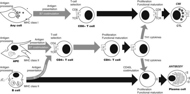
Specific immune responses involve a series of events (Figure 41-3), each of which offers a potential site for immunotherapeutic intervention. Included in this series are antigen processing and presentation, T-cell selection, lymphocyte differentiation, effector function, and termination. These events occur in response to changes in the concentrations of antigens intracellularly and extracellularly.

Antigen processing

Antigen processing is the partial degradation of polymeric antigens into oligomeric units (especially the degradation of a protein into small peptides), which are subsequently bound by MHC I or II molecules. Various hydrophobic peptides and glycolipids can be bound by CD1 antigens, which are related to the MHC I and II molecules.

Intracellular antigens

Processing of intracellular antigens generated within the endoplasmic reticulum or cytosol occurs continuously in all cells. Proteins in the cells become degraded and are replaced by newly synthesized proteins. Degradation of protein occurs in a large, multi-subunit protease-like structure called the proteasome. Peptides can further be trimmed in the endoplasmic reticulum (ER) by the help of aminopeptidases. Some proteasomes accept proteins for degradation only if they are tagged by small polypeptides called ubiquitin. The peptides generated in the cytosol are transported into the ER where they are bound by nascent MHC I molecules.

Viruses and certain types of bacteria are degraded in the cytosol or in the nuclear compartments before they are presented in the context of MHC I. Structurally, MHC I consists of two polypeptide chains, α and β. The α chain is the larger of the two and has three domains, α1, α2 and α3. The smaller, nonpolymorphic β chain, termed β2 microglobulin, is noncovalently attached to the α3 domain. Only the α chain spans the membrane. The α1 and α2 domains form the cleft that binds to the peptide fragment and are highly polymorphic. Peptides that bind to MHC I molecules are transported from the cytosol to the ER. The peptide-binding site of MHC I is formed in the lumen of the ER and is never exposed to the cytosol. Expression of MHC I on the surface of the cells is unstable unless it is bound to peptide. Mutations at a site where peptides bind to the MHC I protein cause significant decreases in the expression of MHC I on the surface of the cells. Mutations in transporters associated with antigen processing (TAPs) may also not allow for the transport of the peptides from the cytosol to the lumen of the ER.

Newly synthesized MHC I α chains bind to a chaperone protein called calnexin, which retains MHC I in a partly folded structure in the ER. When β2 microglobulin binds the α chain the complex dissociates from calnexin and then binds to another chaperone, calreticulin. A third protein, tapasin, forms a bridge between MHC I molecules and TAP, allowing the transport of the suitable peptide from the cytosol. Most of the chaperon proteins play a role in selecting peptides with higher binding affinity.

Viruses have evolved several means of evading recognition by preventing the appearance of peptide:MHC I complexes at the cell surface. For example, the herpes simplex virus prevents the transport of viral peptides into the ER by producing proteins that bind and inhibit TAP, whereas adenoviruses produce a protein that binds MHC I molecules and retains them in the ER. Cytomegaloviruses accelerate retrograde translocation of MHC I back into the cytosol, where they are degraded.

Extracellular antigens

The majority of pathogenic bacteria and some eukaryotic parasites replicate in the endosomes and lysosomes that form the vesicular system. Pathogens such as Leishmania and Mycobacteria are picked up by macrophages through endocytosis. The resultant endocytic vesicles gradually become acidified and finally fuse with lysosomes, forming phagolysosomes. These organelles contain acid proteases that become activated at low pH, resulting in degradation of the protein antigen. Among acid proteases are the cysteine proteases cathepsin B, D, S, and L. Cathepsin L is the most active of the family. Disulfide bonds also need to be cleaved for peptide processing. Degraded peptides from pathogens are then presented in the context of MHC II molecules to T cells.

Each MHC II protein comprises two noncovalently bound chains, α and β, both of which span the membrane. There are two α domains, α1 and α2, and two β domains, β1 and β2. The α1 and β1 domains are very polymorphic and form the peptide-binding locus. The folding of α1 and β1 is more open than the α1 and α2 domain of MHC I, thus the ends of the peptide fragments are exposed.

Binding of nonspecific peptides to MHC II is prevented by the association of MHC II in the ER with a third polypeptide termed the invariant chain. The invariant chain, by forming a trimer with MHC II, covers the antigen-binding groove. Invariant chains also target MHC II to low-pH endosomal compartments where peptide loading can occur. The invariant chain is then cleaved by acid proteases, such as Cathepsin S, to generate a truncated form. The subsequent cleavage releases the MHC II molecule leaving a fragment called CLIP bound to the MHC II.

Most MHC II molecules are brought to the cell surface in vesicles, which at some point fuse with incoming endosomes where they encounter and bind peptides derived from self- or non–self-proteins. HLA-DM assists in the process by stabilizing MHC II molecules, which would otherwise aggregate. It also catalyzes the removal of CLIP and the loading of the peptide into the groove of the MHC II. HLA-DM, closely related to the MHC II molecule, does not bind to the peptide because its groove is closed. HLA-DM also removes weakly bound peptides, allowing for the binding of high affinity peptides. Peptides bound to MHC can remain several days in case the APC does not encounter the target; therefore, increased binding affinity of the peptide to the groove is important.

A second atypical MHC II molecule, called HLA-DO is produced in thymic epithelial cells and B cells. Unlike HLA-DM, which aids in releasing CLIP and binding peptide, HLA-DO inhibits HLA-DM function and subsequently inhibits antigen loading. The activating capacity of HLA-DM is higher than the inhibiting capacity of HLA-DO because secreted IFN-γ can upregulate HLA-DM but not HLA-DO.

T-cell activation

T-cell recognition, activation, and effector function depend on cell-cell contact mediated by cell adhesion molecules. T cells enter the lymph nodes through binding to specialized postcapillary swellings called high endothelial venules. The main classes of adhesion molecules involved in lymphocyte interaction are the selectins, integrins, members of the Ig superfamily, and mucin-like molecules. Selectins are important for leukocyte homing to particular tissues and can be expressed either on leukocytes (L selectin) or on endothelial cells (P and E selectins). Selectins are cell-surface molecules with a common core structure, but different lectin-like domains in their extracellular portions. The lectin domain binds to particular sugar groups on a carbohydrate backbone. L selectin binds to the carbohydrate moiety of mucin-like molecules called vascular addressins, which are expressed in the surface of endothelial cells. The interaction between L selectin and vascular addressins is responsible for the specific homing of naïve T cells to lymphoid tissues, whereas the integrins and the Ig superfamily are involved in their crossing through the endothelial barrier.

Just as binding of neutrophils to endothelial cells is guided by chemokines, migration of naïve T cells into lymphoid tissues is directed by similar molecules, such as secondary lymphoid tissue chemokine. This interaction increases both the affinity and surface expression of integrins on the T cell membranes, which arrests the cells and causes them to move through the endothelial layer to enter the lymphoid parenchyma. The integrin LFA-1, for example, is expressed on all T cells. It binds to Ig superfamily adhesion molecules, such as ICAM-1 and ICAM-2, which are expressed on endothelium and APCs. Binding to LFA-1 allows the leukocytes to migrate through the blood vessel wall in lymph nodes and be able to sample antigen on the surface of APCs. ICAM-3, another adhesion molecule, is expressed only on leukocytes and binds to its partner (DC-sign) on dendritic cells. The binding of CD58 (LFA3) on APCs to CD2 on T cells is yet another example of adhesion molecule interactions.

The initial association of T cells with APCs is mediated by the interplay of multiple adhesion molecules. Binding of LFA-1, ICAM-3 and CD2 on T cells to ICAM-1, ICAM-2, DC-sign and CD-58 on APCs provides enough redundancy that if one pairing is missing the T cells can still bind and recognize the specific antigen on the APCs. The transient binding to APCs allows the T cells to sample a large number of MHC:peptide sequences on APCs, and if it recognizes the antigen, signaling through the TCR will significantl increase the affinity of LFA-1 for ICAM-1.

Both signals for specific antigen and costimulatory molecules are required for the clonal expansion of T cells. In the absence of costimulatory signals, activation of T cells through antigen receptor will lead to anergy or unresponsiveness. Anergic cells are deleted by cell death. The most highly characterized costimulatory molecules are structurally related glycoproteins belonging to the B7 family. The best known are B7.1 (CD80) and B7.2 (CD86). They bind to the CD28 receptor on the surface of naïve T cells and costimulate in the presence of antigen receptor. Once a naïve T cell is activated, it up-regulates CD40 ligand, which binds to the CD40 receptor on APCs and further activates both the T cells and the APCs. Activated T cells will then upregulate cytotoxic T-lymphocyte–associated antigen-4 (CTLA-4 or CD152), which is similar in structure to CD28 and limits further activation by binding to B7 family costimulatory molecules. This way activation and proliferation of T cells are regulated.

Resident dendritic cells such as Langerhans cells are able to pick up antigen by phagocytosis or macropinocytosis. They can travel and take the antigen to the secondary lymphoid tissues where they lose the capability to phagocytize but gain the ability to present antigen to T and B cells. Both dendritic cells and B cells can bind soluble antigens and present them as peptide: MHC II complexes to T cells. B cells do not express costimulatory molecules constitutively; they need to be activated by bacteria to express B7.1 and B7.2.

The actual recognition of antigen by the TCR is a low-affinity reaction. This characteristic enables many TCRs of a given T cell to interact with the few specific antigens presented by the antigen-presenting cell. Multiple interactions are important because T-cell activation depends on the number of TCRs that interact with antigen over time.42 The factors that influence T-cell activation include the number of antigen molecules presented by the antigen-presenting cell, the affinity of the TCR for the antigen, and the number of TCRs. If the interaction with peptide antigen by TCRs is sufficient, the TCRs cluster on the T-cell surface and downregulate (the TCRs are probably internalized). With costimulation, downregulation of approximately 4250 TCRs leads to T-cell activation.

In early stages of exposure to antigen, costimulatory signals permit T cells to become receptive to differentiative signals, allowing them to proliferate or mature in function. These signals also block apoptosis. At later stages, the same signals can permit T cells to differentiate terminally, even to the point of death.

Differentiation

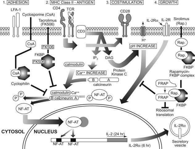
After antigen recognition by TCR and BCR, intracellular signaling events allow the T cell to differentiate into functionally mature cells (Figure 41-4). Much present-day immunotherapy is aimed at this stage of the specific immune response. Binding of antigen to its receptor leads to the clustering of antigen receptor on lymphocytes, which is the first step in signal transduction, Clustering of antigen receptor leads to activation of intracellular signaling molecules. Protein tyrosine kinases are enzymes that affect the function of other proteins by adding a phosphate group to certain tyrosine residues. Specific growth factors such as c-kit have cytoplasmic domains that contain intrinsic tyrosine kinase activity. T- and B-cell signaling is different from c-kit signaling since the TCR and BCR do not have intrinsic kinase domains; rather they rely on the interaction with other tyrosine kinases known as receptor-associated tyrosine kinases.

There are specialized areas within the membrane lipid bilayer that contain high quantities of sphingolipids and cholesterol. These areas are called lipid rafts. Signaling molecules associate with lipid rafts. Disruption of the lipid rafts inhibits T- and B-cell signaling.

Signaling through T and B cells is governed by a complex array of intracellular signaling elements, which can phosphorylate and activate other elements to transduce the signal from the receptor. The receptor-associated protein kinases are localized in the inner surface of the cell membrane and cannot activate their cytosolic targets efficiently unless they themselves are activated. Their activation and subsequent phosphorylation of tyrosines on receptor-associated chains recruits other protein tyrosine kinases, which then transduce the signal. The Src family kinases involved in T and B cell receptor signaling provide an example of this kind of signaling element.

The variant chains of the lymphocyte antigen receptor are associated with invariant accessory chains that mediate the signaling function of the receptor. The BCR is associated with Igα and Igβ invariant chains. The TCR is associated with multiple invariant accessory chains (ε, γ, δ, and ζ). Accessory chains have a structure termed the immunotyrosine activation motif (ITAM), which enables them to signal when the BCR or TCR is bound to the antigen. ITAMs are phosphorylated by src family receptor–associated tyrosine kinases, which gives them the ability to bind to the members of a second family of tyrosine kinases (Syk in B cells and Zap 70 in T cells). The enzyme activity of each src family kinase is regulated by the phosphorylation status of its kinase domain and its carboxy terminal region, each having a regulatory tyrosine residue. Phosphorylation of tyrosine in the kinase domain activates the enzyme, whereas phosphorylation at the carboxy terminal tyrosine is inhibitory. Src family kinases are kept inactive by a tyrosine kinase called CSK (c-terminal src kinase), which phosphorylates the inhibitory domain. Since the function of CSK is constitutive in resting cells, src-family proteins remain quiescent until antigen recognition, which then activates a protein tyrosine phosphatase (CD45 that removes the phosphate block from the inhibitory tyrosine and permits activation of the src-family kinases.

Antigen receptor signaling is enhanced by coreceptors that bind to the same ligand. B cell co-receptors are formed by a complex of CD19, CD21, and CD81 proteins. Src family kinases phosphorylate the ITAMs on Igα and Igβ, and these subsequently recruit Syk to Igβ. Trans-phosphorylation of each kinase by the other occurs, and the activated kinases in turn activate phospholipase C-γ (PLC-γ), guanine exchange factors (GEFs), and Tec kinases.

Similar to BCRs, clustering of TCRs and the co-receptors activates CD45, which removes a phosphate from Src family kinases (e.g., Lck) and activates their function, resulting in the phosphorylation of ITAMs. After phosphorylation, ZAP-70 is able to bind and become activated by Lck, which will initiate a signaling cascade leading to the activation of PLC-γ, GEF, and Tec kinases.

PLC-γ is known to cleave membrane phosphatidylinositol bisphosphate (PIP2), yielding diacylglycerol (DAG) and inositol triphosphate (IP3). DAG activates protein kinase C (PKC), which in turn stimulates nerve factor-κB (NF-κB). NF-κB usually is complexed with inhibitor proteins collectively called I-κB, which prevent its translocation to the nucleus. NF-κB is an important transcription factor for various inflammatory cytokines, and supports inflammatory aspects of the specific immune response. TNF-α, one of the cytokines whose synthesis is stimulated in part by NF-κB, increases the transcription of NF-κB and its dissociation from I-κB.

IP3 increases the intracellular Ca++ concentration, activating calcineurin, a phosphatase that frees NF-AT (nuclear factor of activated T cells) from a phosphate block. GEFs activate Ras, an important GTPase that activates MAP kinases. The Ras-induced MAP kinases then activate Fos and Jun kinases, which are components of the transcription factor AP-1.

In unstimulated cells, NF-AT is rendered inactive and remains in the cytoplasm by phosphorylation on serine/threonine residues. Ca++-activated calcineurin remo/>

Stay updated, free dental videos. Join our Telegram channel

Jan 5, 2015 | Posted by in General Dentistry | Comments Off on 41: Immunotherapy

VIDEdental - Online dental courses

Get VIDEdental app for watching clinical videos