40
Periodontal diseases in children and adolescents
Figure 40.1 Classification of periodontal diseases and conditions for children and adolescents. From Armitage (1999).
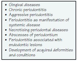
Figure 40.2 Simplified Basic Periodontal Examination (BPE) screening in children and adolescents. WHO, World Health Organisation. From Ainamo et al. (1984); see also Clerehugh (2008).
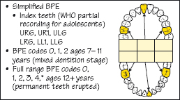
Figure 40.3 Plaque-induced gingivitis in a 16-year-old girl.
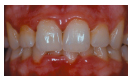
Figure 40.4 Plaque-induced gingivitis in a 15-year-old Asian girl.
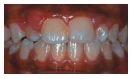
Figure 40.5 (a) Plaque-induced gingivitis and supragingival calculus on the labial of the lower incisors in a 10-year-old Asian girl. (b) Lingual lower anteriors with supragingival calculus.
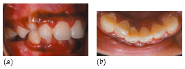
Figure 40.6 Plaque-induced gingivitis and local contributing factors. (a) Localised recession. (b) Frenal attachments near the gingival margin. (c) A 19-year-old girl with amelogenesis imperfecta and calculus. (d) Incompetent lips and mouth breathing exacerbating gingivitis anteriorly in a 12-year-old boy.
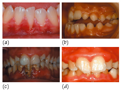
Figure 40.7 Non-plaque-induced gingival condition: primary herpetic gingivostomatitis. Herpes simplex virus 1 vesicles on the gingiva (a, b) and tongue (c). Courtesy of Dr S. Kindelan.
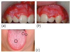
Figure 40.8 Non-plaqueinduced gingival condition: neutropenia in a young child with severe gingival inflammation and mobile primary incisors. Courtesy of Dr S. Kindelan.
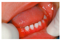
Figure 40.9 Non-plaque-induced gingival condition: histoplasmosis in a young, severely immunocompromised child with bone marrow rejection. There is a fungal infection, deep mycoses and histoplasma capsulatum. Courtesy of Professor I. L. C. Chapple.
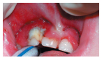
Figure 40.10 (a) The results of a 5-year study of clinical attachment loss in 14–19-year-old adolescents in Rochdale, UK. (b) Incipient chronic periodontitis in a 19-year-old. From Clerehugh et al. (1990).
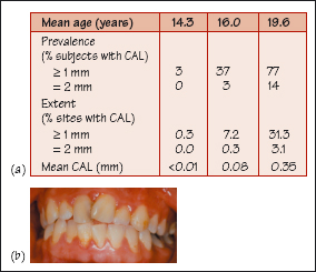
Figure 40.11 (a–c) Localised aggressive periodontitis in a 12-year-old black girl of African origin.
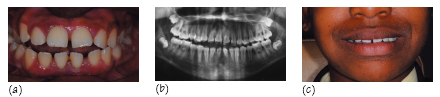
Figure 40.12 Papillon–Lefèvre/>
Stay updated, free dental videos. Join our Telegram channel
VIDEdental - Online dental courses


