39
Periodontal abscess and periodontic–endodontic lesions
Figure 39.1 Common features of periodontal abscesses.
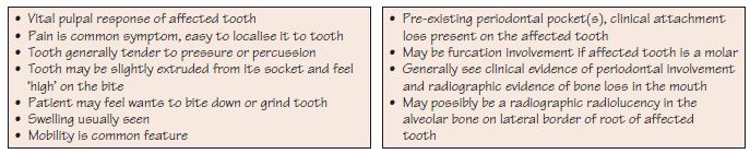
Figure 39.2 (a) Features of early periodontal abscess formation, where pus is not draining. (b) Periodontal abscess with pus not draining through the pocket and not yet ready to point.
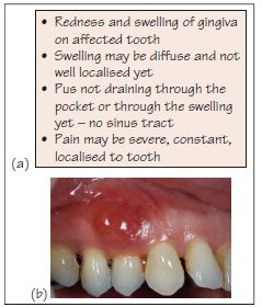
Figure 39.3 (a) Features of a periodontal abscess with pus to drain. (b) Periodontal abscess with pus to drain by LR5. The swelling was fluctuant and pus discharged when the swelling was incised. (c) Panoramic radiograph showing vertical bone loss distal to LR5; note the generalised horizontal and vertical bone loss throughout the mouth. A periodontic–endodontic lesion can be seen on the LL6 distal root and periapical radiolucency on UL5.
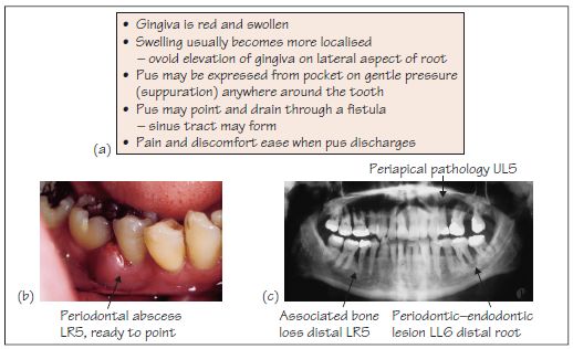
Figure 39.4 (a) Features of a periodontal abscess with systemic involvement. (b) An extra-oral swelling on the lower right angle of the mandible.
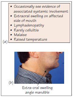
Figure 39.5 (a) Features of a gingival abscess. (b) An abscess on the mesiopalatal marginal gingiva of UR6.
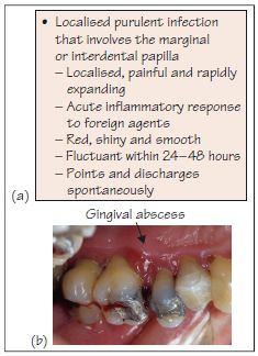
Figure 39.6 Features of a pericoronal abscess.
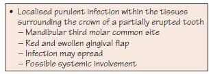
Figure 39.7 (a) Features of a periapical lesion. (b) The buccal sinus at UR6. (c) A diagnostic gutta percha point inserted into the sinus to track the source of infection. (d) A periapical radiograph showing periapical radiolucency around the apices of a root canal treated at UR6. (e) A gutta percha inserted through the buccal sinus extends into the periapical area, indicating that the source of infection is from an endodontic origin not a periodontic origin.
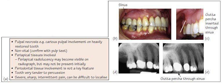
Figure 39.8 (a) Features of a combined periodontic–endodontic lesion.(b) Periodontic–endodontic lesion at LR4 showing a localised but quite large buccal swelling and pus discharging through the pocket. (c) A periapical radiograph showing typical radiographic features of a periodontic–endodontic lesion with vertical bone loss extending to the apex of the tooth and merging with periapical radiolucency due to non-vital pulp.
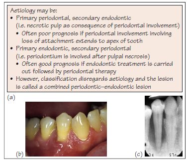
Figure 39.9 Before and after successful endodontic and periodontal treatment of a periodontic–endodontic lesion at LL3. (a) A localised, small, buccal swelling at LL3 (a deep but narrow pocket was detected on periodontal probing distobuccally). (b) A periapical radiograph after obturation of the root canal of LL3. The periapical radiolucency reduced over subsequent months, indicating some healing. (c) Healing of the pocket following successful endodontic and periodontal treatment.
Stay updated, free dental videos. Join our Telegram channel
VIDEdental - Online dental courses


