29
Periodontic-orthodontic interface
Figure 29.1 A Hawley retainer.
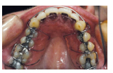
Figure 29.2 (a–c) Fixed orthodontic appliances in a 15-year-old Asian girl.Note the poor plaque control, gingival inflammation and swelling anteriorly and posteriorly.
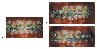
Figure 29.3 (a, b) Interspace brush with a single tufted head for cleaning around fixed appliance brackets, arch wires and elastics.
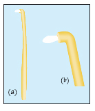
Figure 29.4 (a–c) A 28-year-old female patient (non-smoker) with a history of localised aggressive periodontitis, prior to periodontal therapy. Note inflammation around the lateral incisors, upperleft central incisor and instanding lower left lateral incisor. Also note the position of the upper left central incisor which has drifted and dropped out of line of the arch and is rotated and tilted.
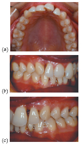
Figure 29.5 Radiographs of the patient in Fig. 29.4 (a, d) Vertical bitewing radiographs showing subgingivalcalculus and bone loss on the distal of the upper first molars and lower left second molar. (b, c, e, f) Periapical radiographs showing subgingival calculus and bone loss on the upper right lateral incisor, upper left central and lateral incisors and lower leftlateral incisor.
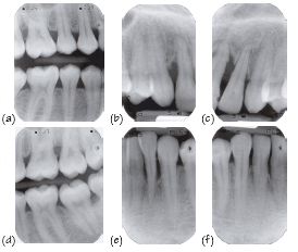
Figure 29.6 Adult orthodontics in periodontally compromised patients.
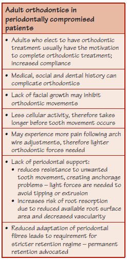
Figure 29.7(a, b) Thepatient in Fig. 29.4 after periodontal therapy. The patient disliked the appearance and position of her upper left central incisor but wanted to retain the tooth if at all possible. Lip trapping occurs and localised recession.
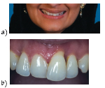
Figure 29.8 Stages of orthodontic treatment for the patient in Fig. 29.7: (a) fixed upper appliance; (b) bonded palatal upper retainer; (c) fixed lower appliance; and (d) aligned (permanently retained) upper anterior teeth and fixed lower appliance. There is improved aesthetics andspontaneous improvement in recession on the upper left central incisor following orthodontic tooth movement.
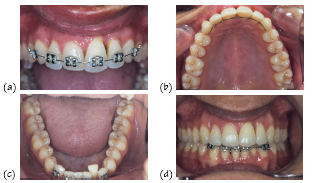
Figure 29.9 (a) Pre-periodontal and orthodontic treatment of drifted upper central incisors in a female adult patient with chronic periodontitis. (b) After treatment. (c) Bonded orthodontic retainer.
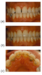
Figure 29.10 (a–c) Pre-periodontal treatment of a 33-year-old female patient with an aggressive form of periodontitis that led to drifting and proclination of the upper left central incisor, wh/>
Stay updated, free dental videos. Join our Telegram channel
VIDEdental - Online dental courses


