Chapter 4 Viruses and prions
Structure
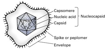
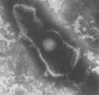
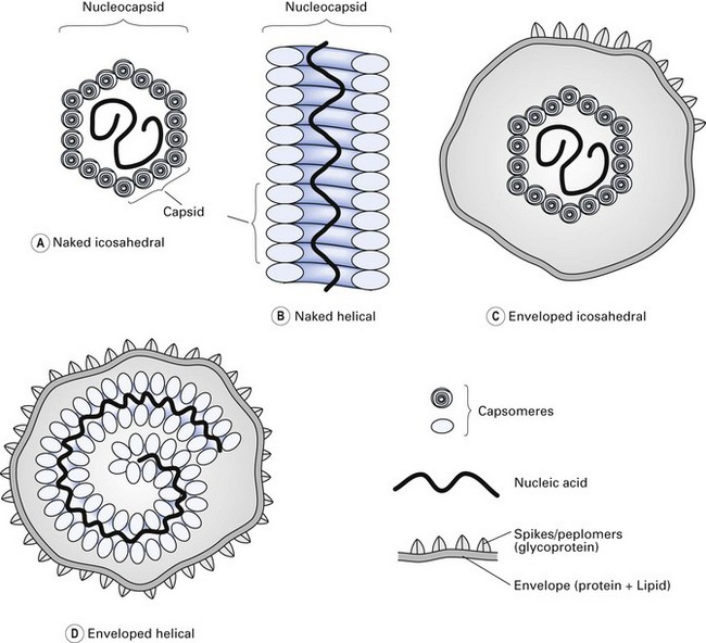
Viruses consist of a nucleic acid core containing the viral genome, surrounded by a protein shell called a capsid (Figs 4.1 and 4.2). The entire structure is referred to as the nucleocapsid. This may be ‘naked’, or it may be ‘enveloped’ within a lipoprotein sheath derived from the host cell membrane. In many viruses (e.g. orthomyxoviruses, paramyxoviruses), the ensheathment begins by a budding process at the plasma membrane of the host cell, while others, such as herpesviruses, ensheath at the membrane of the nucleus or endoplasmic reticulum.
The protein shell or capsid consists of repeating units of one or more protein molecules; these protein units may go on to form structural units, which may be visualized by electron microscopy as morphological units called capsomeres (Fig. 4.1). Genetic economy dictates that the variety of viral proteins be kept to a minimum as viral genomes lack sufficient genetic information to code for a large array of different proteins. In enveloped viruses, the protein units, which comprise the envelopes and are visualized electron microscopically, are called peplomers (loosely referred to as ‘spikes’).
Virus symmetry
The nucleocapsids of viruses are arranged in a highly symmetrical fashion (symmetry refers to the way in which the protein units are arranged). Three kinds of symmetry are recognized (Fig. 4.3):
Taxonomy
Vertebrate viruses are classified into families, genera and species. The attributes used in classification are their symmetry, the presence or absence of an envelope, nucleic acid composition (DNA or RNA), the number of nucleic acid strands and their polarity. Classification of some of the recognized families of RNA and DNA viruses is given in Table 4.1. (Note: to memorize which viruses contain DNA, remember the acronym ‘PHAD’: P is for papova and pox, H for herpes and AD for adenoviruses. Most of the remainder are RNA viruses, including the self-evident picornaviruses.)
Table 4.1 Classification of some of the viruses causing human disease
| Morphology | Virus |
|---|---|
| DNA | |
| Enveloped, double-stranded nucleic acid | Herpesviruses |
| Herpes simplex virus | |
| Varicella-zoster virus | |
| Epstein–Barr virus | |
| Cytomegalovirus | |
| Human herpesvirus 6 | |
| Poxviruses | |
| Vaccinia | |
| Orf | |
| Enveloped, single-stranded | Parvoviruses |
| Non-enveloped, double-stranded | Adenoviruses |
| Papovaviruses | |
| Polyomaviruses | |
| Papillomaviruses | |
| Hepadnaviruses | |
| Hepatitis B virus | |
| RNA | |
| Enveloped, single-stranded | Orthomyxoviruses |
| Influenzavirus | |
| Paramyxoviruses | |
| Parainfluenza | |
| Respiratory syncytial | |
| Mumps | |
| Measles | |
| Togaviruses | |
| Rubella | |
| Retroviruses | |
| Human immunodeficiency viruses HTLV-I, -III | |
| Rhabdoviruses | |
| Rabies | |
| Non-enveloped, double-stranded | Reoviruses |
| Rotavirus | |
| Non-enveloped, single-stranded | Picornaviruses |
| Rhinovirus | |
| Enterovirus | |
| Coxsackievirus | |
| Echovirus | |
| Poliovirus |
HTLV-I, human T cell leukaemia virus type I.
The following is a concise description of the families of mammalian viruses.
DNA viruses
Adenoviruses
Herpesviruses
Structure
Different herpesviruses cause a variety of infectious diseases, some localized and some generalized, often with a vesicular rash. Herpesviruses establish latent infection, which can be readily reactivated by immunosuppression (Table 4.2).
| Virus | Site of latency |
|---|---|
| Herpes simplex virus | Trigeminal ganglion |
| Varicella-zoster virus | Sensory ganglia |
| Epstein–Barr virus | Epithelial cells |
| B lymphocytes | |
| Cytomegalovirus | Salivary gland cells |
| Papillomaviruses | Epi/> |
Stay updated, free dental videos. Join our Telegram channel

VIDEdental - Online dental courses


