4: Problem Solving in the Differential Diagnosis of Bony Defects Resulting from Pulpal and Periodontal Pathosis

Chapter 4

Problem Solving in the Differential Diagnosis of Bony Defects Resulting from Pulpal and Periodontal Pathosis

Problem-Solving List

Problem-solving challenges and dilemmas that deal with the differential diagnosis of bone defects resulting from pulpal and periodontal pathosis addressed in this chapter:

Controversies and Highlights of Disease Interactions
An Endodontic Perspective on Chronic Periodontitis
Periodontal Lesions of Bone that Can Be Confused With Pulpally Induced Bony Lesions

    Acute periodontal abscess
    Lesions of chronic periodontitis
    Periodontal lesions involving the furcation
    Lesions associated with aggressive forms of periodontitis
    External root resorption
    Cemental tears
Pulpally Induced Lesions that Can Be Confused With Periodontal Lesions

    Furcation or lateral lesions without loss of attachment
    Acute periapical abscess
    Chronic sinus tracts of pulpal origin with drainage through the gingival sulcus
    Chronic sinus tracts of pulpal origin with permanent periodontal attachment loss
    Response of the periodontium to mechanical root perforations
Bony Lesions of the Periodontium that Do Not Originate from Either Periodontal or Pulpal Pathosis

    Deep coronal fractures
    Vertical root fractures
    Developmental lingual groove on maxillary lateral incisors and similar lesions
    Other possible rare lesions

“I have never found a pulp removed from a pyorrhetic tooth to be normal … where a pathologic pulp is present, a pyorrhetic condition cannot be cured by treatment applied exclusively to the external surface of the root, with no treatment of the pulp itself.”< ?xml:namespace prefix = "mbp" />12

L.R. Cahn, 1927

“Interradicular periodontal lesions can be initiated and perpetuated by inflamed or necrotic pulps. Extension of the inflammatory lesions from the dental pulp apparently occurs through accessory or lateral canals situated in the furcation regions of premolars and molars.”45

S. Seltzer, I.B. Bender, H. Nazimov, 1967

The relationship of disease processes coming from the pulp of the tooth to the supporting periodontium is a subject that has received much attention in endodontic literature for almost 90 years.10,12,18 The reverse situation has also been controversial: the impact of periodontal pathosis on the dental pulp.* Clinically, two important issues must be kept in focus. Pulpal pathosis and its extension into the periodontium causes a localized periodontitis with the potential for further extension into the oral cavity.26 Periodontal pathosis and its extension have little short-term effect on the dental pulp.26 However, the long-term effect of periodontal pathosis, especially in conjunction with concomitant restorative procedures, must be considered in all diagnostic and treatment planning considerations.

Controversies and Highlights of Disease Interactions

Pulp tissue degenerates after a multitude of insults—caries, restorative procedures, chemical and thermal insults, trauma, and some periodontal treatment. When products from pulp degeneration, in particular inflammatory exudates and bacteria, reach the supporting periodontium, many changes may occur, including a rapid onset of inflammation, lateral or furcation bone loss, tooth mobility, and sinus tract formation through the buccal mucosa or gingival sulcus. If this occurs in the apical region, a periapical lesion forms (see Chapter 3). If this occurs with crestal extension of the inflammation, a periodontitis of pulpal origin is formed.49 However, the lesion formed has little anatomic similarity to a defect of periodontal pathosis.

Periodontal disease is generally a slow-developing process that may have a gradual atrophic effect on the dental pulp. Complete pulpal necrosis caused by periodontal pathosis, however, is uncommon. Changes in the pulp include chronic inflammation, localized tissue death (infarction), fibrosis, decrease in cellular populations, resorptions, local coagulation necrosis, or dystrophic calcification.32,43,46,50 Periodontal procedures such as deep scaling and curettage54 (with the use of localized medicaments) and gingival injury or wounding53 may enhance further pulpal inflammation and perpetuate the interrelated disease process in a small number of cases.

The most intimate and demonstrable relationship of the communication of inflammation between the two tissues is via the vascular system,9,20,31,54 as demonstrated anatomically at the apical foramen and adjacent to aberrant accessory communications.11,18,25,36,43 These channels, when patent, may serve as potential routes of inflammatory interchange. Still other channels, covered by cementum, may be exposed during scaling or other periodontal therapeutic procedures.54 To what extent these vascular communications must be disrupted to cause a resultant overwhelming inflammatory process is unknown and a major concern.32

The main anatomic pathways must be considered as potential pathways in the exchange of inflammatory products and bacteria between the pulp and the periodontium (and vice versa) and include lateral/accessory canals that branch off the main root canal, open dentinal tubules (sometimes due to cemental agenesis), the presence of lingual grooves, removal of cementum during periodontal or restorative procedures or loss due to resorptive defects, and root or tooth fractures.

For intact teeth, the principle avenues of communication would be the main foramen, lateral/accessory canals, and dentinal tubules. In the clinical setting, however, there are few means to determine that these particular avenues of communication are actively involved in the exchange of inflammatory substances and/or bacteria. For our purposes, the pathophysiologic relationships that exist between the pulp and periodontium are less important than an accurate diagnosis or one based on the best clinical evidence available on clinical problems and lesions that present on a daily basis. In general, inflammation or bone loss in the periodontium caused by lesions of pulpal origin will heal following a wide range of endodontic treatments. These types of cases are seen as a radiolucent lesion around the root apex, in an isolated furcation that has no other etiology, or on the lateral surface of a root. Specific periodontal treatment in these cases is rarely if ever warranted. The vast majority of lesions of the periodontium do not affect the dental pulp. The incidence of cases in which a connection between a lesion of pulpal origin and one of periodontal pathosis is even suspected is small.27,42

This chapter has three objectives: (1) to clarify the most important diagnostic characteristics of lesions of the periodontium, with emphasis on those that resemble the extension of pulpal pathosis to the supporting root tissues; (2) to clarify the distinguishing diagnostic characteristics of periradicular lesions of pulpal origin that resemble or are mistaken for periodontal defects; and (3) to discuss the characteristics of lesions that are the result of neither inflammation/infection of the pulp or periodontium, yet may share characteristics of both. Treatment suggestions will accompany the lesions described.

An Endodontic Perspective on Chronic Periodontitis

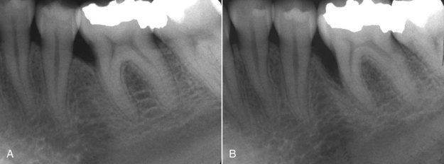
The etiology and pathophysiology of chronic periodontitis is extremely complex. Briefly summarized, lesions of the disease are the result of microbiologic and immunologic effects of a biofilm that forms on the surfaces of roots.4,35,62 Fortunately, an accurate clinical diagnosis of periodontitis can usually be made on the basis of physical examination35 and radiographic findings of bone destruction.39 The clinical characteristic of this disease may be summarized as a “top-down” process, meaning simply that it begins with inflammation of the marginal periodontium and progresses apically with the gradual destruction of the crestal supporting bone (Fig. 4-1, A and B).

image

FIGURE 4-1 A, Typical radiographic presentation of periodontal bone loss on mesial of mandibular first molar. B, 5 years later, bone loss has progressed to a deeper level.

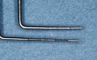
For the dentist/endodontist, the periodontal probe is absolutely indispensable to clinical diagnosis. During assessment of the periodontium, measurement of attachment loss is the standard by which progression or remission of the disease is assessed.62 Within the discipline of endodontics, the discrimination between periodontal lesions and those originating from the dental pulp can usually be made by identifying the physical characteristics of the lesion itself.27,28 The technique of diagnostic periodontal probing should be done using a relatively small-diameter instrument (0.05 mm at the tip) with standard markings (Fig. 4-2).62 Probing should be done with as uniform a pressure as possible, slightly angling the tip of the probe toward the surface of the root.30

image

FIGURE 4-2 Periodontal probes with small-diameter tips are preferable.

The normal periodontium is known to have fairly predictable dimensions.23 The average distance from the gingival margin to crestal bone has been found to be approximately 2.5 mm. Increased probing depths represent the loss of periodontal attachment to the tooth and crestal bone. Since chronic periodontitis is usually generalized, osseous defects are typically found in multiple areas of the dentition, particularly in the posterior sextants. This can be an important finding in the diagnosis of potential endodontic problems. In the diagnosis of a localized defect of unknown origin, begin the diagnostic examination by probing multiple random locations throughout the dentition.27 Findings of osseous defects in several locations well apart from the area of chief concern would tend to support a tentative diagnosis of periodontitis for the area in question. In contrast, random probings that reveal no indication of periodontal bone loss in any other area would suggest a possible nonperiodontal etiology for the localized defect being evaluated.

Traditionally in routine periodontal examinations, probing depths have been recorded in six locations for each tooth: the mesial, midsurface, and distal of both the buccal and lingual surfaces.4 For the purposes of endodontic diagnoses and the differentiation of lesions of pulpal origin from periodontal lesions, probings in the area of principle concern should be performed circumferentially on each tooth in small (1-mm) increments. The intent is to explore the physical morphology or bony architecture of a lesion. There is a distinction between “probing” the attachment levels and “sounding” the location of the crestal bone.27 In the technique of sounding, the probe is inserted past the level of attachment directly to bone. This is a more accurate method of assessment but will usually require some level of local anesthesia. Generally, probing will yield adequate diagnostic information, especially in combination with radiographs.

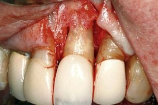
Osseous defects resulting from chronic periodontitis are routinely found to be of varying depth but similar in form. Probing circumferentially across a broad root surface will usually indicate a gradual increase in depth until the deepest area of the defect is reached. Continuing past this point, the probing depths will then gradually decrease. The typical contours of crestal bone found in bony lesions of advanced periodontitis are illustrated in Fig. 4-3. Note the general increase in root exposure from mesial to distal, which would be reflected as a gradually increasing probing depth on a clinical examination.

image

FIGURE 4-3 Surgical exposure of advanced lesion of chronic periodontitis. Note altered contours of the crestal bone.

Periodontal Lesions of Bone that Can Be Confused With Pulpally Induced Bony Lesions

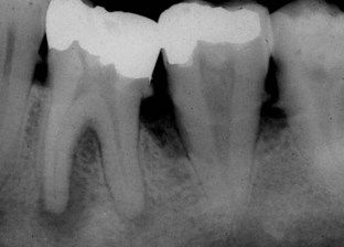
Bony lesions of periodontal disease are usually not difficult to distinguish from bony lesions of pulpal origin. Periodontal defects begin in the marginal periodontium, and even deep periodontal pockets are usually far removed from the root apex (Fig. 4-4). Confusion in diagnosis usually arises in cases of acute periodontal abscess, periodontal infection causing chronic sinus tracts in the mucosa, or localized periodontal bony lesions extending deep enough to involve the apex of a tooth. The severity of periodontal bone destruction, however, varies in the same dentition. At times it may be quite localized and extend to the root apices. The following discussion of pathologic entities will include diagnostic procedures necessary to (1) arrive at the best diagnosis based on the gathered information and (2) determine the appropriate treatment plan.

image

FIGURE 4-4 Lesions of advanced chronic periodontitis with severe bone loss do not generally involve the apex, as seen on the second molar. Lesion on first molar is endodontic, with a drainage tract coursing coronal and exiting near the furcation. Circumferential probings of first molar reflect a level of attachment consistent with bone levels seen on radiograph and do not involve the furcation. Prognosis for root canal treatment of first molar and long-term tooth retention is good. Second molar is hopeless periodontally, regardless of pulpal status.

Acute Periodontal Abscess

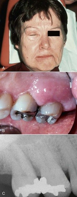
An acute periodontal abscess is clinically identical to many acute periapical abscesses of pulpal origin. The patient may experience severe swelling (Fig. 4-5, A) along with the usual symptoms of acute infection such as pain, fever, and malaise. The location of swelling near the gingival margin is common to both types (see Fig. 4-5, B).

image

FIGURE 4-5 A, Acute facial swelling associated with periodontal abscess is identical to the swelling of acute periapical abscess. B, Clinical view of same acute periodontal abscess. C, Radiograph of periodontally involved teeth. Note bone loss between molars and lack of periapical involvement.

Diagnostic procedures usually begin with a good radiograph. In this case (see Fig. 4-5, C), significant information is immediately apparent: there is loss of crestal bone interproximally, indicating a lesion of periodontitis as opposed to one of pulpal origin. There is no radiolucency at the apices. Since it is possible to have an acute periapical abscess without obvious or significant radiographic evidence of a periapical or lateral lesion, the next step in diagnosis is sensibility testing. In this case, all of the maxillary right posterior teeth respond normally to thermal and electrical sensibility tests. This finding eliminates the possibility of a pulpal etiology. The diagnosis of acute periodontal abscess is confirmed by signs, symptoms, and periodontal probings. Treatment planning will be based on probing depths. Probings that are found to confirm attachment loss to the level of the apical third would support tooth extraction as the treatment of choice. Probings to the level of the midroot might favor periodontal surgery to reduce or eliminate pocket depth.

Lesions of Chronic Periodontitis

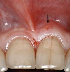
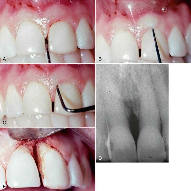
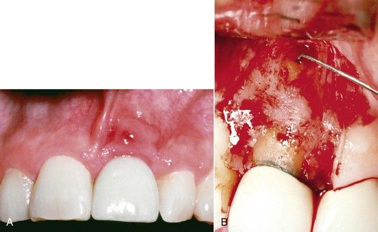
Bony lesions of chronic periodontitis are sometimes confused with lesions of pulpal origin because of a draining sinus tract. In Fig. 4-6, the patient was referred for endodontic evaluation because of the drainage tract in the attached gingiva over the left central incisor (arrow). In a similar case (Fig. 4-7), probings are consistent with a deep periodontal lesion. The radiograph clearly shows no apical rarefaction on either of the central incisors (see Fig. 4-7, D), and sensibility tests elicit normal responses. Surgical exposure illustrates the morphology of the defect (see Fig. 4-7, E). Contrast these cases with the endodontic case presented in Fig. 4-8. Clinically, the sinus tracts appear nearly identical (see Fig. 4-8, A; also see Fig. 4-6), but surgical exposure of the tooth reveals not only the periapical lesion but also intact crestal bone (see Fig. 4-8, B).
image

FIGURE 4-6 Sinus tract of periodontal etiology found in attached gingiva over maxillary left central incisor.
image

FIGURE 4-7 A, Periodontal probing depth normal interproximally. B, Probing depth normal in midlabial area. C, Deep probing pattern associated with periodontal bone loss. D, Radiograph of same lesion. Note absence of periapical involvement. E, Surgical exposure of periodontal lesion.
image

FIGURE 4-8 A, Sinus tract similar to Fig. 4-6 but of pulpal etiology. B, Surgical exposure of apical lesion. Note normal crestal bone contours.

image

CLINICAL PROBLEM

Problem

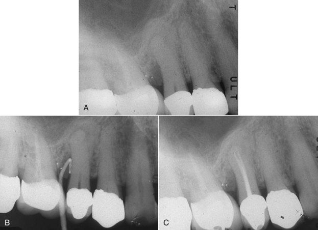
A 52-year-old male was seen for recurrent local swelling and drainage in the area of the maxillary right second premolar. Clinical examination revealed a draining sinus tract in the attached gingiva near the second premolar. A radiograph indicated that there was a widened apical periodontal ligament space consistent with a developing periapical lesion in addition to a deep periodontal defect interproximally on the distal (Fig. 4-9, A). At this point, the origin of the sinus tract was unknown.

image

FIGURE 4-9 A, Maxillary right premolar area with history of recurrent drainage suspected to be of pulpal origin. There is both apical and periodontal pathosis evident on the second premolar. B, Sinus tract exploration with gutta-percha cone, revealing source of drainage is the periodontal lesion. C, Completed root canal treatment will only resolve the periapical lesion. Periodontal surgery is also indicated to eliminate the pocket. This will resolve both associated infection and the drainage tract.

Solution

Sensibility testing was performed. No responses were obtained from the second premolar, and the first molar previously had root canal treatment. A sinus tract exploration was done by placing a gutta-percha cone in the tract and exposing an additional radiograph (see Fig. 4-9, B). This examination indicated that the drainage was of periodontal origin. No communication between the two lesions was evident on the film, and none was found during probing. The diagnosis was concomitant periodontal and periapical lesions.6 Both root canal treatment and periodontal treatment will be required to resolve the infections in this case (see Fig. 4-9, C).

image

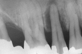
Although there may be ample evidence of a general chronic periodontitis in the oral cavity, some localized areas may have developed extremely severe bone destruction. If periodontal defects extend to the apex, the radiographic lesion present may be confused with a periapical lesion of pulpal origin. The patient in Fig. 4-10 was sent for completion of root canal treatment on a second premolar. Note the endodontic access cavity in the crown of this tooth. The loss of bone radiographically correlated with the loss of attachment circumferentially by clinical probing. The extreme mobility of this tooth was another consequence of bone loss. A root canal procedure would have no effect on this condition, so the tooth was extracted.
image

FIGURE 4-10 Localized lesion of advanced chronic periodontitis. Note tooth has been opened for root canal treatment that will have no effect on this lesion.

image

CLINICAL PROBLEM

Problem

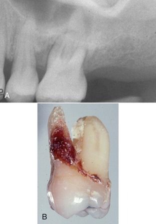
A 57-year-old male with a history of recurrent swelling on the palatal aspect of the maxillary left first molar was referred for root canal treatment. The referring dentist noted a large periapical lesion encompassing the apex of the palatal root (Fig. 4-11, A). The radiograph also shows evidence of severe bone loss around the buccal roots that was not noted by the dentist. The molar had no restorations, and there was no evidence of a fracture in the crown.

image

FIGURE 4-11 A, Radiograph of maxillary left first molar area with apical lesion of chronic periodontitis. Pulp test results are completely normal. B, Following extraction. Note calculus deposits covering entire palatal root.

Solution

Sensibility tests should be performed early in the examination. In this case, results were normal—in fact, the tooth was hypersensitive to a cold stimulus. Periodontal probings indicated that there was bone loss to the apex of the palatal root and confirmed severe attachment loss around the other two roots. As in the previous case, root canal treatment would have no effect on this problem, so the tooth was extracted. In these cases, calculus deposits are commonly seen covering the entire root surface (see Fig. 4-11, B).

image

Occasionally a periodontal bone lesion may resemble a periapical lesion and, at least radiographically, lack other obvious signs of generalized periodontitis.26 At the close of Chapter 3, an excellent example was depicted in Fig. 3-60. What appears to be a typical periapical lesion of pulpal origin is in reality a lesion of severe periodontitis. Once again, root canal treatment would have been of no benefit in this case.

image

CLINICAL PROBLEM

Problem

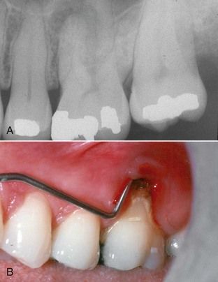
A 38-year-old male presented with a history of recurrent acute abscesses in the buccal vestibule adjacent to the maxillary left first molar. The radiograph showed a “classic” periapical lesion on the apex of the mesial buccal root (Fig. 4-12, A). The lesion was noted to extend coronally along both the mesial and distal surfaces of the root.

image

FIGURE 4-12 A, Maxillary left first molar with a “classic” periapical lesion on mesial buccal root. B, Clinical examination reveals periodontal etiology. Sensibility test results are normal.

Solution

Sensibility tests indicated normal responses on this tooth, confirming that this is not a pulpal problem. Periodontal probing revealed complete loss of attachment over the entire buccal surface of the root (see Fig. 4-12, B). Large deposits of calculus were seen and felt on the root surface with the probe. The diagnosis of periodontal abscess was made. Since there was minimal periodontal involvement of the other two roots, the optimal treatment plan was to resect (amputate) the mesial buccal root; pulp exposure resulting from the amputation would necessitate root canal treatment on the remaining roots.

image

Periodontal Lesions Involving the Furcation

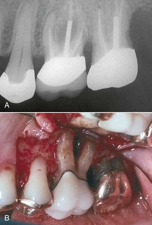
Loss of bone in the furcation of a molar due to periodontal disease is sometimes difficult to distinguish from bone loss due to a necrotic pulp (communication via furcation canals)25 or a sinus tract that is traced to the furcation. As cited earlier, the periodontal probe may reveal a sometimes subtle distinguishing difference. Periodontal defects tend to affect the space in the furcation more or less symmetrically, whereas sinus tracts tend to align with one root and more directly with a lesion at the apex. Therefore periodontal defects will tend to probe both vertically (parallel to the root) and horizontally (buccal-lingually, parallel to the occlusal plane). Sinus tracts of pulp origin tend to probe in a vertical direction only, but in some cases the tract may take a tortuous path, depending on the nature of the bone around the tooth, so straight probes will be of less value in the differential diagnosis of a lesion of this type. This emphasizes the need to have both straight and curved probes (Nabers or Cattoni probes, Hu-Friedy Co., Chicago, IL, USA). The furcation defect in Fig. 4-13, A was suspected to be the result of inadequate root canal treatment of the mesial buccal root. Periodontal probings indicated there were deep vertical and horizontal components to the defect. Surgical exploration showed the extent of the bone loss (see Fig. 4-13, B). The entire furcation was devoid of bone in addition to loss of the buccal plate covering the buccal roots, which accounted for the preoperative probing patterns. The diagnosis of advanced periodontitis was confirmed, and the tooth was extracted.

image

FIGURE 4-13 A, Furcation lesion in bone, suspected to be the result of extension of pulp pathosis into periodontium. B, Surgical exposure confirms the diagnosis of chronic periodontitis.

Lesions Associated With Aggressive Forms of Periodontitis

Aggressive periodontitis in young people, once known as juvenile periodontitis, affects less than 1% of the population.4 This disease process is now associated with a specific pathogen, Aggregatibacter actinomycetemcomitans (formerly Actinobacillus).

Only gold members can continue reading. Log In or Register to continue

Stay updated, free dental videos. Join our Telegram channel

Jan 2, 2015 | Posted by in Endodontics | Comments Off on 4: Problem Solving in the Differential Diagnosis of Bony Defects Resulting from Pulpal and Periodontal Pathosis

VIDEdental - Online dental courses
Get VIDEdental app for watching clinical videos