Head and Neck Anatomy and Physiology
Anatomic Nomenclature
A Location of an anatomic structure is based on the body in the anatomic position; the term anatomic position denotes a body standing erect, head facing directly forward, with arms at the sides and palms facing forward (Figure 4-1)
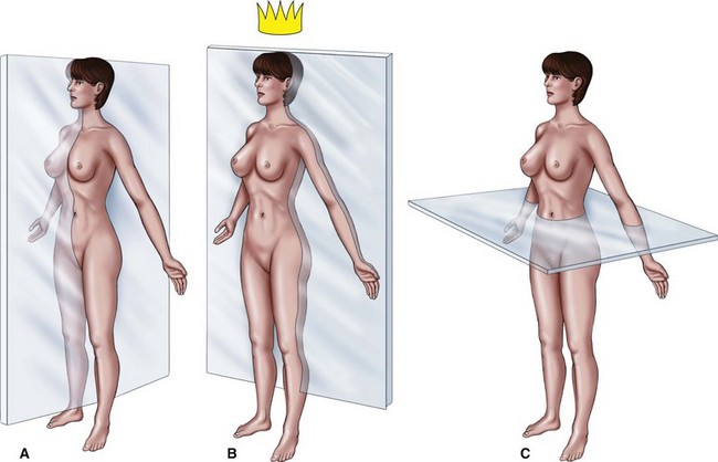
B Planes—the sections of the body are divided into imaginary flat surfaces called planes; a plane is a flat surface determined by the location of these three points in space:
1. Median plane or midsagittal section—passes through the midline, vertically dividing the head and body into right and left sides
2. Frontal or coronal plane—passes through the head and body, vertically dividing it into anterior and posterior sections
3. Horizontal or transverse plane—divides the head and body into upper (superior) and lower (inferior) portions
1. Anterior or ventral—structures nearest the front side of the body or head
2. Posterior or dorsal—structures nearest the back side of the body or head
3. Tongue surfaces—an exception to the previous anatomic positions is the surfaces of the human tongue, which still has the anatomic orientation of a four-footed animal; the dorsal surface of the tongue is the top surface, and the ventral surface of the tongue is the bottom surface
4. Medial—structures closest to the median plane of the body and head
5. Lateral—structures farthest from the median plane of the body and head (for example, ears are lateral to the nose or eyes)
6. Superficial—structures located toward the surface of the body
7. Deep—structures located internally the surface of the body
8. Proximal—near the source of attachment
9. Apex—the tip or pointed end of a structure
10. Contralateral—structures on the opposite side of the body
Osteology
A Definition—the study of bones
B Classification—bone and cartilage are classified as rigid and firm connective tissues; they contain large amounts and various types of intercellular material (or matrix; plural, matrices) and few cells; with the exception of cartilage, connective tissue is highly vascular
C Function—bone supports organs and structures; provides attachments for muscles and ligaments; is involved in movement, body defense, and repair mechanism; protects the soft tissues and organs of the body
D Histology of bone (see the section on “Connective Tissue” in Chapter 2)
1. Intramembranous ossification—osteoblasts are formed from a network of mesenchymal cells; osteoblasts secrete collagen and a matrix of mucoproteins that form osteoids; this matrix initially forms bone
a. Osteoblasts—cells that form bone
b. Osteoclasts—cells that resorb (remove) bone
c. Osteocytes—mature osteoblasts that are entrapped in bone matrix
2. Endochondral ossification—hyaline cartilage “template” becomes mineralized and is replaced by bone; osteoids are formed within cartilage
3. Cartilage—noncalcified, avascular, pliable connective tissue; three types of cartilage:
a. Hyaline cartilage—serves as a template for bones
b. Fibrous cartilage—transitional cartilage found between hyaline cartilage and tendons and ligaments; usually present in joints or articulations
c. Elastic cartilage—contains elastic fibers in its matrix and found in structures such as the external ear, auditory tube, epiglottis, and parts of the larynx
1. Bony prominences—a process is a general term used to describe any prominence on a bony surface
a. Condyle—the large convex, rounded articular end of a bone, usually involved in joints
b. Tuberosity—a large rough prominence that typically serves as an attachment area for muscles or tendons
c. Tubercle or eminence—rounded (small) elevation on the bony surface (e.g., genial tubercles on the mandible)
d. Arch—a length of bone with a bow-like outline; shaped like a bridge
e. Cornu—a horn-like prominence
f. Crest—a thin, wedge-shaped ridge (e.g., crista galli of ethmoid bone)
g. Spine—sharp prominences that serve as attachments for muscles
a. Notch—an indentation at the edge of a bone
c. Fossa—a deep depression in a bone; usually is round in shape; can be an area for muscle attachment (plural, fossae)
d. Sulcus—a shallow depression or groove that usually marks the course of an artery or nerve (plural, sulci)
a. Foramen—a short tube-like opening in a bone (e.g., incisive foramen)
b. Canal—a long, tube-like opening in a bone (e.g., mandibular canal)
c. Meatus—an opening or canal in a bone (e.g., internal acoustic meatus)
d. Fissure—a narrow cleft-like opening; may be a line of fusion between two bones (e.g., superior orbital fissure of the orbit)
4. Skeletal articulations—areas where bones are joined; articulations can be movable or immovable
Axial Skeleton
Bones of the head are grouped into two categories:
A Neurocranium, or cranial bones (eight bones)—bones that surround the brain
1. Frontal bone (single bone) (Figure 4-2)
a. Forms the forehead at the top and front of the skull
b. Contains the frontal sinuses
c. Supraorbital ridge forms the roof of the orbits of the eyes
d. Articulates with the parietal bones to form the coronal suture
e. Articulates with many of the cranial and facial bones
f. Landmarks are the supraorbital notch, the zygomatic process of the frontal bone, and the lacrimal fossa, which contains the tear-producing lacrimal gland
2. Parietal bones (paired bones) (see Figure 4-2)
a. Constitute a large part of the vault and sides of the cranium
b. Articulates with its counterpart and various bones to form the coronal, sagittal, squamosal, and lambdoidal sutures of the skull
3. Temporal bones (paired bones)
a. Form the lateral walls of the skull
b. Articulate on each side of the skull with the zygomatic and parietal bones, and sphenoid and occipital bones
c. Divided into three portions:
(1) Squamous portion—forms the zygomatic process of the temporal bone, which is part of the zygomatic arch; this area provides the cranial portion of the temporomandibular joint (TMJ) containing the articular fossa, articular eminence, and postglenoid process
(2) Tympanic portion—forms most of the external acoustic meatus; it is separated from the petrous portion by the petrotympanic fissure, through which the chorda tympanic nerve emerges
(3) Petrous portion—contains the mastoid process that serves as an attachment for the sternocleidomastoid muscle; other landmarks include the styloid process, stylomastoid foramen, jugular notch, and internal acoustic meatus
4. Occipital bone (single bone) (see Figure 4-2)
a. Forms the posterior portion of the skull
b. Articulates with the parietal, temporal, and sphenoid bones
c. Foramen magnum is formed completely by this bone
d. Jugular notches on both the occipital and temporal bones form the jugular foramen
e. Occipital condyles on each side form a movable articulation with the atlas
f. Occipital bone has paired hypoglossal canals that are openings for cranial nerve XII
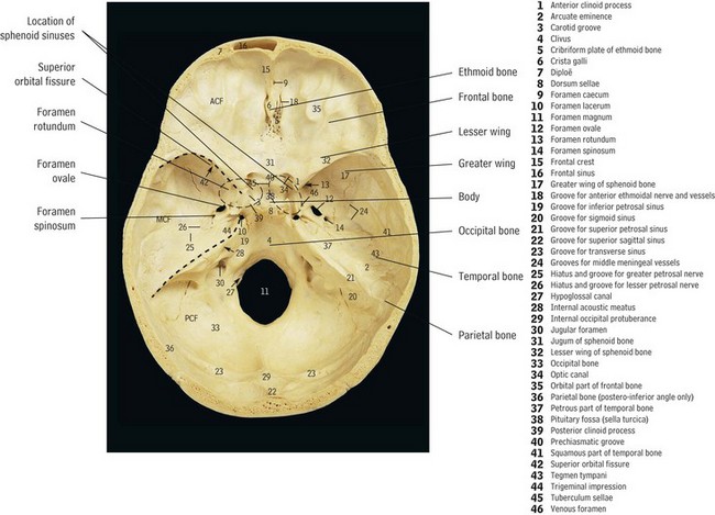
5. Sphenoid bone (single bone)
a. Articulates with the ethmoid and frontal bones anteriorly and the temporal and occipital bones posteriorly
b. Body of the sphenoid bone contains the sphenoid sinuses and the sella turcica, the seat of the pituitary gland, which supplies numerous hormones to the body
c. Pterygoid processes project down from the body of the sphenoid bone in an inferior, backward direction; its landmarks include:
(1) Medial and lateral pterygoid plates—attachment points for important muscles of mastication
(2) Pterygoid fossa—located between the medial and lateral pterygoid plates
(3) Hamulus—inferior termination of the medial pterygoid plate; also provides attachment for the muscles of the soft palate
d. Greater sphenoid wings—lateral projections in the temporal area
(1) Forms the outer wall and floor of the ocular orbits
(2) Infra-temporal crest—divides the temporal and infra-temporal surfaces
(3) Foramen rotundum—maxillary division of cranial nerve V
(4) Foramen ovale—mandibular division of cranial nerve V
(5) Foramen spinosum—opening in the greater wing of the sphenoid that gives passage to the middle meningeal artery
(6) Foramen lacerum—contains the internal carotid artery
(7) Superior orbital fissure—transmits cranial nerves III, IV, VI, and the ophthalmic division of cranial nerve V
6. Ethmoid bone (single bone) (see Figure 4-2)
a. Single midline bone of the skull; forms the base of the cranium and orbits
b. Articulates with the frontal, sphenoid, lacrimal, and maxillary bones; joins the vomer inferoposteriorly
d. Cribriform plate—perforated to allow the passage of olfactory nerves
e. Crista galli—an extension of the perpendicular plate that serves as an attachment for the meninges of the brain
f. Perpendicular plate—along with the vomer and nasal septal cartilage, forms the nasal septum
g. Superior nasal conchae and the middle nasal conchae are formed from the lateral portions of the ethmoid bone; their purpose along with the inferior nasal conchae is to increase the surface area of the respiratory epithelium
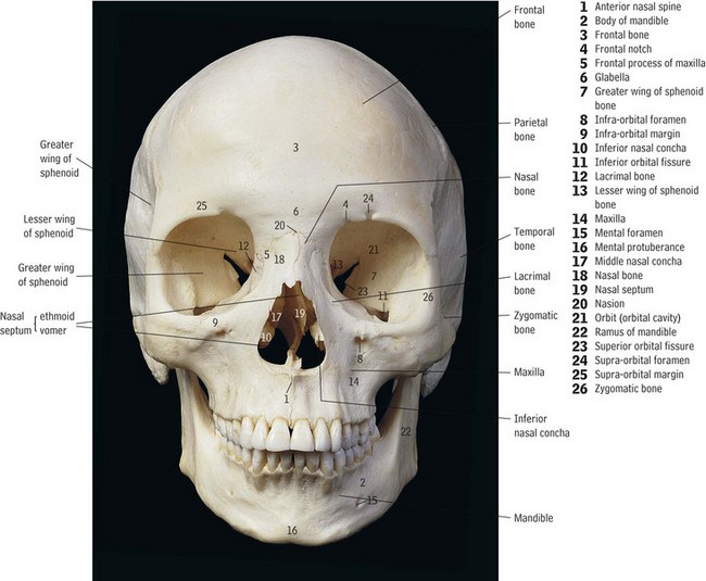
B Viscerocranium, or facial bones (14 bones)—surround the face (Figure 4-3)
1. Inferior nasal conchae (paired bones)
a. Project from the maxilla to form part of the lateral walls of the nasal cavity
b. Are separate from the ethmoid bone, but articulate with it
2. Nasal bones (paired bones) (see Figure 4-3)
a. Forms the posterior portion of the nasal septum, articulating with the perpendicular plate of the ethmoid bone
4. Lacrimal bones (paired bones) (see Figure 4-3)
a. Thin bones that form part of the anterior medial wall of the orbit of the eye
b. The nasolacrimal duct is located at the junction of the lacrimal and maxillary bones
c. Fluid (tears) from the lacrimal gland is drained through this duct into the inferior nasal meatus
5. Zygomatic bones (paired bones)—(see Figure 4-3)
a. Zygomatic arch (cheekbone)—composed of the temporal process of the zygomatic bone and the zygomatic process of the temporal bone
b. Three processes are named for the bones with which the zygomatic bones articulate: frontal process, maxillary process, and temporal process
6. Maxillae (paired bones)—have a body and four processes (see Figure 4-3)
a. The body contains the maxillary sinus and forms the lower and medial rims of the orbits and the borders of the nasal cavity (piriform aperture); other landmarks are:
(1) Infraorbital foramen—originates as the inferior orbital fissure and becomes the infraorbital canal that carries the infraorbital nerve, the inferior ophthalmic vein, and the infraorbital artery; it is a landmark for the administration of local anesthesia to the maxillary premolars, canines, and incisors
(2) Canine fossa—inferior to the infraorbital foramen and distal to the roots of the maxillary canines
b. The frontal process articulates with the frontal bone, forming the medial orbital rim; it articulates with the lacrimal bone within the orbit
c. The alveolar process (see the section on “Supporting Tissues” in Chapter 2)
(1) Less dense bone containing the roots of the maxillary teeth
(2) Sockets (alveoli) for maxillary teeth
(3) Canine eminence—a protuberance over the root of the maxillary canine
(4) Maxillary tuberosity—contains the posterosuperior alveolar fifth cranial nerve foramina, a soft tissue depression distal to the last maxillary molar; is a landmark for the posterosuperior alveolar injection to achieve anesthesia for the maxillary molar teeth
7. Palatine bones (paired bones)
a. The horizontal plates of the palatine bones articulate with each other to form the posterior portion of the hard palate, a continuation of the median palatine suture
b. Articulate with the maxillary and sphenoid bones
c. The vertical plates form part of the lateral walls of the nasal cavity and a small part of the orbital apex
d. Contain the greater palatine foramen, a landmark for the administration of local anesthetic agent, and the lesser palatine foramen, where nerve and blood vessels pass through to the soft palate and tonsils
8. Mandible (single bone) (see Figure 4-3)
a. Largest, strongest, and only movable facial bone; articulates with the temporal bones on both sides
b. Body—horizontal portion runs from the anterior to the lateral aspects; landmarks include:
(1) Mental protuberance—the chin
(2) Symphysis—midline, nonmovable suture
(3) Alveolar process of the mandible
(4) Sockets (alveoli) for mandibular teeth
(5) Mental foramen—located bilaterally on the external aspect, below and between the first and second premolars; the mental nerve transmits to the inferior alveolar nerve; the mental artery transmits to the inferior alveolar artery, the landmark for the administration of local anesthesia to premolars and anterior teeth
(6) Genial tubercles—form the midline on the internal surface and provide points of muscle attachment
(7) Retromolar triangle—distal to the third mandibular molar
(8) Sublingual and submandibular fossae—on the internal aspect, these fossae contain their corresponding salivary glands
c. Ramus—projects vertically and backward from the body of the mandible
(1) External oblique line—located on the external surface; forms a crest where the ramus joins the body of the mandible
(2) Mandibular foramen—located bilaterally on the internal surface; forms the opening of the mandibular canal and the exit for blood vessels and the inferior alveolar nerve
(3) Lingual—a raised bony prominence; anterior to the mandibular foramen
(4) Condyle—articulates with temporal bone, forming the movable part of the TMJ
(5) Coronoid process—the superior margin, which forms the anterior border of the ramus and provides points of muscle attachment
(6) Mandibular notch—concave area between the condyle and the coronoid process
1. Hyoid bone—U-shaped bone suspended in the neck; located superior and anterior to the thyroid cartilage; landmarks are the greater and lesser horns for the attachment for many muscles and ligaments of the tongue and throat
2. Atlas—first cervical vertebra; its lateral masses articulate superiorly with the occipital condyles of the skull and inferiorly with the axis
3. Axis—second cervical vertebra; along with the atlas, the axis provides attachment points for many muscles responsible for the movement of the head
Paranasal Sinuses
A Air-filled, mucus-lined cavities or openings in the bones of the skull that function to lighten the weight of the skull and act as sound resonators; the four paired sinuses are:
1. Frontal sinuses—frontal bone above the orbit; drain into the middle nasal meatus
2. Sphenoid sinuses—body of sphenoid bone; drain into the superior nasal meatus
3. Ethmoid sinuses—consist of three small compartments: anterior and middle compartments, which drain into the middle meatus; posterior compartment, which drains into the superior meatus of the nasal cavity
4. Maxillary sinuses—triangle shaped, largest, and most complicated of the sinus cavities; drain into the middle meatus; infection of these sinuses can cause discomfort and complications of the maxillary posterior teeth
The Muscular System
1. Muscle tissue, one of the four classifications of body tissue, consists of specialized fibers for contraction; three types of muscle tissue are voluntary skeletal muscles, involuntary cardiac, and involuntary smooth muscles; muscles in the head and neck region are skeletal muscles
2. Movement—muscles are under neural control to shorten or contract; contraction is the action of the muscle fibers
3. Origin—attachment to a relatively immovable structure (e.g., sternocleidomastoid muscle originates on the clavicle and sternum)
4. Insertion—attachment to the more movable structure; insertion moves toward the origin (e.g., sternocleidomastoid muscle inserts on the mastoid process of the temporal bone and the muscles flex, moving the head downward)
B Muscles of facial expression—most facial muscles are superficial, paired muscles originating in bone and inserting into skin; they are innervated by the seventh cranial or facial nerve and are responsible for functions related to speech, emotional expression, and mastication
1. Epicranial or occipitofrontalis muscle—scalp region; composed of two bellies (the fleshy, contractile part of a muscle), the frontal and occipital, which are connected by the epicranial aponeurosis; raise the eyebrows and scalp
2. Orbicularis oculi muscle—surrounds the eye; closes the eyelid
3. Corrugator supercilii muscle—superior to the orbicularis; wrinkles the forehead
4. Orbicularis oris muscle—encircles the mouth; closes lips
5. Buccinator muscle—anterior part of the cheek; originates on the maxilla, mandible, and pterygomandibular raphe and inserts into the angle of the mouth; functions to pull the mouth laterally, thereby shortening the cheek, and as an aid in keeping food on the chewing surfaces of the teeth
6. Risorius muscle—mouth region; acts in smiling and widening the mouth
7. Levator labii superioris muscle—upper lip; raises the upper lip
8. Levator labii superioris alaeque nasi muscle—upper lip; raises the upper lip and dilates the nose, as in sneering
9. Zygomaticus minor muscle—upper lip; raises the upper lip
10. Zygomaticus major muscle—angle of the mouth; pulls the angle of the mouth laterally, causing the appearance of a smile
11. Levator anguli oris muscle—angle of the mouth; elevates the corner of the mouth, as in smiling
12. Depressor labii inferioris muscle—lower lip; lowers the lower lip to expose lower teeth
13. Mentalis muscle—chin area; raises the chin, narrows the vestibule near mandibular incisors
14. Platysma muscle—neck region; originates in the clavicle fascia and inserts in the region of the mandible and facial muscles of the mouth; pulls down the corners of the mouth, raising the skin of the neck
C Muscles of mastication—four paired muscles, all inserting on the mandible, innervated by the fifth cranial or trigeminal nerve; they are responsible for movement of the jaw
1. Masseter muscle—most superficial, largest, and strongest of the four muscles
a. Originates on the zygomatic arch; originates from two heads, one superficial and one deep, and inserts on the lateral surface of the angle of the mandible
a. Originates from a fan-like attachment on the temporal fossa; inserts on the coronoid process of the mandible
b. Action—when the entire muscle contracts, it elevates the mandible, raising the jaw; contraction of only the posterior portion causes retraction of the mandible
3. Medial (internal) pterygoid muscle (Figure 4-4)
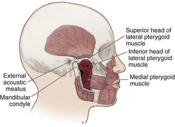
4. Lateral pterygoid muscle; lies within the infra-temporal fossa (see Figure 4-4)
a. Originates from two heads: the superior head originates from the greater wing of the sphenoid, the inferior head originates from the lateral pterygoid plate of the sphenoid; both insert on the mandibular condyle
b. Action—both heads working together depress (opens) and protrude the jaw; when only one side is contracted, the jaw shifts to the opposite side, causing lateral deviation of the mandible
D Cervical muscles—paired superficial, large, easily palpated muscles, both innervated by cranial nerve XI (accessory nerve)
1. Sternocleidomastoid muscle—well-defined, large muscle; important landmark for palpating lymph nodes
E Hyoid muscles—all are attached to the hyoid bone; usually grouped as suprahyoid or infrahyoid muscles, depending on their relationship to the hyoid; they aid in mastication and swallowing
1. Suprahyoid muscle group—located superior to the hyoid; acts to raise the hyoid and the larynx
(1) Two separate bellies—anterior belly originates from the intermediate tendon on the hyoid bone and inserts near the symphysis of the mandible; posterior belly originates from the mastoid notch and inserts on the intermediate tendon of the hyoid
(2) Action—pulls back the jaw; anteriorly innervated by the mylohyoid nerve; posteriorly by the posterior digastric nerve
b. Mylohyoid muscle—forms the floor of the mouth
(1) Originates from the inner surface of the mandible; unites medially with its counterpart and inserts on the body of the hyoid
(2) Helps elevate the tongue and depress the mandible; innervated by the mylohyoid nerve, a division of cranial nerve V (trigeminal nerve)
Stay updated, free dental videos. Join our Telegram channel
VIDEdental - Online dental courses


