Development of the face and palate
Learning objectives
■ Describe prenatal facial development during the fourth to seventh weeks of gestation.
■ Describe palatal development during the seventh to ninth weeks of gestation.
■ Explain how the tongue and thyroid develop.
■ Discuss development of facial and palatal clefts and other facial defects.
Key terms
Auditory tube
Auricular hillocks
Cheeks
Cleft lip
Foramen cecum
Forehead
Frontal process
Frontonasal process
Hyoid arch
Internasal area
Lateral lingual swellings
Lateral nasal process
Lateral palatine processes
Lingual tonsil
Lower jaw
Mandibular arch
Maxillary processes
Medial nasal process
Nasal fin
Nasolacrimal duct
Orbicularis oris
Oronasal optic groove
Palatal shelf elevation
Palatine and pharyngeal tonsils
Palatine shelf closure or fusion
Philtrum
Primary palate
Terminal sulcus
Thyroglossal cyst
Thyroglossal duct
Thyroglossal fistula
Tuberculum impar
Overview
This chapter describes the development of the human face and palate and defects that may occur during development. An understanding of this subject is important to the dental health professional for two reasons. First, the professional must understand the variability that can occur in facial form, and, second, he or she must be aware that the human face and palate are among the areas in the body most likely to develop malformations.
The human face develops early in gestation, during the fourth through seventh weeks, and the palatal processes begin to close during the eighth week. These two structures are closely related in time of development and sometimes have related malformations. The face develops from the tissues immediately surrounding the oral pit, but the forehead develops from the frontal area that lies above the pit (Fig. 4-1). The nose later develops from this area as well, so the name changes from frontal area to frontonasal area (Fig. 4-2). Below the oral pit is the mandibular arch, from which the mandible arises and articulates with the temporal bone. Lateral to the oral pit are the right and left maxillary processes, which develop from the mandibular arch. Cheek tissues come from these processes. Intraorally, the palate forms the mouth’s roof, which separates the oral and nasal cavities. First, the medial palatal segment forms as part of the medial nasal segment. This segment provides the first separation of the oral and nasal cavities. Next, two lateral palatal shelves close anteriorly (not posteriorly) to the pharynx (see Fig. 4-12, p. 55). At the same time, the tongue develops in the floor of the oral cavity but grows rapidly and expands into the nasal cavity. The tongue functions in palatine shelf closure because the shelves must override it before closure can be accomplished.
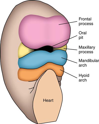
Around the centrally located oral pit are grouped the frontal and maxillary processes and the mandibular arch. Although appearing unrelated at this time, these processes and the first arch form the human face.
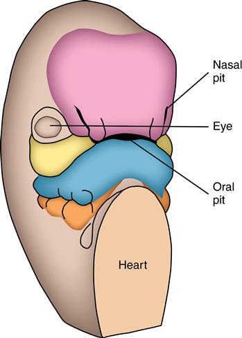
The nasal pits develop and appear on the sides of the face. The frontal process now becomes the frontonasal process.
Many environmental factors can cause clefts of the face, palate, or both. These defects of the lip or palate may be unilateral or bilateral and also incomplete or complete.
Facial development: Weeks 4 to 7
Tissue organization
The face develops primarily from tissues surrounding the oral pit. Above the oral pit is the covering of the brain termed the frontal process, from which develops the forehead. Lateral to the oral pit are the right and left maxillary processes, from which develop the cheeks, and below the oral pit is the mandibular arch, from which the lower jaw is formed. In the fourth week, when the facial tissues have just begun to organize, they measure only a few millimeters in height and width and are only as thick as a sheet of paper. Further growth of the face from this minute assembly of tissue sites is anterior to the brain. Lying inferior to the mandibular arch is the second pharyngeal or hyoid arch, and its muscles expand into and contribute to the face. The hyoid arch also forms part of the external and middle ear.
Development of the human face is most easily described in terms of the changes that occur at weekly intervals from the fourth to the seventh prenatal weeks.
 CLINICAL COMMENT
Syndromes associated with the pharyngeal arches are seen commonly as a group of defects. They can appear as a malformed mandible, defective ear, small mouth, enlarged tongue or unequal growth of the sides of the tongue, cleft lip or palate, and swelling, cysts, or clefts of the front or sides of the neck. Usually, several or more defects appear simultaneously.
Fourth week
At 4 weeks’ gestation, the oral pit is surrounded by several masses of tissue. Pharyngeal arches are also evident below the pit and on the sides of the neck. The frontal processes of the brain bulge forward and laterally to dominate the facial area. Below the frontal processes are two small wedge-shaped tissues termed the maxillary processes that lie lateral to the oral pit. Beneath the maxillary processes is the mandibular arch, which appears divided or constricted in the midline (see Fig. 4-1). The heart lies immediately below the face and is one of the fastest growing organs. During the fourth week, the heart begins to pump blood throughout the body.
 CLINICAL COMMENT
Environmental factors play an important role in facial and palatal malformations. The period before the fifth week is the critical time during which these factors can affect facial development.
Fifth week
During the fifth week, the bilateral nasal placodes, or thickened areas of epithelium, appear in the upper border of the lip. They develop into nostrils as the tissues around these placodes grow, resulting in two slits opening into the oral pit. At this point, the frontal area becomes known as the frontonasal process. The nostrils deepen as the tissues around them continue to grow anteriorly, and the internasal area, the distance between the nostrils, represents the width of the face. Gradually the frontal prominence diminishes and the face broadens. The eyes become prominent on the sides of the head. Throughout the fifth week, the mandibular arch loses its midline constriction (see Fig. 4-2).
Sixth week
At the beginning of the sixth week, the lateral parts of the face expand, broadening the face. This is also caused by lateral growth of the brain. The eyes and maxillary processes, which were located on the sides of the face in the fifth week, come to the front of the face. The mouth slit widens to the point at which the maxillary and mandibular tissues merge. The nasal processes are limited to the middle of the upper lip, which causes the face to appear more human. The upper lip is now composed of a medial nasal process and two lateral maxillary segments (Fig. 4-3). The medial nasal process is called the philtrum. A ridge of tissue surrounds each nasal pit. The tissue lateral to the pits is the lateral nasal process, and the tissue medial to the pits is the medial nasal process.
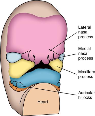
Nasal pits appear more centrally located in the medial nasal process. This is the result of growth of the lateral face, which also causes the eyes to approach the front of the face. The enlarged maxillary processes are near contact with the medial nasal process. Nasal pits may be sites of cleft lips. Auricular hillocks bordering the ear canal have merged.
The medial nasal process is in close contact with the medial aspect of the maxillary process, and the lateral nasal process is above the maxillary process. The border of the lip consists of two maxillary processes, and the medial third is the medial nasal. A lack of contact or fusion of the medial nasal and maxillary processes results in either a unilateral or bilateral cleft lip. The epithelial coverings of the medial nasal and maxillary processes normally contact and create a zone of fusion termed the nasal fin (Fig. 4-4). This epithelial fin is soon penetrated by connective tissue growth, which binds together the two maxillary and medial nasal parts of the lip. If this penetration were not to occur, the lip could pull apart. Soon the orbicularis oris muscle grows around the oral pit to provide support to the upper lip. The nasal pits continue behind the nasal fin to open into the roof of the mouth at 6 week/>
Stay updated, free dental videos. Join our Telegram channel
VIDEdental - Online dental courses


