36
Periodontal management of patients who smoke
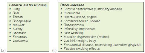
Figure 36.1 Health problems related to cigarette smoking.

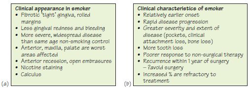
Figure 36.2 Details of clinical appearance and characteristics in a smoker.

Figure 36.3 A male cigarette smoker with smoking-related chronic periodontitis. (a) Drifting and diastema between UR1 and UL1. (b) Palatal effects of smoking. (c) Nicotine-stained supragingival calculus on the lingual of the lower incisors. (d) A panoramic radiograph showing generalised bone loss and a periodontic–endodontic lesion on LR5 which was extracted. (e) Anterior view showing fibrotic, pale gingiva. (f) Right view following extraction of LR5. (g) Upper palatal surfaces showing nicotine staining and inflamed, rolled gingival margins. (h) Left side showing recession and nicotine staining.

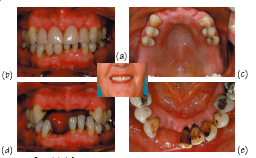
Figure 36.4 (a) A female cigarette smoker with smoking-related chronic periodontitis. (b) Anterior view with the upper and lower partial dentures in place; note the staining and generalised recession. (c) Palatal view showing a shortened dental arch following several extractions and also denture-related stomatitis. (d) Marked recession/loss of attachment anteriorly. (e) Heavily nicotine-stained supragingival calculus on the lingual of the lower incisors.

Figure 36.5 Quit smoking!

Figure 36.6 The five ‘A’s: ask, advise, assess, assist and arrange.

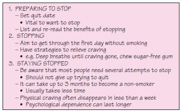
Figure 36.7 Three steps to stop smoking.

Figure 36.8 Resources and support to stop smoking in the UK and the USA (all websites accessed 14 April 2009).

Table 36.1 Direct benefits of stopping smoking.
| Time after cessation | Direct benefits |
| 2days | Sense of taste and smell improved |
| 1 month | Skin clearer, more hydrated |
| 3 months | Improved breathing, no cough or wheeze Improved lung function (up to 10%) Risk of mouth and throat cancer reduced |
| 6 months | Most smoking-related oral white patches will have disappeared |
| 1 year | Gingival circulation improved |
| 10 years | Risk of heart attack reduced to half that of a smoker |
Stay updated, free dental videos. Join our Telegram channel
VIDEdental - Online dental courses
 Get VIDEdental app for watching clinical videos

|