35
Differential Diagnosis of Pathology of the Jaws
Guides to Diagnosis
Examination of the radiographic appearance of a particular lesion will aid in the development of a differential diagnosis. Consider the:
- location of the lesion;
- association with other anatomic structures;
- definition of margins;
- internal nature of the lesion.
Is the lesion rapidly expanding where there is resorption of adjacent structures or is it slowly growing with associated soft tissue expansion and movement of the teeth? The possible diagnoses listed below are representative of pathology found in children and are not meant to be all inclusive.
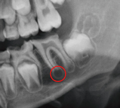
Periapical Radiolucencies (Fig. 35.1 and Box 35.1)
The majority of these lesions are associated with the loss of vitality of a tooth. While a tooth may have been treated with root canal therapy, a residual defect or healing area may remain for many months and the isolated presence of a periapical area may not necessarily indicate ongoing pathology.
Figure 35.1 Periapical area associated with a necrotic lower first permanent molar.

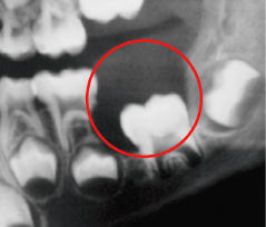
Radiolucencies Associated with the Crowns of Unerupted Teeth (Fig. 35.2 and Box 35.2)
These lesions are odontogenic and are related to changes in the follicle around the developing tooth. Associated teeth tend to be displaced away from the lesion and consequently away from the occlusal plane. Cysts associated with mandibular teeth commonly result in migration of those teeth inferiorly and posteriorly, while in the maxilla, the teeth move superiorly and inferiorly.
Figure 35.2 A dentigerous cyst associated with lower left first permanent molar that has resorbed the roots of the second primary molar.

Stay updated, free dental videos. Join our Telegram channel

VIDEdental - Online dental courses


