34
Chronic periodontitis
Figure 34.1 (a) Section through a tooth with incipient (initial) chronic periodontitis. (b) Section through a tooth with chronic periodontitis.

Figure 34.2 Incipient chronic periodontitis. (a) Calculus on mesiobuccal UR6 (arrow). (b) Anterior view. (c) Supragingival calculus on UL7 and subgingival calculus on mesiobuccal UL6 (arrows). (d) Right horizontal bitewing radiograph showing early crestal changes (slight horizontal bone loss) and calculus on mesiobuccal UR6 (arrow). (e) Left horizontal bitewing radiograph showing early crestal changes (slight horizontal bone loss) and calculus on mesiobuccal UL6 (arrow).
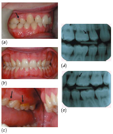
Figure 34.3 Smoking-related chronic periodontitis. (a) Calculus on the labial lower left central incisor. (b) Subgingival calculus visible as a dark shadow on the palatal upper incisors (arrows). (c) Supragingival calculus on the lingual lower anteriors (arrow). (d) Left horizontal bitewing radiograph showing a mesial overhang and vertical bone loss at UL8 (arrow), with furcation involvement at LL8 (arrow). (e) Right horizontal bitewing radiograph showing subgingival calculus.
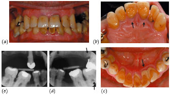
Figure 34.4 (a–c) Slight drifting of incisors, with diastemas present between the upper incisors. (d) Right vertical bitewing radiograph. (e) Left vertical bitewing radiograph showing vertical bone loss between UL6 and UL7 (arrow)
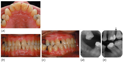
Figure 34.5 (a–c) Severe drifting of UL1 due to bone loss leading to lip trapping. (d) Scanora dentition only panoramic radiograph showing bone loss and periodontic–endodontic lesions on UL6 and LR2, which were both extracted.
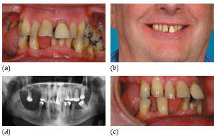
Figure 34.6 Inflammation and blunting of papilla between UL2 and UL3.
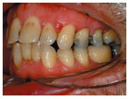
Figure 34.7 Suppuration: (a) palatal suppuration, and (b, c) periapicals of crowned incisor teeth showing horizontal bone loss.
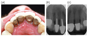
Figure 34.8 (a, b) Pre-treatment inflammation and swelling. (c) Post-treatment showing resolution.

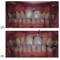
Figure 34.9 (a) Pre-treatment inflammation and swelling around the upper anterior teeth. (b) Post-treatment (non-surgical) resolution of inflammation with recession and exposure of the crown margin of UL1 which is now supragingival. Due to poor aesthetics, the crown could now be replaced (arrow).
Stay updated, free dental videos. Join our Telegram channel

VIDEdental - Online dental courses


