32
Gingival recession
Figure 32.1 Aetiological factors associated with gingival recession.

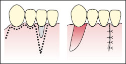
Figure 32.2 Pedicle grafts: laterally repositioned rotational flap. The flap is taken from an adjacent site to cover the localised area of recession.

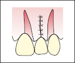
Figure 32.3 Pedicle grafts: double papilla rotational flap. Coverage is achieved from adjacent donor sites on either side of the area of recession.

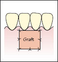
Figure 32.4 Free grafts: free epithelialised gingival graft. At the recipient site the epithelium and outer part of the connective tissue are removed by split dissection around and below the recession defect. The graft is taken from the remote donor site and sutured securely at the recipient site.

Figure 32.5 Free grafts: free epithelialised gingival graft. (a) Site shown preoperatively. (b) The tissue graft sutured in place. (c) The site shown 4 months’ postoperatively. Courtesy of Mr P. J. Nixon.

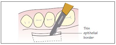
Figure 32.6 Free grafts: subepithelial connective tissue graft. A free graft is taken from the palate, removing underlying connective tissue with only a thin epithelial border. The tissue is sutured at the prepared recipient site. The graft is then covered by a flap (commonly a coronally advanced flap) so that the graft gains its nutrition from the underlying periosteum and deep connective tissue and the overlying flap.

Gingival recession is defined as the location of marginal tissue apical to the cement–enamel junction resulting in exposure of the root surface. Aetiological factors associated with gingival recession are shown in Fig. 32.1.
Consequencesof gingival recession
Unlike many other oral problems, patients are very often aware of gingival recession. Patients may potentially experience the following problems associated with recession:
A history and examinatio/>
Stay updated, free dental videos. Join our Telegram channel

VIDEdental - Online dental courses


