31
Non-plaque-induced gingival conditions and lesions
Figure 31.1 Classification of non-plaque-induced gingival conditions resulting from the 1999 International Workshop for the Classification of Periodontal Diseases and Conditions (Armitage, 1999).
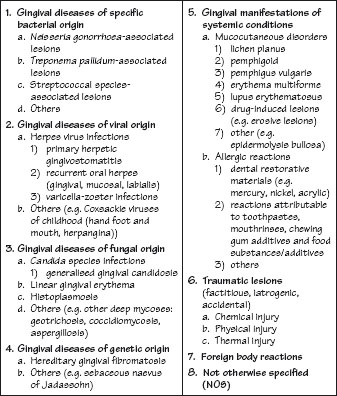
Figure 31.2 The surgical sieve: arriving at a correct differential diagnosis.
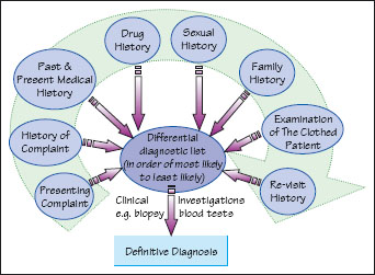
Figure 31.3 (a, b) Drug-induced gingival overgrowth.
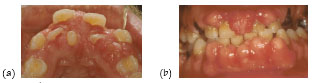
Figure 31.4 The normal gingival anatomy.
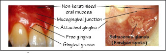
Figure 31.5 Sarcoidosis.
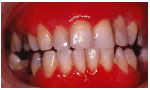
Figure 31.6 Varicella zoster.
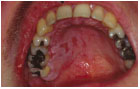
Figure 31.7 Lichen planus: (a) desquamative gingivitis; (b) plaque-like lichen planus; (c) reticular lichen planus; and (d) erosive lichen planus.
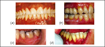
Figure 31.8 Leukoplakia: (a) localised veruccous leukoplakia; and (b) generalised homogenous leukoplakia.
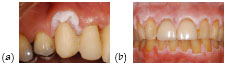
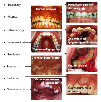
Figure 31.9 A surgical sieve utilising a mnemonic: ‘HINTED’.
Non-plaque-induced gingival conditions were classified for the first time in 1999 by an international workshop in periodontology (Fig. 31.1), but the system is by no means comprehensive. Given the heterogeneity of the constituent cells of the gingivae (epithelial, endothelial, fibroblasts, adipocytes, inflammatory-immune cells) and the various disorders associated with mixed tissues (ranging from immune-mediated to traumatic ulceration, to granulomatous inflammation, atopy and infections to tumour formation), it is estimated that well over 100 systemic or local non-plaque-induced conditions may involve the gingivae.
Establishing a differential diagnosis for such a large number of cond/>
Stay updated, free dental videos. Join our Telegram channel
VIDEdental - Online dental courses


