30
Plaque-induced gingivitis
Figure 30.1 Classification of plaque-induced gingivitis from the 1999 International Workshop.
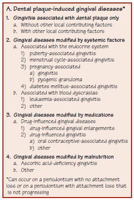
Figure 30.2 Section through a tooth with established, clinically evident, plaque-induced gingivitis.
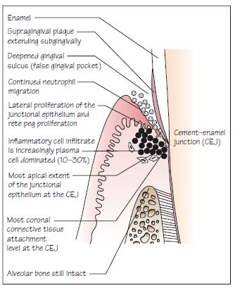
Figure 30.3 Plaque-induced gingival inflammation. (a) Left and (b) right views showing plaque visible at the gingival margin, blunted papillae and red, swollen gingiva.
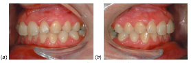
Figure 30.4 BPE codes, criteria and management. Plaque-induced gingivitis would typically be associated with a BPE code 1 or 2. Note that if the BPE code is 3, there is a need to differentiate between false gingival pockets and true periodontal pockets.
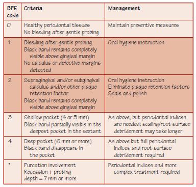
Figure 30.5 A false gingival pocket.
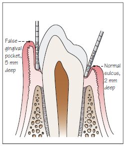
Figure 30.6 (a) Pre-treatment: a false pocket is associated with a subgingival cavity (arrow) and plaque retention on the mesial UL2. The papilla is swollen and red. (b) Post-treatment for gingivitis: the inflammation is diminished. The cavity on UL2 is now supragingival and accessible for restoration.
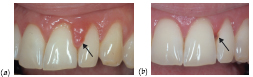
Figure 30.7 Plaque accumulation around a fixed orthodontic appliance in a 17-year-old female. There is gingival inflammation and a hyperplastic response to plaque.
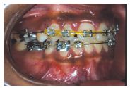
Figure 30.8 A 13-year-old boy with pronounced gingivitis anteriorly which is associated with poor plaque control and lack of saliva there due to incompetent lips, a high lip line and being a mouth breather in relation to nasal blockages.
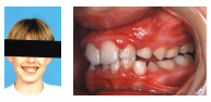
Figure 30.9 Pregnancy-associated gingivitis. (a) A 21-year-old pregnant patient with inflamed anterior gingivae, which bled on gentle probing. (b) A close up of the lower anterior gingivae which are inflamed and swollen. (c) A different pregnant patient with an even more marked response to plaque and severe gingival overgrowth, especially on the lower incisors.
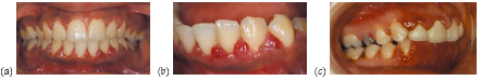
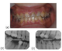
Figure 30.10 (a) Diabetes-associated gingivitis and uncontrolled caries in a young woman with poorly controlled type 1 diabetes mellitus. (b, c) Right and left bitewing radiographs showing caries but good bone levels. Courtesy of Mr P. J. Nixon.
Stay updated, free dental videos. Join our Telegram channel
VIDEdental - Online dental courses


