3: White Lesions

White Lesions

Lesions of the oral mucosa, which are white, result from the scattering of light through a thickened layer of keratin, epithelial hyperplasia, intracellular epithelial edema, and/or reduced vascularity of subjacent connective tissue. White or yellow-white lesions may also be due to fibrinous exudate covering an ulcer, submucosal deposits, surface debris, or fungal colonies.

Hereditary Conditions
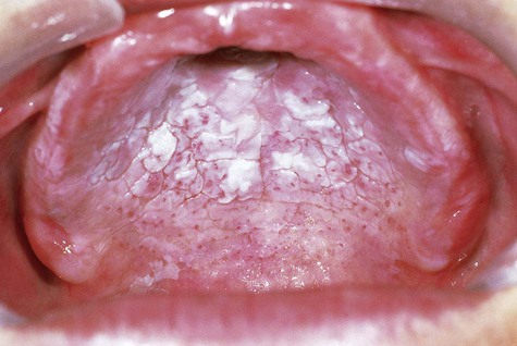
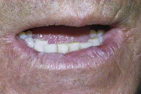
Leukoedema

Leukoedema is a generalized mild opacification of the buccal mucosa that is regarded as a variation of normal. It can be identified in the majority of the population.

Clinical Features

Leukoedema is usually discovered as an incidental finding. It is asymptomatic and symmetrically distributed in the buccal mucosa, and to a lesser extent over the labial mucosa. It appears as a gray-white, diffuse, filmy, or milky surface alteration (Fig. 3-1). In exaggerated cases, a whitish cast with surface textural changes, including wrinkling or corrugation, may be seen. With stretching of the buccal mucosa, the opaque changes dissipate. It is more apparent in nonwhites, especially African Americans.

image
FIGURE 3-1 Leukoedema.

White Sponge Nevus

White sponge nevus (WSN) is an autosomal-dominant condition that is due to point mutations for genes coding for keratin 4 and/or 13. It affects oral mucosa bilaterally and symmetrically, and no treatment is generally required.

Clinical Features

WSN presents as an asymptomatic, folded, white lesion that may affect several mucosal sites (Fig. 3-2; Box 3-1). Lesions tend to be thickened and have a spongy consistency. The presentation intraorally is almost always bilateral and symmetric and usually appears early in life, typically before puberty. The characteristic clinical manifestations of this particular form of keratosis are usually best observed on the buccal mucosa, although other areas such as the tongue and vestibular mucosa may also be involved. The conjunctival mucosa is usually spared, but mucosa of the esophagus, anus, vulva, and vagina may be affected.
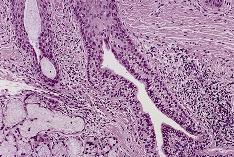
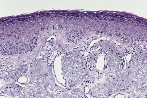
Histopathology

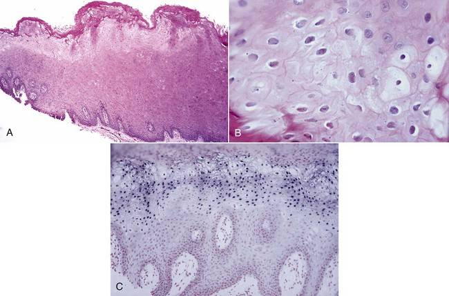
Microscopically, the epithelium is greatly thickened, with marked spongiosis, acanthosis, and parakeratosis (Fig. 3-3). Within the stratum spinosum, marked hydropic or clear cell change may be noted, often beginning in the parabasal region and extending very close to the surface. Perinuclear eosinophilic condensation of cytoplasm is characteristic of prickle cells in WSN. It is often possible to see columns of parakeratin extending from the spinous layer to the surface.

Differential Diagnosis

The differential diagnosis includes hereditary benign epithelial dyskeratosis, lichen planus, lichenoid drug reaction, lupus erythematosus (LE), cheek chewing, and possibly candidiasis (Table 3-1). Once tissue diagnosis is confirmed, no additional biopsies are necessary.

TABLE 3-1

BILATERAL BUCCAL MUCOSAL WHITE LESIONS: DIFFERENTIAL DIAGNOSIS

Disease Features/Action
White sponge nevus and HBID Hereditary; does not disappear when stretched; biopsy to confirm; HBID may also involve conjunctiva
Lichen planus Look for white reticulations (striae) and skin lesions; biopsy
Lichenoid drug reaction Look for white lesions in context of new drug history
Cheek chewing White shaggy lesions along occlusal plane or trauma sites
Lupus erythematosus Delicate radiating striae; biopsy
Candidiasis Look for predisposing factors; can rub off; responds to antifungal therapy

HBID, Hereditary benign intraepithelial dyskeratosis.

Hereditary Benign Intraepithelial Dyskeratosis

Etiology

Hereditary benign intraepithelial dyskeratosis (HBID), also known as Witkop’s disease, is a rare, hereditary condition (autosomal dominant). It was noted within a tri-racial isolate of white, Indian, and African American composition in Halifax County, North Carolina. The initial cohort of 75 patients was traced to a single common female ancestor who lived nearly 130 years earlier. With the use of genetic linkage and molecular analyses in two large families affected by HBID, one group localized the candidate causative genetic region to a telomeric region of chromosome 4q35, where three alleles for two linked markers exist. The precise gene that causes the condition has not been characterized.

Clinical Features

HBID presentation includes early onset (usually within the first year of life) of bulbar conjunctivitis, conjunctival plaques at the corneal limbus, and oral white lesions. Preceding the bulbar conjunctivitis are foamy gelatinous plaques that represent the ocular counterpart of the oral mucosal lesions.

Oral lesions consist of soft, asymptomatic, white folds and plaques of spongy mucosa. Areas characteristically involved include the buccal and labial mucosa and the labial commissures, as well as the floor of the mouth and lateral surfaces of the tongue, gingiva, and palate. The dorsum of the tongue is usually spared. Oral lesions are generally detected within the first year of life, with a gradual increase in intensity until mid-adolescence.

In some patients ocular lesions may vary seasonally, with spontaneous shedding of conjunctival plaques. Patients may complain of photophobia, especially in early life. Blindness, resulting from corneal vascularization, has been reported.

Follicular Keratosis

Etiology and Pathogenesis

Follicular keratosis (Darier’s disease, Darier-White disease) is an autosomal-dominant disorder that results in desmosomal defects and dysfunction by way of altered epithelial cell adhesion. Many cases appear sporadically as new mutations. Screening of candidate genes has led to the discovery that mutations in ATP2A2, a gene that encodes the sarcoplasmic/endoplasmic reticulum calcium–adenosine triphosphatase (Ca2-ATPase) isoform 2, cause this condition. It has been proposed that abnormalities in this calcium pump function interfere with cell growth and differentiation of calcium-dependent processes. To date at least 140 different mutations of the ATP2A2 gene have been identified as the genetic basis of Darier’s disease.

Clinical Features

Onset occurs between the ages of 6 and 20 years. The disease has a predilection for the skin, with 13% of patients demonstrating oral lesions. Skin manifestations are characterized by small, skin-colored papular lesions symmetrically distributed over the face, trunk, and intertriginous areas. The papules eventually coalesce and feel greasy because of excessive keratin production. The coalesced areas subsequently form patches of vegetating to verrucous growths that have a tendency to become infected and malodorous. Lesions may also occur unilaterally or in a zosteriform pattern. Thickening of the palms and soles (hyperkeratosis palmaris et plantaris) by excessive keratotic tissue is not uncommon. Fingernail changes may include fragility, splintering, and subungual keratosis. Nail changes are often helpful in establishing a diagnosis.

The extent of the oral lesions may parallel the extent of skin involvement. Favored oral mucosal sites include the attached gingiva and hard palate. Lesions typically appear as small, whitish papules, producing an overall cobblestone appearance. Papules range from 2 to 3 mm in diameter and may become coalescent. Extension beyond the oral cavity into the oropharynx and pharynx may occur.

Reactive Lesions

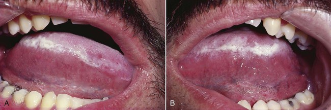
Focal (Frictional) Hyperkeratosis

Etiology

Focal (frictional) hyperkeratosis is a white lesion that is related to chronic rubbing or friction against an oral mucosal surface. This results in a presumably protective hyperkeratotic white lesion that is analogous to a callus on the skin.

Clinical Features

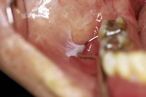
Friction-induced hyperkeratoses occur in areas that are commonly traumatized, such as the lips, lateral margins of the tongue, buccal mucosa along the occlusal line, and edentulous alveolar ridges (Figs. 3-4 to 3-7; Box 3-2). Chronic cheek or lip chewing may result in opacification (keratinization) of the affected area. Chewing on edentulous alveolar ridges produces the same effect.

Diagnosis

Careful history taking and examination should indicate the nature of this lesion. If the practitioner is clinically confident of a traumatic cause, no biopsy may be required. Patients should be advised to discontinue the causative habit, or the offending tooth or denture should be smoothed. The lesion should resolve or at least should be reduced in intensity over time, confirming the clinical diagnosis. Resolution of the lesion would allow unmasking of any underlying lesion that may not be related to trauma (Table 3-2). If the clinical diagnosis is in doubt, a biopsy should be taken.

TABLE 3-2

SOLITARY WHITE LESION: DIFFERENTIAL DIAGNOSIS

Disease Features/Action
Frictional keratosis Look for cause (e.g., ill-fitting denture, trauma); biopsy
Dysplasia, in situ carcinoma, squamous cell carcinoma Assess risk factors; biopsy
Burn History of aspirin or other agent application at site of lesion—discontinue use
Lupus erythematosus Delicate radiating striae; biopsy
Hairy leukoplakia Lateral borders of tongue; look for irregular surface architecture; often bilateral; biopsy

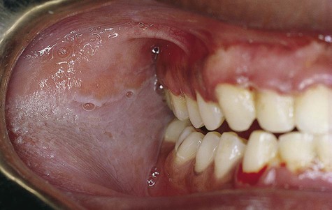
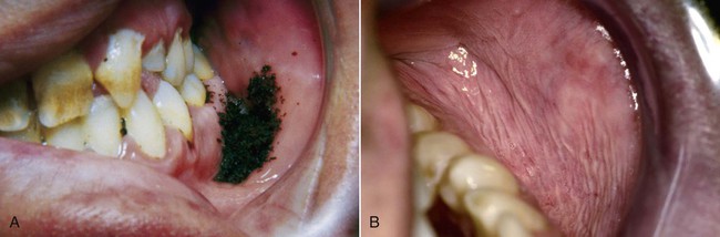
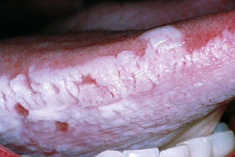
White Lesions Associated With Smokeless Tobacco

Marked geographic and gender differences in tobacco use have been identified. In the United States, a relatively high prevalence of smokeless tobacco users is found in the southern and western states. Use by men in New York and Rhode Island is less than 1% of the population, but in West Virginia, use exceeds 20%. Among teenagers, whites are the predominant users of smokeless tobacco, with males making up nearly this entire group. Smokeless tobacco is also used in Sweden in the form of snus, a nonfermented type of tobacco with lower concentrations of harmful nicotine and tobacco derivatives versus those types of fermented smokeless tobaccos traditionally used in the United States. In regions such as the Indian subcontinent and Southeast Asia, use is even more common and the materials more destructive. The tobacco-containing preparations generally are of a higher (alkaline) pH and often are mixed with other ingredients, including shredded areca (betel) nut; they may also contain lime, camphor, and spices.

The general increase in smokeless tobacco consumption has been related to both peer pressure and increased media advertising, which often glamorizes the use of smokeless tobacco, or snuff dipping. In addition, individuals who have been intense smokers and those who wish to avoid smoking may gravitate to this alternative. The clinical results of long-term exposure to smokeless tobacco include the development of oral mucosal white patches with a slightly increased malignant potential, dependence, alterations of taste, acceleration of periodontal disease, and significant amounts of dental abrasion.

Etiology

A causal relationship has been documented between smokeless tobacco and white tissue changes. Although all forms of smokeless tobacco may cause alterations in the oral mucosa, snuff (particulate, finely divided, or shredded tobacco) appears to be much more likely to cause oral lesions than does chewing tobacco. Oral mucosa responds to the topically-induced effects of tobacco with inflammation and keratosis. At the molecular level, altered cell signaling and subsequent cell damage have been demonstrated. Dysplastic changes may follow, with a low potential risk of malignant change. Smokeless tobacco–induced alterations in tissues are thought to be a response to tobacco constituents and perhaps other agents that are added for flavoring or moisture retention. Carcinogens such as nitrosonornicotine, an organic component of chewing tobacco and snuff, have been identified in smokeless tobacco. The pH of snuff, which ranges between 8.2 and 9.3, may be another factor that contributes to the alteration of mucosa.

Duration of exposure to smokeless tobacco that is necessary to produce mucosal damage is measured in terms of years. It has been demonstrated that leukoplakia can be predicted with the use of three tins of tobacco per week or duration of the habit of longer than 2 years.

Clinical Features

White lesions associated with smokeless tobacco develop in the immediate area where the tobacco is habitually placed (Figs. 3-9 and 3-10; Box 3-3). The most common area of involvement is the mucobuccal fold of the mandible in the incisor or the molar region. The mucosa develops a granular to wrinkled appearance. In advanced cases, a heavy, folded character may be seen. Less often, an erythroplakic or red component may be admixed with the white keratotic component. The lesions are generally painless and asymptomatic, and their discovery is often incidental to routine oral examination.
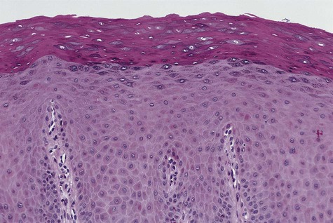
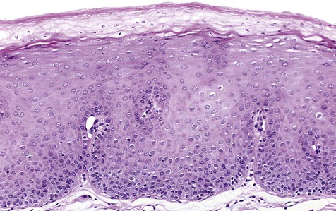
Histopathology

Slight to moderate parakeratosis, often in the form of spires or chevrons, is noted over the surface of the affected mucosa (Fig. 3-11). Superficial epithelium may demonstrate vacuolization or edema. A slight to moderate chronic inflammatory cell infiltrate is typically present. Epithelial dysplasia may occasionally develop in these lesions, especially among long-time users of smokeless tobacco. On occasion, a diffuse zone of basophilic stromal alteration may be seen, usually adjacent to inflamed minor salivary glands.
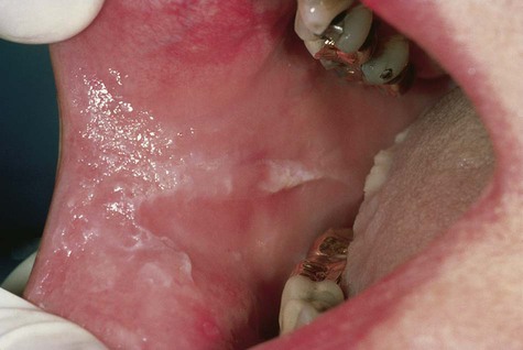
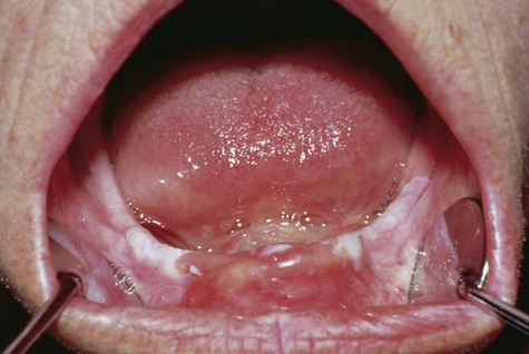
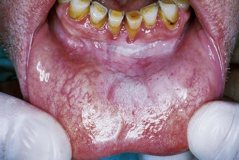
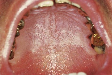
Nicotine Stomatitis

Etiology

Nicotine stomatitis is a common tobacco-related form of keratosis. It is typically associated with pipe and cigar smoking, with a positive correlation between intensity of smoking and severity of the condition. The importance of the direct topical effect of smoke can be appreciated in instances in which the hard palate is covered by a removable prosthesis, resulting in sparing of the mucosa beneath the appliance and hyperkeratosis of exposed areas. The combination of tobacco carcinogens and heat is markedly intensified in reverse smoking (lit end positioned inside the mouth), adding significant risk for malignant conversion (see Fig. 3-13).

Clinical Features

The palatal mucosa initially responds with an erythematous change followed by keratinization (Box 3-4). Subsequent to opacification or keratinization of the palate, red dots surrounded by white keratotic rings appear (Figs. 3-12 and 3-13). The dots represent inflammation surrounding the minor salivary gland excretory ducts.
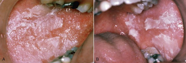
image
FIGURE 3-12 Nicotine stomatitis.

Histopathology

Nicotine stomatitis is characterized by epithelial hyperplasia and hyperkeratosis (Fig. 3-14). The minor salivary glands in the area show inflammatory change, and excretory ducts may show squamous metaplasia.

Hairy Leukoplakia

Etiology and Pathogenesis

In 1984 an unusual white lesion along the lateral margins of the tongue, predominantly in gay men, was first described. Evidence indicates that this particular form of leukoplakia, known as hairy leukoplakia, represents an opportunistic infection that is related to the presence of Epstein-Barr virus (EBV) and is found almost exclusively in human immunodeficiency virus (HIV)–infected individuals. In a small percentage of cases, hairy leukoplakia may be seen in patients with other forms of immunosuppression, particularly those associated with organ transplantation (medically-induced immunosuppression), hematologic malignancy, and long-term use of systemic corticosteroids. A few cases have been reported in patients who are taking corticosteroids, and a few patients who are purported to be otherwise healthy.

The prevalence of hairy leukoplakia in HIV-infected patients has been declining as a result of new chemotherapeutic regimens for HIV. Of importance is that this lesion has been associated with subsequent or concomitant development of clinical and laboratory features of acquired immunodeficiency syndrome (AIDS) in as many as 80% of cases. A positive correlation with depletion of peripheral CD4 cells and with the presence of hairy leukoplakia has been noted. Several other oral conditions have been described as having greater than expected frequency in patients with AIDS (Box 3-5).

The presence of EBV in hairy leukoplakia, as well as in the normal epithelium of patients with AIDS, has been confirmed. Through the use of molecular methods and ultrastructural analysis, viral particles have been localized within the nuclei and cytoplasm of the oral epithelial cells of hairy leukoplakia. Studies further indicate that this particular virus replicates within the oral hairy leukoplakia lesion. It is not understood why the lateral surface of the tongue is the favored site.

Clinical Features

Hairy leukoplakia presents as a well-demarcated white lesion that varies in architecture from a flat and plaquelike to a papillary/filiform or corrugated lesion (Figs. 3-15 and 3-16; Box 3-6). It may be unilateral or bilateral. A vast majority of cases have been located along the lateral margins of the tongue, with occasional extension onto the dorsal surface. Rarely, hairy leukoplakia may be seen on the buccal mucosa, the floor of the mouth, or the palate. Lesions have not been seen in the vaginal or anal mucosa.
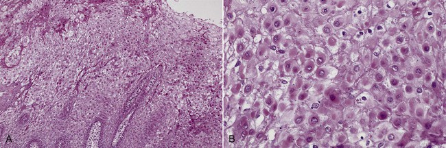
In general, no associated symptoms have been reported, although associated infection with Candida albicans might call attention to the presence of this condition. In more severe cases, the patient may become visually aware of the lesion.

Histopathology

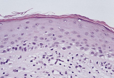
The characteristic microscopic feature of hairy leukoplakia is found in the nuclei of upper level keratinocytes (Fig. 3-17). Viral inclusions or peripheral displacement of chromatin with a resultant smudgy nucleus is evident. This is seen in the context of a markedly hyperparakeratotic surface, often with the formation of keratotic surface irregularities and ridges. C. albicans hyphae are often seen extending into the superficial epithelial cell layers. Beneath the surface, within the spinous cell layer, cells show ballooning degeneration and perinuclear clearing. A general paucity of subepithelial inflammatory cells has been noted, and Langerhans cells are scant.
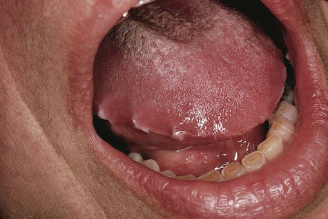
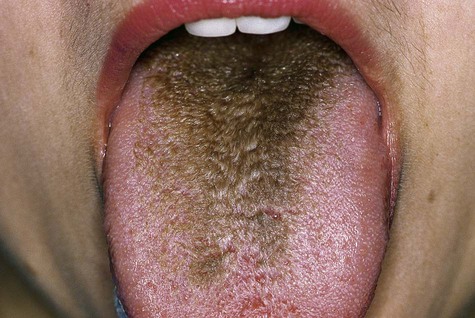
In situ hybridization studies have demonstrated the presence of EBV within cells, showing nuclear inclusions and basophilic homogenization. Further confirmation has been accomplished by ultrastructural demonstration of intranuclear virions of EBV.

Treatment and Prognosis

No specific treatment is available for hairy leukoplakia. For patients whose immune status is unknown and in whom biopsy findings indicate hairy leukoplakia, investigation for HIV infection or other causes of immunosuppression should be undertaken. Some discretion is required because a small percentage of patients with hairy leukoplakia is not associated with AIDS.

For cosmetic reasons, patients may request treatment of their lesions. Responses to acyclovir, ganciclovir, famciclovir, tretinoin, and podophyllum have been reported, with return of lesions often noted on discontinuation of therapy.

Approximately 10% of individuals with diagnosed hairy leukoplakia have AIDS at the time of diagnosis, and an additional 20% develop this disease in the following year. The probability that AIDS will develop in untreated HIV-positive individuals with HIV-associated hairy leukoplakia is approximately 50% within 1.5 years; within 2.5 years the probability is 80%.

Hairy Tongue

Hairy tongue is a clinical term referring to a condition of filiform papillary overgrowth on the dorsal surface of the tongue of variable color.

Etiology

Numerous initiating or predisposing factors for hairy tongue have been identified. Broad-spectrum antibiotics, such as penicillin, and systemic corticosteroids are often identified in the clinical history of patients with this condition. In addition, oxygenating mouth rinses containing hydrogen peroxide, sodium perborate, and carbamide peroxide have been cited as possible etiologic agents in this condition. Hairy tongue may also be seen in individuals who are intense smokers, in those who have undergone radiotherapy to the head and neck region for malignant disease, and in patients who have undergone hematopoietic stem cell transplantation. The basic problem is believed to be related to an alteration in microbial flora, with attendant proliferation of fungi and chromogenic bacteria, along with papillary overgrowth.

Clinical Features

The clinical alteration translates to asymptomatic hyperplasia of the filiform papillae, with concomitant retardation of the normal rate of desquamation. The result is a thick, matted surface that serves to trap bacteria, fungi, cellular debris, and foreign material (Fig. 3-18; Box 3-7).

image
FIGURE 3-18 Hairy tongue.

Hairy tongue is predominantly a cosmetic problem because symptoms are generally minimal. However, when extensive elongation of the papillae occurs, a gagging or a tickling sensation may be felt. The color may range from white to tan to deep brown or black, depending on diet, oral hygiene, oral medications, and the composition of the bacteria inhabiting the papillary surface.

Treatment and Prognosis

Identification of a possible etiologic factor, such as antibiotics or oxygenating mouth rinses, is helpful. Discontinuing one of these agents should result in improvement within a few weeks. In other patients, brushing with a slurry of sodium bicarbonate (baking soda) in water or gentle once-daily scraping of the dorsum of the tongue may be beneficial. In individuals who have undergone radiotherapy with resultant xerostomia and altered bacterial flora, management is more difficult. Brushing the tongue and maintaining fastidious oral hygiene should be of some benefit (application of a 1% solution of podophyllum resin with thorough rinsing has also been described as a useful treatment). It is important to emphasize to patients that this process is entirely benign and self-limiting, and that the tongue should return to normal after physical debridement and proper oral hygiene have been instituted.

Dentifrice-Associated Slough

Dentifrice-associated slough is a relatively common phenomenon that has been associated with the use of several different brands of toothpaste. It is believed to be a superficial chemical burn or a reaction to a component in the dentifrice, possibly the detergent or flavoring compounds. This process may be related to the use of essential oil–containing mouth rinses. Clinically, it appears as a superficial whitish slough of the buccal mucosa, typically detected by the patient as oral peeling that easily swipes away (Fig. 3-19). The condition is painless and is not known to progress to anything significant. The problem resolves with a switch to another, blander toothpaste or mouth rinse.

White mucosal changes have been described in association with the use of toothpaste and mouthwashes containing the substance sanguinaria (Fig. 3-20). The alteration is typically seen in the maxillary vestibule, although other sites may be affected.

Preneoplastic and Neoplastic Lesions
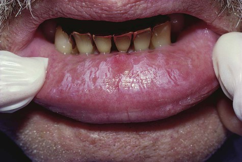
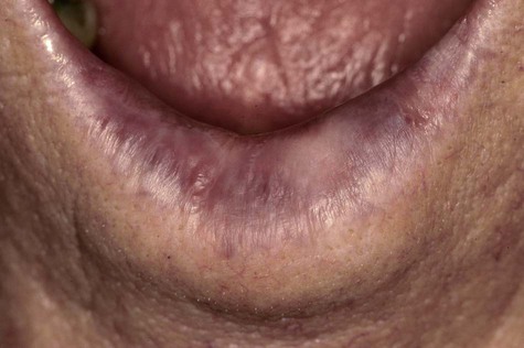
Actinic Cheilitis

Actinic, or solar, cheilitis represents accelerated tissue degeneration of the vermilion (dry mucous membrane) of the lips, especially the lower lip, as a result of chronic exposure to sunlight; it is considered to represent a potentially premalignant condition. This condition occurs almost exclusively in whites and is especially prevalent in those with fair skin.

Clinical Features

The affected vermilion of the lips takes on an atrophic, pale to silvery gray, glossy appearance, often with fissuring and wrinkling at right angles to the cutaneous-vermilion junction (Fig. 3-21; Box 3-8). Slightly firm, bilateral swelling of the lower lip is common. In advanced cases, the junction is irregular or totally effaced, with a degree of epidermization of the vermilion. Mottled areas of hyperpigmentation and keratosis are often noted, as well as superficial scaling, cracking, erosion, ulceration, and crusting (Fig. 3-22).

image
FIGURE 3-21 Actinic cheilitis.

Histopathology

The overlying epithelium is typically atrophic and hyperkeratotic (Fig. 3-23). Basophilic changes in the submucosa (altered elastin that replaces normal collagen) and telangiectasia are also seen.

Stay updated, free dental videos. Join our Telegram channel

Jan 12, 2015 | Posted by in Oral and Maxillofacial Pathology | Comments Off on 3: White Lesions

VIDEdental - Online dental courses

Get VIDEdental app for watching clinical videos