3
Maximising Mini-implant Success: Design Factors
It may be a confusing task for orthodontists to select a mini-implant kit since a large number and variety of systems are now available. These differ from one another in terms of their design features, technique steps, ease of insertion and use (e.g. how traction auxiliaries or wires are attached), versatility, the size and complexity of the kit, clinical recommendations, and the clinical instruction material. It is unlikely that one system provides dramatically better mini-implant stability rates than all of the others and key principles are widely applicable. However, since the treatments and illustrations in this book involve the Infinitas Mini-implant System (DB Orthodontics Ltd, UK, www.infinitas-miniimplant.com [accessed 5th December 2012]; Dentsply GAC, USA, www.gacintl.com) it is worth describing both its generic and distinctive features for clarity and reference purposes. The Infinitas mini-implant is unique in several aspects, especially in its head design and customised three dimensional (3D) guidance stent facility.1 This chapter describes the key design and clinical features of the Infinitas system’s clinical and guidance kits.
Infinitas mini-implant design features
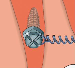
Many mini-implant head designs have two separate tiers represented by (1) a channel or ‘X’ shaped cross-slots at the top of the head for wire engagement and (2) an external circumferential undercut at a more apical level for the application of traction auxiliaries. In contrast, the Infinitas design has a unique, multi-purpose head which combines cross-slots with both external and internal undercuts, all on one vertical level (Fig. 3.1). This gives the head a very low profile (intra-oral prominence) whilst enabling the direct attachment of all forms of traction auxiliaries and wires (up to 0.021 × 0.025 inch dimensions). In particular, standard nickel titanium (NiTi) coil springs may be directly engaged, within the internal undercut, on one corner of the bracket-like Infinitas head (Fig. 3.2). Aside from patient comfort, this low profile is biomechanically favourable since it reduces the ratio of the head and neck (extra-bony section) to body length, and hence the risk of adverse tipping moments.2
Figure 3.1 Diagram of the Infinitas mini-implant head showing its four bracket-like wings, divided by perpendicular cross-slots, and the external and internal undercuts. A rectangular wire is illustrated within the transverse cross-slot.
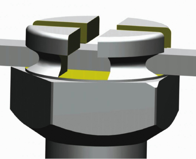
Figure 3.2 Diagram of an obliquely inserted Infinitas mini-implant with a coil spring engaged within the internal undercut of a single wing of its head.
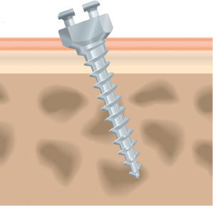
The Infinitas head’s precise engineering means that it is a relatively delicate structure. Therefore the insertion screwdriver achieves a secure connection by engaging the neck section, and hence avoids large and potentially deformational forces on the mini-implant head. Consequently the internal shape of the screwdriver closely matches the pentagonal shape of the coronal neck section (Fig. 3.1). The more apical part of the neck is tapered to enable mini-implant insertion at both perpendicular and oblique angles to the cortical plate, maximising cortical engagement yet minimising compression of the adjacent mucosa (Fig. 3.3). There are short (1.5 mm) and long (2.5 mm) neck length versions, to allow for the mucosal depth at typical buccal and palatal insertion sites, respectively.3 Whilst buccal insertions are routinely performed with a direct transmucosal technique, a re-usable circular mucotome (soft tissue punch) is used to remove thick (palatal) or loose mucosa prior to insertion of a long neck mini-implant.
Figure 3.3 Diagram of an obliquely inserted mini-implant where the body traverses the cortical layer at an angle, facilitated by the tapered shape of the neck. Notably, one side of the head is closer to the mucosa than the opposite side.
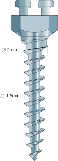
The Infinitas body has only four size variables: 1.5 and 2.0 mm diameters (as measured in the mid-body region), and 6 and 9 mm lengths. In combination with the two neck variables and the multi-purpose head design, this means that a range of only five mini-implants provides for all alveolar and palatal insertion options (Table 3.1). This helps to simplify both the clinical decision-making process and stock keeping. All of the Infinitas bodies feature a self-drilling tip and threads (Fig. 3.4), since self-drilling insertion preserves more original bone than a pre-drilled technique.4,5 Engagement of the cortical bone plate, in order to maximise primary stability, is also enhanced by two specific Infinitas design features. First, the thread continues to the coronal end of the body such that it may be fully seated in bone. Second, the 1.5 mm (narrow) Infinitas body version has an additional tapered feature at its coronal end, such that the thread diameter gradually widens from 1.5 to 2 mm before the junction with the neck (Fig. 3.4). This creates a clinically noticeable increase in torque during the final stage of insertion as the body part engaging the cortex widens from the cylindrical to tapered form, with the effect of increasing primary stability.6,7,8 Another benefit of this additional body taper is that it greatly increases strength in this critical coronal area since a 0.2 mm increase in diameter can increase strength by 50%.8,9 This helps to minimise the risk of fracture of the body during insertion and removal.10,11 So why not have a 2 mm diameter over most of the body length? Unfortunately this would greatly increase the risk of close root proximity in narrow interproximal sites (compared to the 1.5 mm mid-body size). Hence, this tapered body design provides a 2 mm diameter superficially where it is most beneficial for strength and stability, but avoids root proximity problems since the narrowest inter-root space is frequently 4 mm in from the external bone surface.12
Figure 3.4 Diagram showing how the body’s coronal section tapers out from a 1.5 to 2.0 mm thread diameter.
Stay updated, free dental videos. Join our Telegram channel
VIDEdental - Online dental courses


