Anatomy and Physiology
Basic Concepts
Anatomy
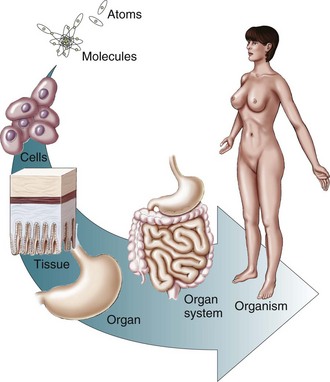
Levels of Organization
A Chemical level—organization of chemical structure separates living and nonliving material; atoms, molecules, and macromolecules result in living matter
B Organelle level—organelles are structures made of molecules and organized to perform specific functions; allow the cell to perform vital functions; types include:
C Cellular level—cells comprise the basic structural and functional units of an organism; the smallest living units in the human body
D Tissue level—groups of cells and materials surrounding them that work together to perform a particular function
E Organ level—different types of tissues joined together to form body structures
1. Each organ has a unique size, shape, appearance, and placement in the body (e.g., stomach, heart, liver, lungs, brain)
F System level—related organs that have a common function (e.g., digestive system breaks down and absorbs molecules in food; organs include the mouth, salivary glands, pharynx, esophagus, stomach, liver, gallbladder, pancreas, small intestine, and large intestine)
G Organism level—all the systems of the body combine to make up an organism
Anatomic Nomenclature
(See the section on “Anatomic Nomenclature” in Chapter 4.)
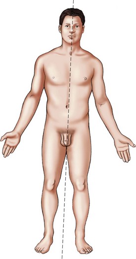
A Anatomic position—erect body position with arms at the sides and palms upward (Figure 3-2)
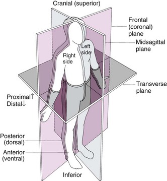
B Plane or section—imaginary flat surfaces that pass through the body (Figure 3-3)
1. Sagittal plane—vertical plane dividing the body into right and left sides; midsagittal plane bisects the body at the exact midline
2. Coronal or frontal plane—divides the body or organ into anterior and posterior portions
3. Transverse plane—divides the body or organ into superior and inferior portions (may also be called cross-sectional or horizontal plane)
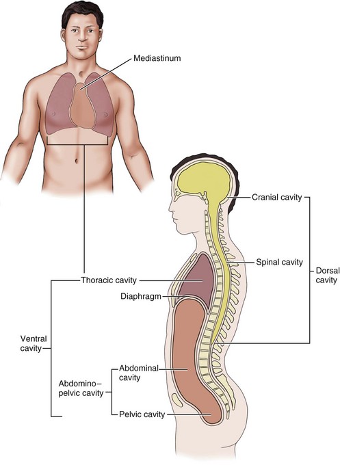
Body Cavities
1. Cranial cavity—formed by the cranial bones of the skull; contains the brain
2. Vertebral cavity—formed by the vertebrae; contains the spinal cord
Cells
(See the section on “General Histology” in Chapter 2.)
Cellular Structure
1. Surrounds and contains the cytoplasm of a cell; composed of proteins and lipids
2. Selective permeability characteristics
a. Protects cell from external environment
b. Permits the entrance and exit of selected substrates
c. Membrane proteins have several functions—channels and transporters are integral proteins that help specific solutes across the membrane; receptors serve as cellular recognition sites; some membrane proteins are enzymes
3. Basic framework—lipid bilayer; two layers of phospholipids, cholesterol, and glycolipids
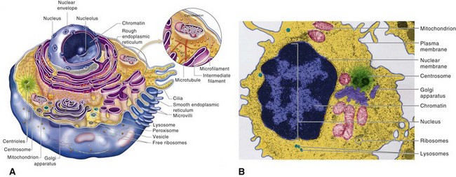
B Cytoplasm—all cellular contents between the plasma membrane and the nucleus; includes:
1. Cytosol—fluid portion of cytoplasm; site of many chemical reactions for the cell’s existence
2. Cytoskeleton—network of several kinds of protein filaments that extend throughout the cytoplasm; structural framework for the cell; generates movement
3. Organelles—specialized cellular structures with characteristic shapes and specific functions
1. Free ribosomes—not attached to other organelles; synthesize proteins used inside the cell
2. Bound ribosomes—attached to the endoplasmic reticulum (ER); form rough ER; synthesize proteins destined for use in the plasma membrane or for export from cell
D Endoplasmic reticulum—network of membranes that form flattened sacs called cisterns; arranged in parallel rows within the cytoplasm of a cell; contains enzymes involved in a variety of metabolic activities
1. Stack of 3 to 20 flattened membranous sacs (cisterns)
2. Within the cisterns, proteins are modified, sorted, and packaged into vesicles for transport to different destinations
1. Membrane-enclosed vesicles that form in the Golgi complex
3. Function in the digestion of worn-out organelles (autophagy) and self (autolysis)
1. Ellipsoid bodies that consist of two membranes that contain enzyme complexes in a particular array (e.g., tricarboxylic acid cycle enzymes)
2. Function as the powerhouse of the cell by transforming the chemical energy bond of nutrients into the high-energy phosphate bonds of adenosine triphosphate (ATP)
3. A single cell may contain 50 to 2500 of these organelles, depending on the cell’s energy needs
Movement of Substances Through Cell Membranes
TABLE 3-1
Some Important Transport Processes
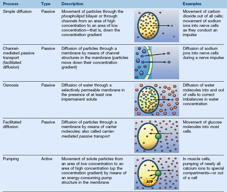
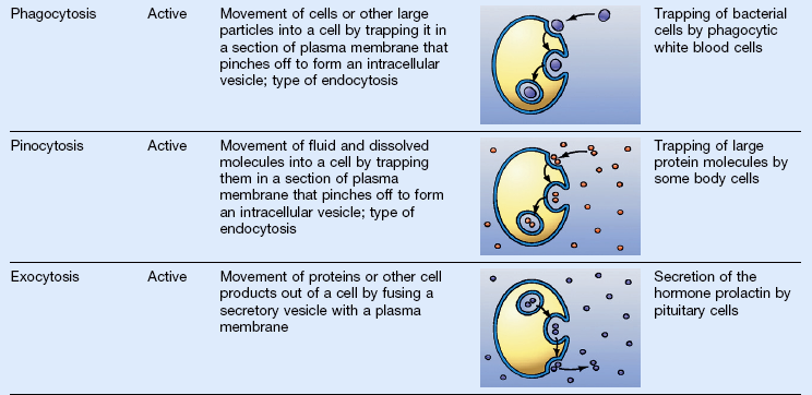
(Art from Patton KT, Thibodeau GA: Anthony’s textbook of anatomy and physiology, ed 19, St Louis, 2010, Mosby.)
A Passive transport processes—do not require energy expenditure of the cell membrane
1. Diffusion—a passive process
a. Molecules spread through the membranes
b. Molecules move from an area of high concentration to an area of low concentration (down a concentration gradient)
c. Eventually a state of equilibrium is reached
d. Membrane channels—pores in cell membranes through which specific ions or small water-soluble molecules can pass
2. Simple diffusion—substances diffuse across a membrane in one of two ways: lipid-soluble substances diffuse through the lipid bilayer, and ions diffuse through pores
a. Diffusion of water through a selectively permeable membrane (limits diffusion of at least some solute particles); results in gain of volume on one side of the membrane and loss of volume on the other side of the membrane
b. A solution containing solute particles that cannot pass through a membrane exerts osmotic pressure on the membrane
c. Potential osmotic pressure—maximum pressure that could develop in a solution when it is separated from pure water by a selectively permeable membrane; knowledge of potential osmotic pressure allows the prediction of the direction of osmosis and resulting change of pressure
(1) Isotonic—when two fluids have the same potential osmotic pressure
(2) Hypertonic (higher pressure)—cells placed in solutions that are hypertonic to intracellular fluid shrivel as water flows out of cells by osmosis faster than it enters
(3) Hypotonic (lower pressure)—a solution that has a lower concentration of solutes than the cytosol inside the cell; water molecules enter the cells by osmosis faster than they leave
4. Facilitated diffusion (carrier-mediated passive transport)
a. Movement of molecules made more efficient by the action of specific transport mechanisms in the plasma membrane; facilitated by channel proteins or carrier proteins
b. Transports substances down a concentration gradient
c. Substances moved by facilitated diffusion include glucose, fructose, galactose, urea, and some vitamins
B Active transport processes—require the expenditure of metabolic energy by the cell
a. Process that moves substances against a concentration gradient (from an area of low concentration to an area of high concentration)
c. Substances moved by “pumps,” for example, calcium pumps and sodium–potassium pumps
2. Endocytosis and exocytosis—allow substances to enter or leave the interior of a cell without actually moving through its plasma membrane
a. Endocytosis—process by which the plasma membrane “traps” some extracellular material and brings it into the cell in a vesicle; the two basic types are:
(1) Phagocytosis (cell eating)—large particles are engulfed by the plasma membrane and enter the cell in vesicles; vesicles fuse with lysosomes, where particles are digested
(2) Pinocytosis (cell drinking)—the plasma membrane folds inward, forming a pinocytic vesicle containing a droplet of extracellular fluid; the vesicle detaches from the plasma membrane and enters the cytosol
b. Exocytosis—process by which large molecules, notably proteins, can leave the cell, even though they are too large to move through the plasma membrane; large molecules are enclosed in membranous vesicles and then pulled to the plasma membrane by the cytoskeleton, where the contents are released
Cell Metabolism
A Metabolism—chemical reactions in a cell
1. Catabolism—breaking of large molecules into smaller ones; usually releases energy
2. Anabolism—building of large molecules from smaller ones; usually consumes energy
1. Enzymes—chemical catalysts, reducing activation energy needed for a reaction
3. Chemical structure of enzymes
a. Proteins of a complex shape
b. Active site—where the enzyme molecule fits the substrate molecule; lock-and-key model
a. Enzymes usually have an “-ase” suffix; the first part of the word often signifies the substrate or the type of reaction catalyzed
b. Oxidation-reduction enzymes—known as oxidases, hydrogenases, and dehydrogenases; energy release depends on these enzymes
c. Hydrolyzing enzymes—hydrolases, for example, digestive enzymes
d. Phosphorylating enzymes—phosphorylases or phosphatases; add or remove phosphate groups
e. Carboxylases and decarboxylases—add or remove carbon dioxide
a. Regulate cell functions by regulating metabolic pathways; specific in their actions
b. Chemical and physical agents called allosteric effectors alter enzyme action by changing the shape of the enzyme molecule, for example:
c. Most catalyze chemical reactions in both directions
1. Cellular respiration—pathway in which glucose is broken down to yield its stored energy; an important example of cell catabolism; has three chemically linked pathways:
(1) Pathway in which glucose is broken apart into two pyruvic acid molecules to yield a small amount of energy (which is transferred to ATP and nicotinamide adenine dinucleotide [NADH])
(2) Includes many chemical steps (reactions that follow one another), each regulated by specific enzymes
b. Citric acid cycle (Krebs cycle)
(1) Pyruvic acid (from glycolysis) is converted into acetyl coenzyme A (CoA) and enters the citric acid cycle after losing carbon dioxide (CO2) and transferring some energy to NADH
(2) A cyclic sequence of reactions that occurs inside the inner chamber of a mitochondrion. The acetyl splits from the CoA and is broken down, yielding CO2 and energy (in the form of energized electrons), which is transferred to ATP, NADH, and flavin adenine dinucleotide (FADH2)
c. The electron transport system (ETS)
(1) Energized electrons are carried by NADH and FADH2 from glycolysis and the citric acid cycle to electron acceptors embedded in the cristae of the mitochondrion
(2) As electrons are shuttled along a chain of electron-accepting molecules in the cristae, their energy is used to pump accompanying protons (H+) into the space between mitochondrial membranes
(3) Protons flow back into the inner chamber through carrier molecules in the cristae; their energy of movement is transferred to ATP
(4) Low-energy electrons coming off the ETS bind to oxygen and rejoin their protons, forming water (H2O)
1. Protein synthesis is a central anabolic pathway in cells
2. DNA (see the section on “Genetics” in Chapter 7)
a. A double-helix polymer (composed of nucleotides); functions to transfer the information encoded in genes, which directs protein synthesis
b. Gene—a segment of a DNA molecule that consists of approximately 1000 pairs of nucleotides; contains the code for synthesizing one polypeptide
a. Messenger ribonucleic acid (mRNA) forms along a segment of one strand of DNA
b. Noncoding introns are removed, and the remaining exons are spliced together to form the final edited version of the mRNA copy of the DNA segment
a. After leaving the nucleus and being processed, mRNA associates with a ribosome in the cytoplasm
b. Transfer ribonucleic acid (tRNA) molecules bring specific amino acids to the mRNA at the ribosome; the type of amino acid is determined by the fit of a specific tRNA’s anticodon with an mRNA’s codon
c. As amino acids are brought into place, peptide bonds join them, eventually producing an entire polypeptide chain
5. Processing—enzymes in the ER and Golgi apparatus link polypeptides into whole protein molecules or process them in other ways
Cell Growth and Reproduction
A Cell growth and reproduction of cells are the most fundamental of all functions in a living being; together they constitute the life cycle of the cell
1. Cell growth—depends on the use of the genetic information in DNA to make structural and functional proteins for cell survival
2. Cell reproduction—ensures that genetic information is passed from one generation to the next
1. Production of cytoplasm—more cell material is made, including growth and replication of organelles and plasma membrane; a largely anabolic process
3. Growth phase of the cell’s life cycle—subdivided into the first phase (G1), the DNA synthesis phase (S), and the second growth phase (G2)
1. Mitosis—process of organizing and distributing nuclear DNA during cell division; cells reproduce by splitting themselves into two smaller daughter cells (see the section on “Cell Replication” and Figure 2-6 in Chapter 2)
2. Meiosis—germ cell division; produces gametes (sperm and oocytes), the cells needed to form the next generation of sexually reproducing organisms
Tissues
(See the sections on “Concepts Relating to Dental Tissues,” “Basic Tissues,” “Epithelial Tissue,” “Connective Tissue,” “Blood and Lymph,” “Nerve Tissue,” and “Muscle Tissue” in Chapter 2.)
Body Membranes
A Thin tissue layers that cover surfaces, line cavities, and divide spaces or organs
B Epithelial membranes are the most common
a. Parietal membranes—line closed body cavities
b. Visceral membranes—cover visceral organs
c. Pleura—surround the lung and line the thoracic cavity
d. Peritoneum—covers the abdominal viscera and lines the abdominal cavity
3. Mucous membranes (see the section on “Soft Tissue of the Oral Cavity” and Figure 2-21 in Chapter 2)
Systems of the Body and Their Components
The Integumentary System
A Functions—regulation of body temperature, protection, sensation, excretion, immunity, synthesis of vitamin D
1. Epidermis—thin outer portion composed of keratinized stratified squamous epithelium
C Skin color—from melanin, carotene, and hemoglobin pigments
D Accessory structures—hair, skin glands, and nails
1. Hair—threads of fused, dead keratinized cells that function in protection
a. Consist of a shaft above the surface, a root that penetrates the dermis and subcutaneous layer, and a hair follicle
2. Sebaceous glands—usually connected to hair follicles; absent in palms and soles of feet; produce sebum, which moistens hair and waterproofs skin
3. Sudoriferous glands—produce perspiration; carry waste to the skin’s surface; assist in maintaining body temperature
4. Nails—hard keratinized epidermal cells covering terminal portions of fingers and toes; the principal parts are body, free edge, root, lunula, cuticle, and matrix
Tissues and Membranes of the Body
The Skeletal System
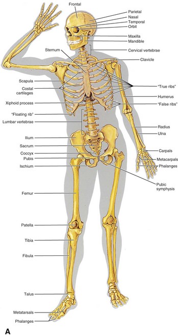
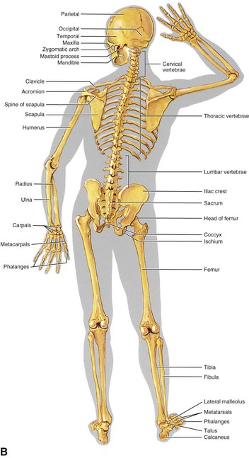
(See Figure 3-6 and the section on “Connective Tissue” in Chapter 2.)
1. Provides rigid support system
2. Provides protection, for example, cranial bones protect the brain
3. Serves as a source and a sink for calcium; involved in the formation of blood cells (hemopoiesis)
3. Articular cartilage—layer of hyaline cartilage that covers the articular surfaces of epiphyses; cushions jolts and blows to bone
4. Periosteum—white fibrous membrane that covers bone; contains cells that form and destroy bone, blood vessels; point of attachment for ligaments and tendons
6. Endosteum—epithelial membrane that lines the medullary cavity
1. Most distinctive form of connective tissue
2. Extracellular components are hard and calcified
3. Rigidity allows its supportive and protective functions
4. Tensile strength nearly equal to cast iron at less than one third the weight
(1) Hydroxyapatite—highly specialized chemical crystals of calcium and phosphate contribute to the hardness of bone
(2) Slender needle-like crystals oriented to resist stress and mechanical deformation
Stay updated, free dental videos. Join our Telegram channel

VIDEdental - Online dental courses


