28: Multidisciplinary Team Approach to Cleft Lip and Palate Management

CHAPTER 28 Multidisciplinary Team Approach to Cleft Lip and Palate Management

Cleft lip and palate, the most common of the craniofacial anomalies, are severe congenital anomalies that have an incidence of 0.28 to 3.74 per 1000 live births globally. In the United States, cleft lip and palate occur in approximately 1 in 1000 newborns. The incidence varies widely among races. Cleft lip and palate occur in about 1 in 800 white newborns, 1 in 2000 black newborns, and 1 in 500 Japanese or Navaho Indian newborns. Isolated cleft palate occurs in about 1 in 2000 newborns and demonstrates less racial variation. Cleft lip and palate together account for approximately 50% of all cases, whereas isolated cleft lip and isolated cleft palate each occur in about 25% of cases. Many of these congenital anomalies appear to be genetically determined, although the majority are of unknown cause or are attributable to teratogenic influences (see Chapter 6).

CLASSIFICATION OF CLEFT LIP AND PALATE

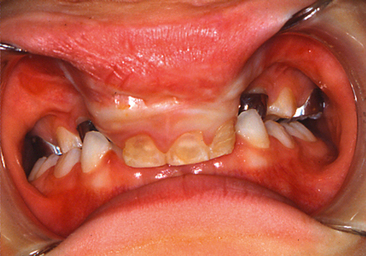
There is a tendency to conceptualize cleft lip and palate as a homogenous anomaly. If that was true, a treatment plan applicable to all cases could be formulated. However, the reality is that clefts vary widely in their clinical presentations (Fig. 28-1).

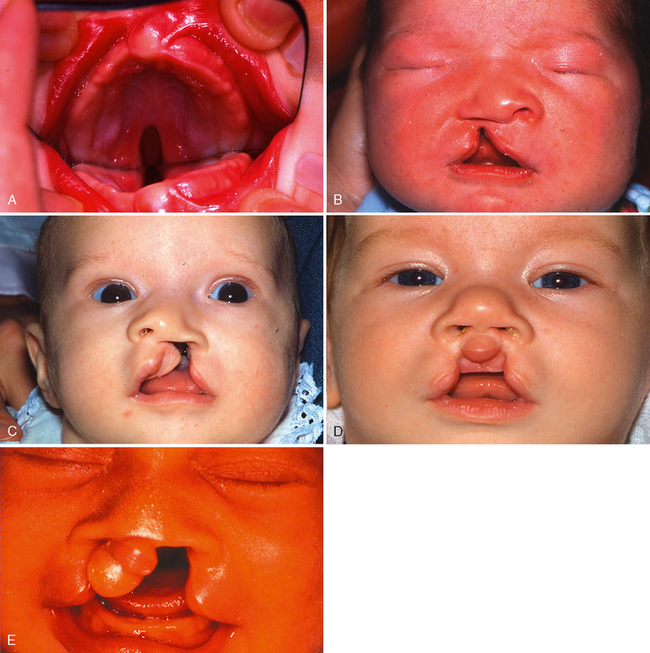
To standardize reporting of cleft lip and palate, the Nomenclature Committee of the American Association of Cleft Palate Rehabilitation devised a classification system that later was adopted by the Cleft Palate Association. The complexity of this system, however, has made its acceptance less than overwhelming. Veau proposed the most frequently used system.1 He classified clefts of the lip as follows:

Veau divided palatal clefts into four classes as follows (Fig. 28-2):

Veau did not include submucous clefts of the palate in his classification system. Submucous clefts may frequently be diagnosed by the following physical findings: bifid uvula, palpable notching at the posterior portion of the hard palate, and a zona pellucida (thin, translucent membrane). Submucous clefts of the palate may be associated with an incomplete velopharyngeal mechanism or eustachian tube dysfunction.

MULTIDISCIPLINARY CLEFT LIP AND PALATE TEAM

Children born with cleft lips and palates have many problems that need to be solved for successful habilitation. The complexity of these problems requires that numerous health care practitioners cooperate in providing the specialized knowledge and skills necessary to ensure comprehensive care. The cleft palate team concept has evolved from that need.

To address the many treatment regimens and different care protocols, the American Cleft Palate– Craniofacial Association (http://www.cleftpalate-craniofacial.org) convened a consensus conference on recommended practices for the care of patients with craniofacial anomalies. This conference produced the document “Parameters for Evaluation and Treatment of Patients with Cleft Lip/Palate or other Craniofacial Anomalies.”2 This serves as a guide for implementing the multidisciplinary approach to cleft and craniofacial care and is used by teams in the United States and Canada.

Because optimal care is best achieved by multiple types of clinical expertise, the teams may be composed of individuals in (1) the dental specialties (orthodontics, oral surgery, pediatric dentistry, and prosthodontics), (2) the medical specialties (genetics, otolaryngology, pediatrics, plastic surgery, and psychiatry), and (3) allied health care fields (audiology, nursing, psychology, social work, and speech pathology).

These care providers assess the patient’s medical status and general development, dental development, facial esthetics, psychological well-being, hearing, and speech development (Fig. 28-3). Team members must communicate effectively among themselves, with the child and parents, and with the primary care physician and dentist. Individuals on the team must respect one another’s opinions and be flexible in planning and carrying out therapy. Periodic evaluation is necessary to assess the effect of previous therapy and to determine whether an alternative approach may be necessary. A team conference immediately after patient examination is a desirable way to discuss current problems and plan timely therapy.

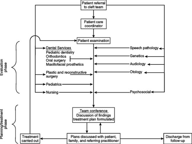
Whitehouse describes the clinical team as a “close, cooperative, democratic, multiprofessional union devoted to a common purpose—the best treatment of the fundamental needs of the patient.”3

GENERAL RESPONSIBILITIES OF TEAM MEMBERS

DENTAL SPECIALTIES

The pediatric dentist is responsible for the overall dental care of the patient. Numerous dental anomalies and malocclusions occur with a cleft lip or palate. These may be attributed to the congenital clefting itself or may be secondary to the surgical correction of the primary defects. A high correlation is found between the number and severity of dental problems and the type and severity of the cleft.

The pediatric dentist should discuss with the patient and parents the traditional dental problems associated with clefting. Any one, or several, of the following conditions may occur with a significantly greater frequency than in the general population:

3. There is a significant increase in the frequency of supernumerary teeth (Fig. 28-5), which are often seen in those with complete unilateral or bilateral clefts. Again, the primary lateral incisors are most frequently absent. Occasionally, there can be supernumerary teeth and congenitally missing teeth in patients with cleft lip only with or without notching of the alveolus.
10. In a patient with a complete unilateral or bilateral cleft of the palate, the lateral facial profile may appear noticeably convex (Fig. 28-7). This may become more perceptible as the child grows older. The appearance may be attributed to a true mandibular or pseudomandibular prognathism. In pseudomandibular prognathism, the maxilla is in spatial disharmony with the mandible. This may be caused by a retrognathic maxilla or an attenuation of the anteroposterior and vertical growth of the maxilla.

Parents are often so overwhelmed by other aspects of the cleft that they give dental care a low priority or even neglect it altogether. Preventive dental care is extremely important in these cases. The intact dental occlusion is the foundation around which future orthodontic therapy takes place. For this reason, optimum dental health is essential for total habilitation of the patient. Any compromise will lead to a less than optimal result. Routine prophylaxis and fluoride treatments are mandatory. Referral for preventive dental care should be made during the first year of life. Fluoride supplements, dentifrices, and rinses are indicated if the patient lives in a nonfluoridated community. The parents and patient should be instructed in proper dental hygiene techniques, especially around the defect. Close communication between the primary care dentist and the cleft team is important to ensure the continuity of care necessary during the extended treatment of such patients. Routine periodic reports from the cleft team should be forwarded to the child’s primary care dentist, especially during orthodontic or surgical treatment. Pediatric dentists often are involved in the presurgical and postsurgical treatment phase of maxillary orthopedics. Both active and passive appliances are used to bring the cleft segments into a more ideal alignment and thereby promote a more favorable initial surgical outcome.

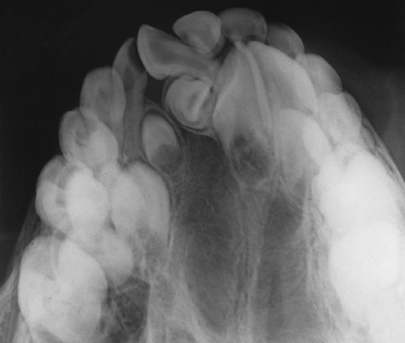
The orthodontist plays a key role in the diagnosis and treatment of a cleft condition by obtaining records necessary for diagnosis and treatment planning. These include cephalometric and panoramic radiographs, study models, and diagnostic photographs. Analysis of these records enables the orthodontist to describe and quantitate the facial skeleton and soft tissue deformities. Using expertise in the growth and development of the facial skeleton, this specialist can identify problem areas and, with some limitations, predict growth and development. Many team members depend on the orthodontist’s analysis and quantitations of the cleft anomaly for treatment planning.

The orthodontist also provides comprehensive orthodontic care for patients. Most orthodontic care can be considered conventional, but for difficult dental configurations, innovation and imagination are required for treatment. If surgical treatment is indicated, the orthodontist works closely with the surgeon to plan the most appropriate procedure. Immediate postoperative function, esthetic result, and long-term stability are factors considered before surgery.

The ability to surgically alter skeletal relationships of the maxillomandibular complex is the basis for participation by the oral and maxillofacial surgeon on the cleft team. This specialist evaluates all patients for facial form and function and jaw position. Many patients have significant skeletal malocclusions that cannot be treated by conventional orthodontics and require surgical correction.

The surgical placement of primary and secondary alveolar cleft bone grafts is another important role of the oral and maxillofacial surgeon. These grafts aid in dental habilitation. The grafted bone supports the teeth adjacent to the cleft site and provides bone through which teeth may erupt. A detailed discussion of these grafts follows later in this chapter.

The maxillofacial prosthodontist replaces, restores, or rehabilitates orofacial structures that may be congenitally missing or malformed. Nonliving materials are used to restore and enhance form and anatomy. There is a special commitment to the oral cavity because this specialist fabricates prosthetic appliances to rehabilitate mastication, deglutition, speech, and oral esthetics.

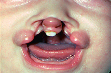
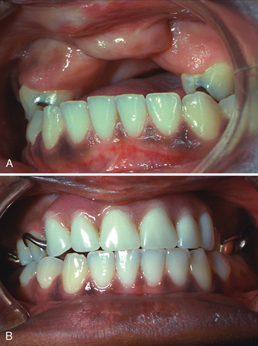
Many patients with clefts have congenitally missing teeth or malformed teeth that may need to be removed. In these cases, masticatory function, speech, and orofacial esthetics are compromised, and successful habilitation dictates that these missing teeth be replaced to achieve as near normal a condition as possible (Fig. 28-8). The maxillofacial prosthodontist may do this with fixed or removable appliances or with a combination of the two.

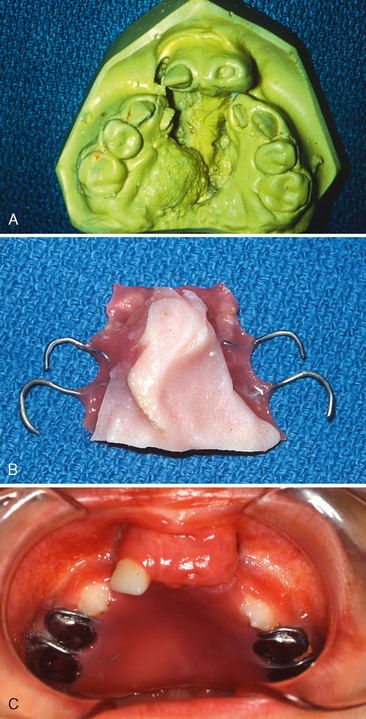
Occasionally, patients demonstrate aberrant speech patterns caused by failure of the soft palate to elevate properly. In such cases, a palatal lift appliance is fabricated to aid the speech mechanism. In other cases the maxillofacial prosthodontist may fabricate a speech bulb prosthesis to aid or augment the velopharyngeal mechanism. In patients with considerable escape of air through persistent palatal fistulas, the fistulas can be obturated (Fig. 28-9).

MEDICAL AND ALLIED HEALTH SPECIALTIES

The patient care coordinator arranges appointments, maintains patient records, and monitors the interaction of the patient and family with the various team members. The coordinator corresponds with health and school personnel near the patient’s home to help provide continuity of care for the cleft patients within the community. The coordinator is the most convenient point of contact with team members for the patients, their families, and health care practitioners outside the medical center complex.

The pediatrician, often the patient’s own pediatrician or family physician, is responsible for maintenance of the patient’s overall health. This specialist performs complete physical evaluations and helps assess the patient’s physiologic status. Close attention is also directed to growth status and other developmental milestones.

The medical geneticist examines the patient to find characteristics of syndromes associated with cleft lip and palate. Consideration is given to the genetic basis for the anomaly, and this information is related to the parents. Genetic counseling is a very important function of the geneticist. Parents are vitally interested in risk assessment relative to future offspring, and other family members who may be at risk are often counseled (see Chapter 6).

The role of the plastic and reconstructive surgeon usually begins with a determination of the timing and method of lip closure. With complete clefts, the plastic surgeon may next be responsible for obtaining bone grafts to be used in closing defects of the maxillary dental alveolus. This specialist is also involved in cleft palate repair.

An additional responsibility may be to conduct a nasopharyngoscopic examination of the speech mechanism. If a defect is identified, the plastic surgeon may perform a pharyngoplasty to improve velopharyngeal function. The final role is to correct internal or external cleft nasal deformities.

The social worker acts as the patient’s advocate in many cases and aids in psychosocial assessment. This team member assists the family by making referrals to persons or agencies at the local, county, and state levels for guidance regarding financial resources for medical care. During hospitalization, the social worker provides supportive counseling and facilitates communication between the family and medical or hospital personnel. The focus is on helping the family cope with stress during and after surgery and on dealing with emotional factors involved in forming realistic expectations of surgical outcomes and in adapting to problems of body image.

The psychiatrist and psychologist evaluate the patient for strengths and weaknesses in cognitive, interpersonal, emotional, behavioral, and social development. Emphasis is placed on the patient’s ability to cope with the emotional and physical stress created by the cleft defect. Consultation with the parents and school regarding educational or behavioral management occurs when indicated.

The speech pathologist functions essentially as a monitor of speech output. All speech sounds are analyzed to determine deviations from normal, and the cause of any deviation is evaluated. To the extent that anatomic variations are corrected, the speech pathologist offers therapeutic options to enhance maturation of speech or to achieve satisfactory compensation in motor production for optimal speech.

The audiologist performs tests to identify any hearing difficulties. When neither the child nor the parents recognize hearing problems, the result can be a delay in speech and language development and poor performance in school. When hearing tests or impedance measures are abnormal, a referral is made to an otolaryngologist for an ear examination. The otolaryngologist coordinates audiologic tests and any special studies that may be needed to evaluate middle ear structures. Any middle ear surgery that is to be done is performed by this member of the team. He or she also may perform a nasopharyngoscopic examination in coordination with the speech pathologist.

Nurses provide varying functions that are valuable to the cleft lip and palate team. They actively communicate with other disciplines in passing on information regarding the special needs of each child and family. Nurses emphasize total family involvement, not just treatment of the patient. They prepare patients and families for either outpatient or inpatient surgery and assist in the overall management process. Above all, they are extremely important in assessing the initial feeding issues and advising parents with ongoing nutritional concerns.

The presence of more complex craniofacial anomalies, such as Crouzon syndrome, Treacher-Collins syndrome, or hemifacial microsomia, require additional specialists on the team because there is more deformity. Some of these care providers include anesthesiologists, diagnostic medical imaging radiologists, neurologists, neurosurgeons, and ophthalmologists in addition to those cleft team providers already mentioned.

MULTIDISCIPLINARY SEQUENCING OF TREATMENT IN CLEFTS

The following discussion focuses on major treatment procedures performed by members of the cleft team. For convenience, treatment is divided into four stages, which generally correspond to stages in the child’s dental development.

STAGE I (MAXILLARY ORTHOPEDIC STAGE: BIRTH TO 18 MONTHS)

Management of the patient with a cleft begins with immediate attention to the needs of the newborn. Feeding problems are often associated with cleft anomalies, which make it difficult for the infant to maintain adequate nutrition. These problems include insufficient suction to pull milk from the nipple, excessive air intake during feeding (requiring several burpings), choking, nasal discharge, and excessive time required to take nourishment.

McNeil in the 1950s4,5 and other authors since then have advocated various prosthetic appliances, both active and passive, for the treatment of infants born with unilateral and bilateral clefts of the lip and palate. One such prosthesis, an intraoral maxillary obturator, has proved beneficial by providing an artificial palate. The advantages of this prosthetic therapy include the following:

In a study by Jones, maxillary obturators were constructed to facilitate feeding for 51 infants with unilateral or bilateral cleft lip and palate.6 From birth, each infant had continuously experienced feeding difficulties before obturator therapy. After the infants had worn the obturator for at least 8 months, parents reported that they were more comfortable while feeding their infants and that nasal discharge was reduced. The time required for feeding and the difficulty experienced by the parents were also reduced. Of particular importance was the reported reduction of parental apprehension during feeding. All parents recommended the obturator for others who have infants with cleft lips and palates. It was also reported that the weights of the infants at 1, 3, and 6 months of age consistently remained at, or above, the 50th percentile compared to normative growth data. No fluctuation in weight was noted even after primary lip closure at about 3 months of age.

Not all clinicians who work with infants with cleft lips and palates advocate use of prosthetic feeding appliances. Some believe that such appliances are not effective in facilitating feeding. Pashayan and McNab recommend using a standa/>

Stay updated, free dental videos. Join our Telegram channel

Jan 14, 2015 | Posted by in Pedodontics | Comments Off on 28: Multidisciplinary Team Approach to Cleft Lip and Palate Management

VIDEdental - Online dental courses

Get VIDEdental app for watching clinical videos