24
Hypodontia
INTRODUCTION
Hypodontia is the term used to describe the developmental absence of one or more primary or secondary teeth, excluding the third molars. The third molars are excluded as they are commonly missing to varying degrees in 20–25% of people.1 The term anodontia is used to describe the total absence of teeth.
Hypodontia may be classified according to its severity, as mild (1–2 missing teeth), moderate (3–5 missing teeth) or severe (≥6 missing teeth). More than 80% of patients with hypodontia have mild, ≤10% moderate and ≤1% severe hypodontia. The prevalence of hypodontia in the primary dentition is 0.3–0.9% with the maxillary and mandibular lateral incisors being most commonly missing2. The prevalence of hypodontia in the permanent dentition is 4.5–6.5%.3 Ethnic variation exists, with the common missing tooth types in Caucasians being lower second premolars > upper lateral incisors > upper second premolars > lower central incisors.3 In some Asian populations, lower central incisors are reported to be commonly missing.4 Overall, females are more commonly (×1.37) affected by hypodontia than males.3
AETIOLOGY OF HYPODONTIA
The aetiology of hypodontia is multifactorial with both genetic and environmental influences. Genetics is important as there is often a family history of hypodontia.5 Several genes have been identified where mutations may be associated with hypodontia in humans, including MSX1, PAX9 and AXIN2.6 An example of an environmental factor is the absence of a maxillary lateral incisor associated with a cleft palate where the cleft causes a localised disruption of the dental lamina and a failure in formation of the lateral incisor tooth bud. Hypodontia may also be associated with childhood chemotherapy and radiotherapy.7
ORAL ANOMALIES ASSOCIATED WITH HYPODONTIA
Hypodontia can be associated with a number of dental anomalies and some medical conditions. Some dental anomalies include the following.
- Delayed dental development. The second premolars are particularly prone to delays (Figure 24.1) in dental development and may not be visible radiographically until the age of 9 years. Hence, a diagnosis of their absence should be made with caution before this age.
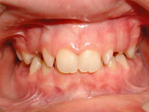
- Microdontia. This may be localised or generalised and its severity is often correlated with the severity of hypodontia. A common clinical example of microdontia is the presence of diminutive or peg-shaped lateral incisors (Figure 24.2).
- Maxillary canine impaction. Impaction is associated with the absence, or the presence of a diminutive, maxillary lateral incisor. The root of the lateral incisor may be important in guiding the canine into position (guidance theory). Up to 5% of those with absent lateral incisors may be affected by maxillary canine impaction.8
- Abnormal tooth position. It is common for permanent teeth to migrate into any spacing present, for overeruption of teeth opposing edentulous spaces, and for teeth, particularly premolars, to be severely rotated.
- Infraocclusion of the retained primary molar when the successor is absent. This can result in significant occlusal disruption when the infraocclusion is severe.
- Transposition between the maxillary canine and first premolars.
- Taurodontism. This is a developmental anomaly where the roots of the molars are shortened at the expense of an elongated pulp chamber. The roots of taurodont teeth may be more prone to orthodontically related root resorption and they offer less anchorage because of their reduced surface area. Additionally, endodontic treatment and extractions may be complicated by the abnormal root morphology.9
- Alveolar atrophy. When a permanent tooth is absent and the deciduous tooth is lost, there will be localised alveolar atrophy which can complicate orthodontic space closure or later implant therapy.
Figure 24.1 A late-forming upper right second premolar.
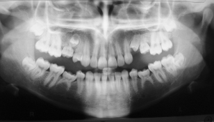
Figure 24.2 Peg-shaped maxillary lateral incisors.
MEDICAL CONDITIONS ASSOCIATED WITH HYPODONTIA
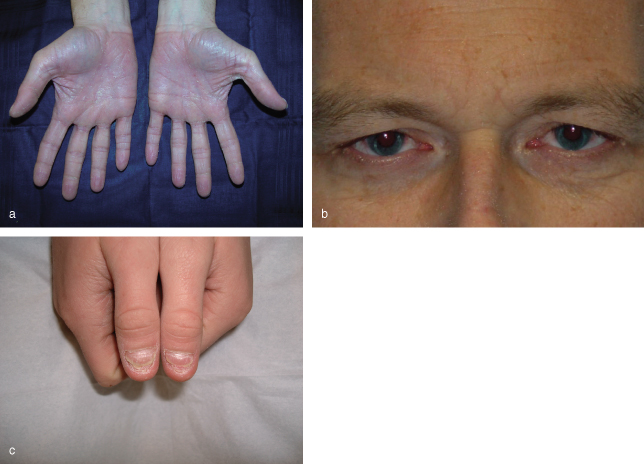
The ectodermal dysplasias are commonly associated with varying degrees of hypodontia or anodontia. These are a group of genetically transmitted conditions in which there is a defect within ectodermal tissues. Apart from hypodontia or anodontia, clinical features include hypohidrosis (failure to sweat leading to heat intolerance and dry erythematous skin, Figure 24.3a), hypotrichosis (sparse hair, Figure 24.3b), nail defects and xerostomia. Other medical conditions associated with hypodontia include Down syndrome, cleft lip and palate and hemifacial microsomia.
Figure 24.3 Signs of ectodermal dysplasia: (a) dry skin, (b) absence of eye lashes and (c) nail defects.
MANAGEMENT OF HYPODONTIA
The management of hypodontia involves a multidisciplinary team approach including the general dental pract/>
Stay updated, free dental videos. Join our Telegram channel
VIDEdental - Online dental courses


