23 File Systems
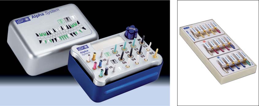
 Alpha System
 FlexMaster
 K3
K3
 LightSpeed
 Mtwo
Mtwo
 ProFile
 ProSystem GT
 ProTaper
 ProTaper Universal
 RaCe and S-Apex
 Summary and Conclusions
Alpha System
The Alpha system, developed in 2005, consists of NiTi instruments with tapers of 2%, 4%, 6%, and 10%. The ISO sizes range from 20 to 50. The Alpha files are commercially available in two different cross-sectional conformations.
The files for coronal widening are kite-shaped in cross section with 10% taper. Their large chip space makes them a good choice as the first instrument to work with. This “pre-enlargement” provides improved tactile control and makes possible an improved insertion path with the prebent size 10 K file. This makes the introduction of other files into the canal less complicated by reducing the chances of coronal sticking of the files and permits a larger quantity of irrigating fluid to be used to improve the cleaning and antibacterial effect, thus reducing postoperative complications to a minimum.
For the apical finish, instruments with a pentagonal cross-section should be used. This design aims to prevent instrument fracture; the tested torque values are the highest in comparison to all other instruments. The negative cutting angle achieves passive dentin removal and prevents excessive cutting into the root canal wall.
23.1 Principles of manufacture
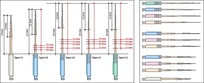
Design principles of the Alpha file. The 19-mm long orifice widener exhibits a taper of 10% (.06), making the entire instrument length 22 mm; all other files are 25 mm long with a working portion of 13 mm.
Right: The color coding corresponds to ISO recommendations. The taper is indicated by notches on the instrument shaft: 2% taper file has three notches, 4% taper file has two, and 6% taper file has one notch.
23.2 Working end and cross section
Left: Working end of the orifice widener with a large chip space for necessary dentin removal from the canal.
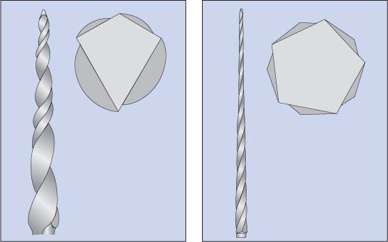
Left above: Kite-shaped cross section of the orifice-opening file.
Right: Working end of files with tapers of 2%, 4%, and 6%.
Right above: Pentagonal cross section of the Alpha file. This instrument cross section is designed for a high level of fracture resistance.
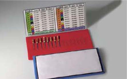
23.3 Instruments
Instrument set with additional sizes indicated for larger root canals. Using the green sequence, canals are instrumented with an apical scouting file of size 20.
Right: Instruments for basic treatment of small (yellow sequence), medium (red sequence), and larger root canals (blue sequence).
Size selection is carried out with the apical “scouting” file: Using the size 8 file, the canal is instrumented using the yellow sequence, increasing the file size by one number at a time until the final apical opening corresponds to a size 25 file tip.
23.4 Crown-down technique
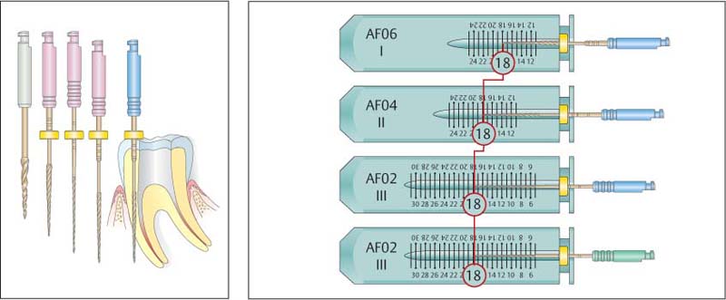
Alpha files for estimating the working length. The estimated working length is transferred to the various files, resulting in a crown-down instrumentation technique.
Left: Crown-down technique. Following coronal widening of the canal orifice, the initial depth is prepared with a file with 6% taper. The file with a taper of 4% can be used up to 2 mm above the apex; the 2% taper file is then used to the full working length.
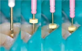
23.5 Clinical procedure, step-by-step
Left: The procedure is initiated using the orifice-widener and instrumentation to just short of the curvature of the canal.
Middle left: Instrumentation of the middle third of the canal using a file with 6% taper.
Middle right: Instrumentation to just short of the apical foramen.
Right: Instrumentation to full working length using a file with 2% taper.
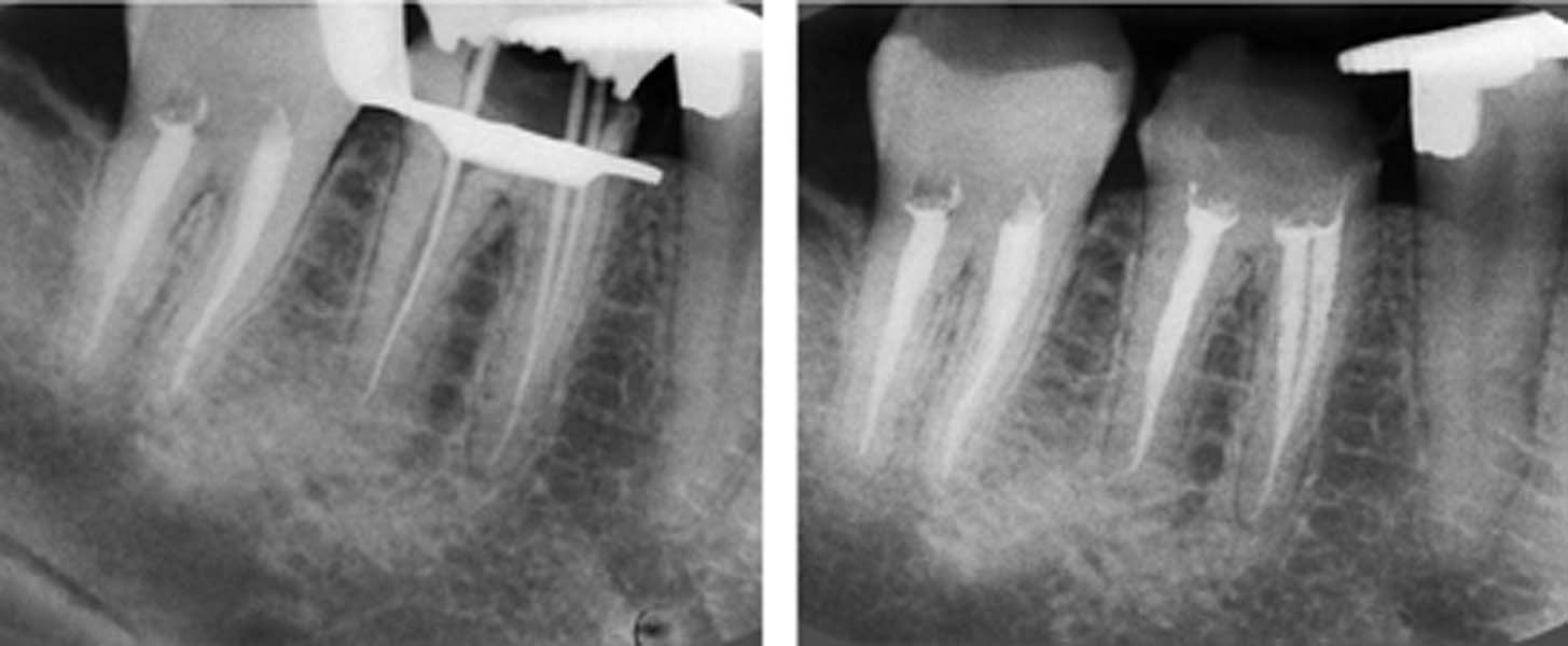
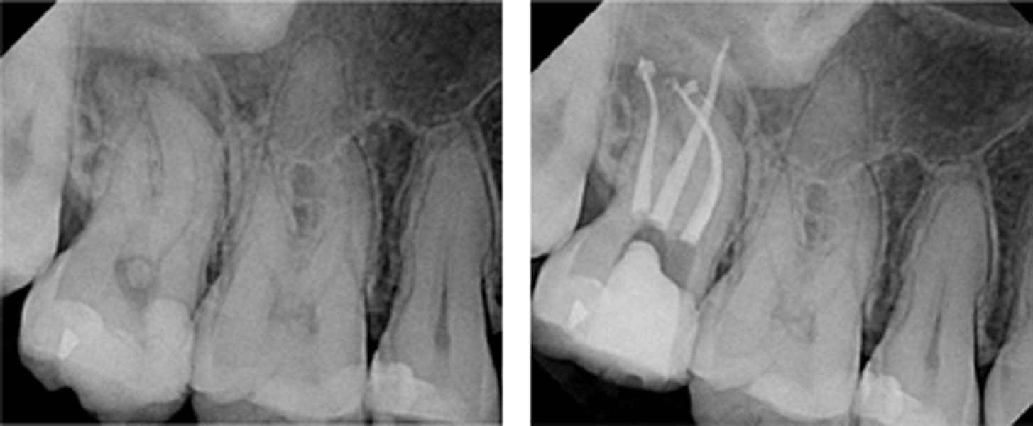
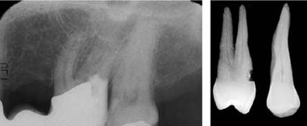
23.6 Clinical case
Left: Master-point radiograph following complete cleaning and shaping of tooth 46.
Right: Follow-up radiograph of the definitive root canal filling.
FlexMaster
The FlexMaster system (VDW, Munich, 2000) is characterized by a large assortment of root canal instruments of various sizes (15–70) and tapers (2%, 4%, 6%). The instruments are fabricated from a 55-nitinol alloy. The convex instruments exhibit an increased resistance to torsion and therefore reduced danger of fracture within the canal.
FlexMaster instruments are used at a constant rotation speed of 150–300 rpm. The instrumentation of the root canal is carried out using the crown-down technique. As with all other nickel titanium (NiTi) systems, the FlexMaster files should only be used in a torque-controlled handpiece to avoid the danger of the file getting stuck in the canal and possibly fracturing because of torsion.
Depending on the size of the root canal, instrument manufacturers recommend a variety of file sequences for instrumentation as a guide for narrow, middle, and broad canals, which are represented by color coding in the instrument set. For example, the start of the sequence for a broad root canal involves the use of an Introfile following exposure of the root canal orifice for instrumentation of the coronal segment of the canal with a.06/30 instrument.
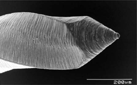
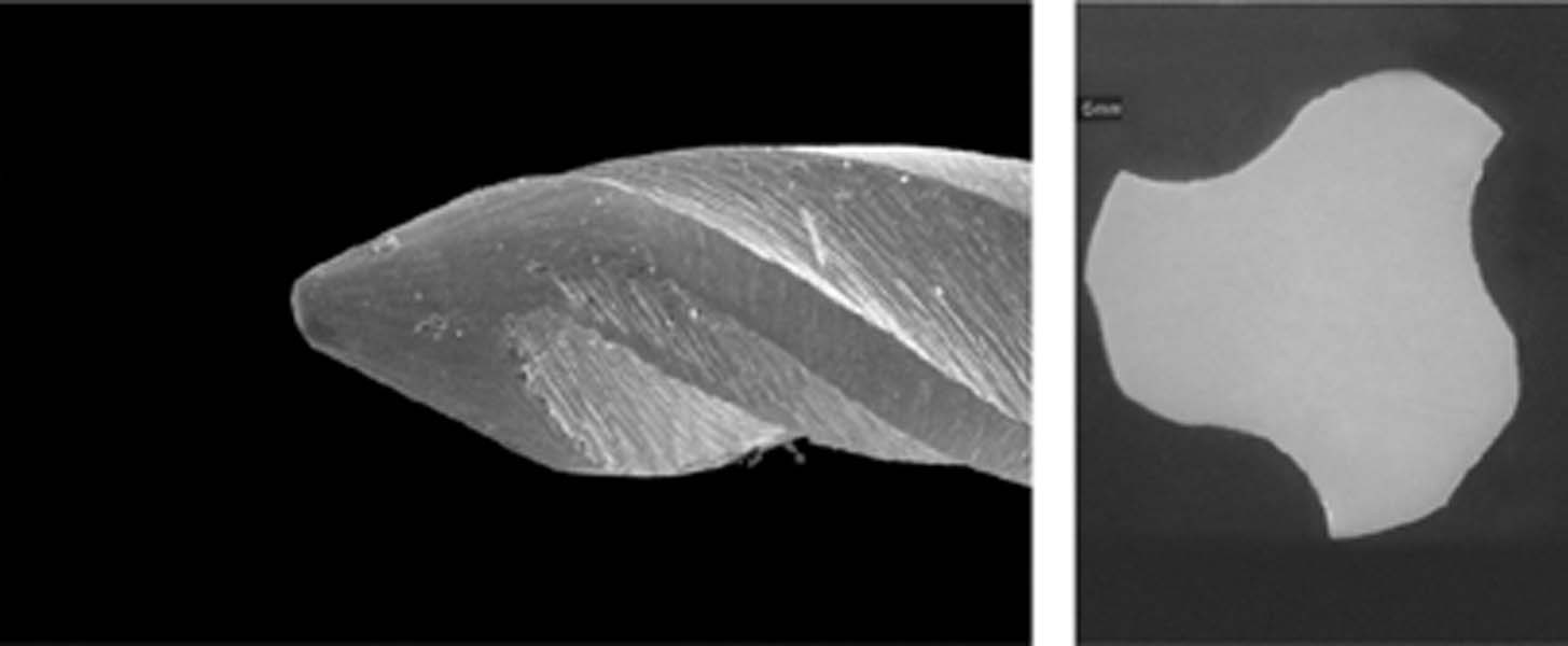
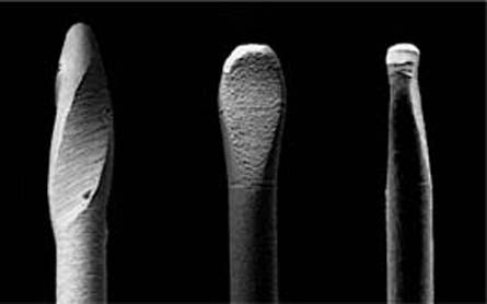
23.7 Instrument tip
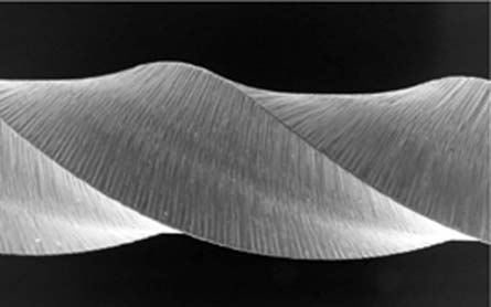
Similar to the majority of current NiTi file systems, the FlexMaster instruments also have a passive, noncutting tip with rounded transition angles. In addition to tapers of.06 and.04, the FlexMaster system also includes files with.02 taper. Because of their low taper, these files make it possible to make the preparation significantly wider at the apex, up to ISO size 70 (scanning electron microscopy [SEM] ×100).
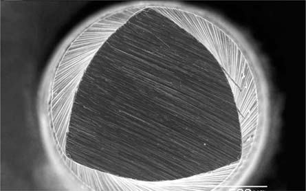
23.8 Instrument cross section
The convex triangular cross section of the FlexMaster instruments is designed to increase the resistance to torsion, therefore also reducing the risk of fracture. In contrast to instruments exhibiting a U-shaped cross section, the chip space width is relatively small with the FlexMaster files. Despite the relatively broad file cross section, these instruments possess sufficient flexibility to prepare even severely curved canals (SEM ×100).
23.9 Cutting edges
All FlexMaster instruments have three cutting edges. These files do not have broad radial lands on the lateral aspect of the cutting edges to ensure the highest possible cutting efficiency. Because the use of rotary instruments with sharp cutting edges is associated with the potential danger of “screwing” the instrument into the canal, such instruments should only be used with torque-controlled motor systems (SEM ×100).
The cleaning efficiency of FlexMaster files is universally viewed as positive (Hülsmann, 2003). However, in comparison to purely hand instrumentation, canals instrumented with the FlexMaster files exhibit a larger smear layer as well as residual debris (Schäfer, 2002).
With regard to the shaping of the root canal and to the original root canal axis, the FlexMaster system is clearly superior to conventional, manual instrumentation (Schäfer, 2002). Even narrow and severally dilacerated canals can be almost perfectly instrumented and prepared for root canal filling (Hübscher, 2003).
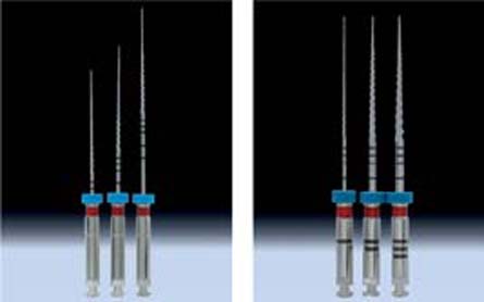
23.10 FlexMaster files
Left: These files are available in lengths of 21 mm, 25 mm, and 31 mm.
Right: The taper of each file is indicated by grooves in the shaft (1 ring =.02, 2 rings =.04, 3 rings =.06). The length of the working portion, incorporating the cutting edges, is 16 mm in all of the files.
Note the black circular markings above the cutting edges, which permit reliable determination of working length.
23.11 Preparation
Instrumentation is carried out using the crown-down technique, with decreasing file size. For the mandibular molar depicted here, a sequence for the mesial root would proceed from .06/20 to.04/30,.04/25, and.04/20. After reaching the working length, apical preparation is carried out using .02/30 files.
Instrumentation up to ISO size 30 should be the goal in every canal to ensure adequate irrigation of the prepared canal.
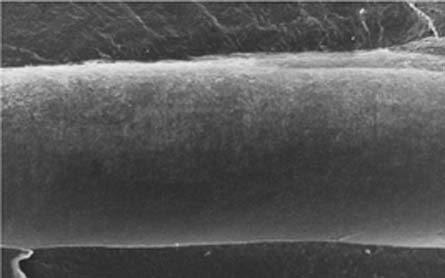
23.12 Prepared canal wall
This SEM image depicts a section of the canal wall in the middle segment of a root, prepared using FlexMaster instruments. This section is free of debris, the dentin appears smooth and free of gross scratches or other evidence of instrumentation. The prepared dentin surface is coated by a thin and homogeneous smear layer (SEM ×100).
K3
The rotary NiTi K3 file system (K3 RNT; Sybron-Endo, Orange, CA, USA) differs from other available systems because of its asymmetric design, which gives it extraordinary flexibility, cutting efficiency, tactile control, and fracture resistance. The K3 file system should be used clinically based on the following prerequisites:
• K3 files must be passively and gently inserted;
• each time the file is inserted only 1–2 mm of dentin should be removed;
• irrigation and recapitulation should follow each K3 file insertion.
After traversing the full length of the canal and manual widening with a K file of at least size 15, the K3 file system is used with the crown-down technique.
The variety of file types in the K3 file system is large: for coronal enlargement, files with a taper of.08,.10, and.12 and a tip diameter of size 25 are available. For canal shaping, files of taper .02 and sizes 15–45 as well as tapers of.04 and.06 in sizes 15–60 are available. Corresponding gutta-percha points of taper.04 and .06 in sizes 15–45 complete the K3 set-up.
23.13 The K3 files
The NiTi files from Kerr (SybronEndo) are available in a broad range of tapers and diameters for all root canal configurations.
23.14 SEM images
The K3 files have a broad cutting area, which is also tapered. The cutting edges merge smoothly at the precisely formed torpedo-shaped, round edge file tip.
Right: This cross-sectional SEM view reveals the complex cutting geometry with its U-shaped cut-outs.
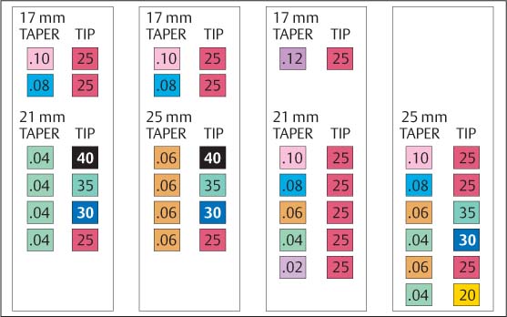
23.15 File sequence
Depending on canal shape and length, instrumentation using NiTi files requires selection from a variety of file sequences. The manufacturer’s guidelines should be adhered to in this regard.
The Procedure Pack is used with the crown-down technique,.04 taper and 21 mm (left),.06 and 25 mm (middle, left). In the G-Pack only the taper varies (middle, right). In the VTVT Pack, tip diameter and taper vary alternately (right).
The manufacturer recommends three file sequences:
• The Procedure Pack:.10/25 and.08/25 should be used for enlarging the access cavity, and sizes 40–25 (taper.04/.06) for the crown-down preparation.
• With the G-Pack, the tip diameter remains constant at size 25, and the taper varies from.12 to.02.
• With the VTVT Pack there is a change in taper (variable taper) and tip diameter (variable tip), which is generally regarded today as an adequate and fracture-reducing clinical model.
K3 has to date been reported in 33 publications. K3 files are much more resistant to cyclic fatigue than ProFile (Yao et al., 2006) in the 40/.04 and 40/.06 sizes, and also more resistant to fracture (Ankrum et al., 2004 [although not statistically significant], Troian et al., 2006). On the basis of its effectiveness in removal of gutta-percha, the K3 file system has been recommended for endodontic retreatment (Masiero et al., 2005; de Oliveira et al., 2006):
K3 files also help to precisely prepare the apical anatomy in complex root curvatures and minimize compression of the smear layer (Yoshimine et al., 2005; Kum et al., 2006).
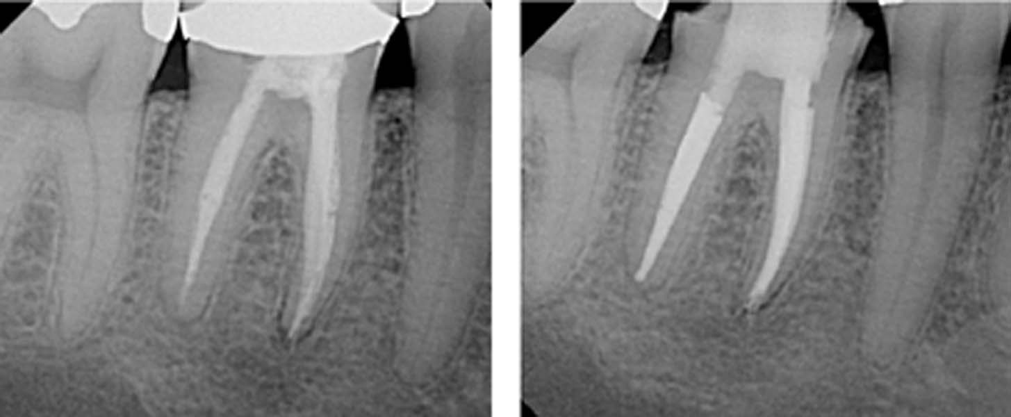
23.16 Clinical case I
Left: Preoperative radiograph of a mandibular molar showing a broad coronal pulp and “normal” root canals within a fused root situation.
Right: In the apical third, the three root canals are severely curved, and the distal canal has a double-S shape. However, a perfect root canal filling with preservation tooth substance was accomplished.
23.17 Clinical case II
Left: Preoperative radiograph of the maxillary second molar showing an almost 90° dilaceration of the mesial root.
Right: In order to achieve straight-line access, considerable tooth substance had to be sacrificed. This radiographic view of the dense root canal filling is testament to the success of the clinical procedure in the severely dilacerated canal.
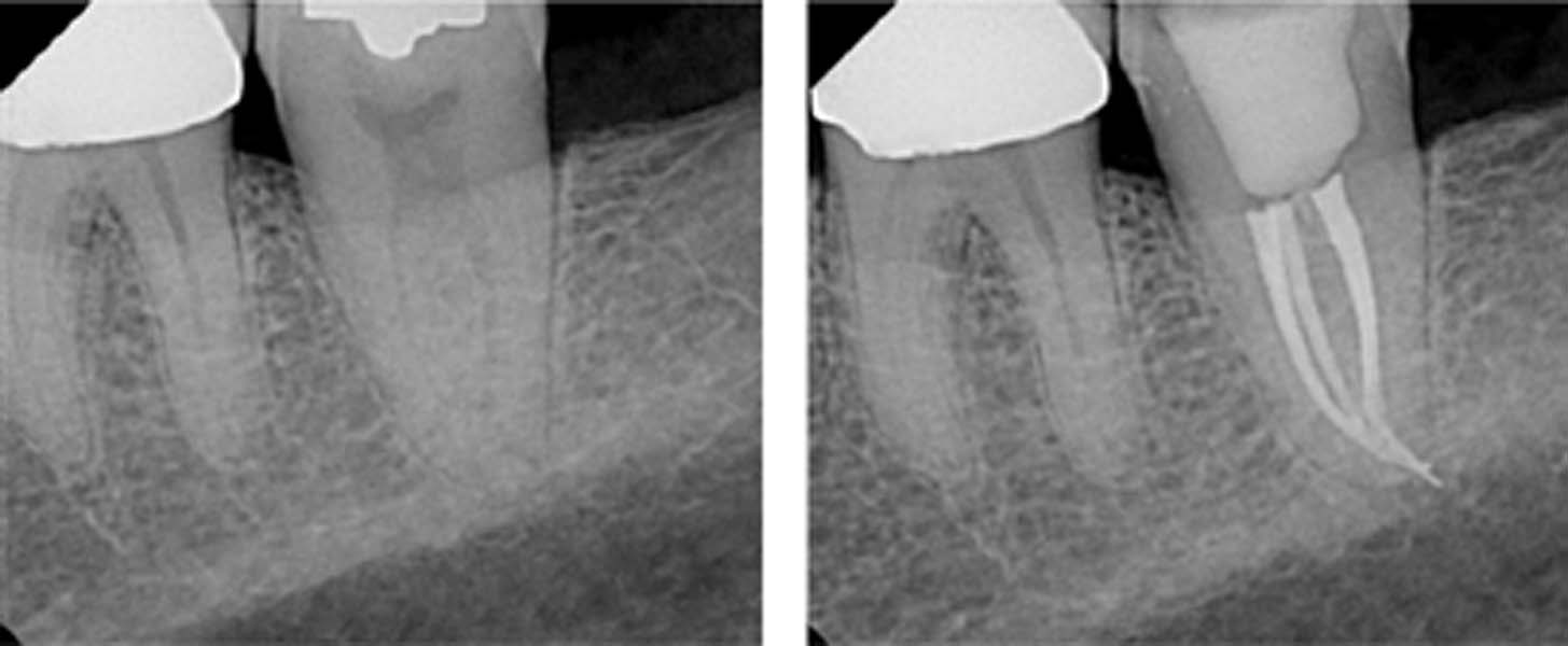
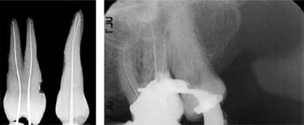
23.18 Clinical case III
Left: A previous endodontic “attempt” resulted in an inadequate root canal filling. The radiograph shows persistence of periapical inflammation, and the patient continued to experience pain and discomfort.
Right: Following endodontic retreatment of the tooth, with dense, warm vertical compaction of Resilon, a tight seal was obtained resulting in complete healing and resolution of the apical lesions.
LightSpeed
LightSpeed instruments are made of NiTi indicated for root canal instrumentation. They were developed by Wiley and Senia and entered the commercial dental market in 1993. A classic Light-Speed set consists of 22 instruments of ISO sizes 20–100. Between sizes 20 and 70, half-sizes are also provided (22.5, 27.5–65). The root canal is instrumented at a speed of 2000 rpm.
In contrast to the conventional endodontic instruments, which have a 16-mm long working section, LightSpeed files have a long, thin shaft and a short working end with a blunt tip.
The length of the working section depends on the ISO size. For example, the size 20 file has a 0.25-mm long cutting section whereas the size 100 file has a cutting section that is 1.75 mm long.
During root canal preparation, the noncutting tip points away from the canal wall and the file is thus retained in a more central position. Because of the high flexibility of the instrument shaft, the danger of “straightening” the canal is significantly reduced (Glosson et al., 1995; Shadid et al., 1998; Hülsmann et al., 2003).
23.19 LightSpeed instruments
The 22 LightSpeed files are color coded according to the ISO norm. The half-sizes are marked with a point toward the head end of the handle. Each of the 12 instruments of an LSX instrument set has the appropriate ISO size on the handle. Special sizes up to 140 are available in LightSpeed and LSX files.
Right: Because of the very thin shaft, LS and LSX instruments are extremely flexible.
23.20 Geometry of the working section
SEM of LS file size 35 and an LSX file size 35 seen from the front and the side. The working head of the LS has U-shaped grooves separated from each other by radial lands (left). The working end of the LSX (middle and right) is in contrast flat and spade-shaped and has only two radial lands (original magnification ×60, 15 kV).
23.21 Initial radiograph
Tooth 26 was clinically symptomatic and the radiograph revealed apical periodontitis in relation to the palatal root. The canal orifices of the distobuccal and palatal root canals were completely obliterated.
Right: Mesiodistal and bucco-palatal views of an extracted maxillary first premolar.
Since mid-2006 the LightSpeed eXtra (LSX) system has been available on the commercial market; it will eventually replace the LS system. LSX consists of only 12 instruments of sizes 20–80. The new instruments are not manufactured using the grinding process because this mechanical process can lead to imperfections of the surface (Eggert et al., 1999), which can later lead to instrument fracture.
LSX files are punched out of raw stock. The head of the instrument has a short and flat working area with two radial lands and a blunt tip. The long, thin instrument shaft of the LightSpeed files is unchanged in the LSX system. The recommendation is to utilize these instruments at 2500–3000 rpm and a higher level of torque. These modifications of the LightSpeed system have been designed to simplify the technique and reduce the risk of instrument fracture. The advantages of LS with regard to canal straightening and loss of working length remain unchanged (Iqbal et al., 2007).
23.22 Measurement radiograph
The working length was determined using size 15 K files. Thereafter, the canals were instrumented using LS files up to size 20, until the apical stop was prepared. Note that no size should be skipped during the preparation of the root canal. The stepback preparation technique was used, with shortening of the working length by 1 mm with each subsequent file.
Left: Determination of working length using size 15 K files.
Stay updated, free dental videos. Join our Telegram channel
VIDEdental - Online dental courses


