19
Principles of periodontal diagnosis and treatment planning
Figure 19.1 Flow diagram showing diagnosis and treatment plan options.
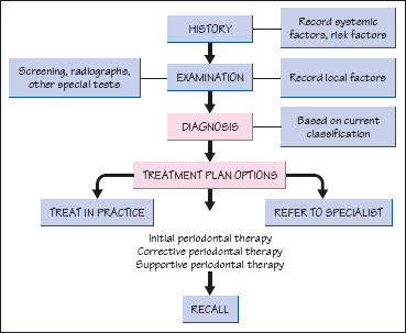
Figure 19.2 Dental examination of a patient.
Figure 19.3 Classification of periodontal diseases based on the 1999 International Workshop for the Classification of Periodontal Diseases and Conditions (Armitage,1999).
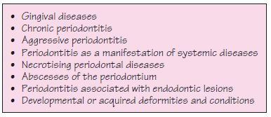
Figure 19.4 Periodontal diagnosis and descriptors. CAL, clinical attachment loss.
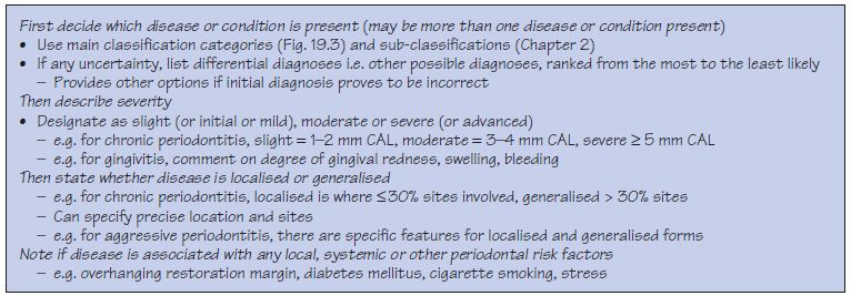
Figure 19.5 Initial periodontal therapy.
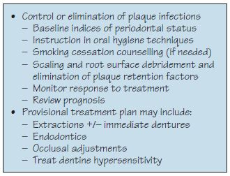
Figure 19.6 Corrective periodontal therapy.
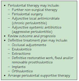
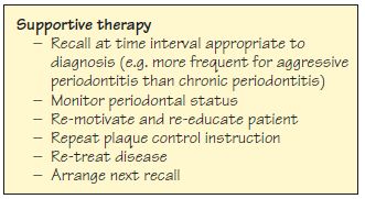
Figure 19.7 Supportive periodontal therapy.
The periodontal diagnosis follows a thorough history and examination (Figs 19.1, 19.2; Chapter 16) and should be based on the current classification derived from the 1999 International Workshop for the Classification of Periodontal Diseases and Conditions (Fig. 19.3; Chapter 2). If more than one disease is present, more than one diagnosis should be made. If there is any uncertainty, a differential diagnosis can be made. For each condition diagnosed additional descriptors can be used (Fig. 19.4), such as the severity, extent and location of disease or description of associated risk factors.
It is also important for other non-periodontal diagnoses to be made based on the extra-oral and intra-oral examination of soft and hard oral tissues, e.g.temporomandibular joint disorders, oral mucosal diseases, caries/ root caries, deficient, fractured or overhanging restorations, apical pathology, retained roots, unerupted or impacted teeth,/>
Stay updated, free dental videos. Join our Telegram channel
VIDEdental - Online dental courses


