19
Facial asymmetry
INTRODUCTION
Symmetry is defined as correspondence in size, shape and relative position of parts on opposite sides of a dividing line or median plane. Asymmetry is described as a lack or absence of symmetry. The facial midline is usually taken as the dividing line of the face and is a line passing through soft tissue nasion and the midpoint of the upper lip.
A degree of facial asymmetry is normal and acceptable within the face.1 It can be caused by an asymmetry in the facial skeleton and/or overlying soft tissue drape. The point at which an asymmetry becomes unacceptable is when an individual begins to experience aesthetic concerns and/or functional limitations. Although asymmetries can occur at many levels of the face, this chapter will focus on developmental asymmetries affecting the mandible and maxilla as these are most relevant to the dental profession. Table 19.1 provides a classification of facial asymmetries.
Table 19.1 A classification of facial asymmetry
| Cause | Examples |
| Developmental | Hemi-mandibular elongation |
| Hemi-mandibular hyperplasia | |
| Hemifacial microsomia | |
| Hemifacial hypertrophy | |
| Torticollis | |
| Hemifacial atrophy (Parry–Romberg syndrome) | |
| Pathological | Tumours and cysts |
| Infection | |
| Condylar resorption | |
| Condylar fractures | |
| Traumatic | Post irradiation |
| Functional | Mandibular displacement |
DEVELOPMENTAL CAUSES
Hemi-mandibular Elongation and Hyperplasia
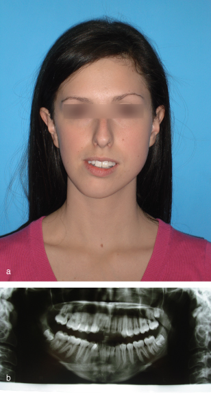
Hemi-mandibular elongation was first described by Obwegeser and Makek2 and is a developmental deformity of unknown aetiology affecting the mandible. It presents with a progressively increasing transverse displacement of the chin, away from the affected side, which usually becomes apparent during or after the adolescent growth spurt (Figure 19.1a,b). The mandibular dentition follows the skeletal displacement which predisposes to buccal crossbite and centreline displacement away from the affected side and a scissor bite on the affected side. Since there is a minimal vertical component to the abnormal growth pattern, there is typically no overeruption of the maxillary dentition on the affected side. Radiographically, there is clear elongation of the affected side of the mandible, principally located in the condylar region and the body of the mandible (Figure 19.1b).
Figure 19.1a,b A case of left-sided hemi-mandibular elongation with the corresponding dental panoramic tomograph.

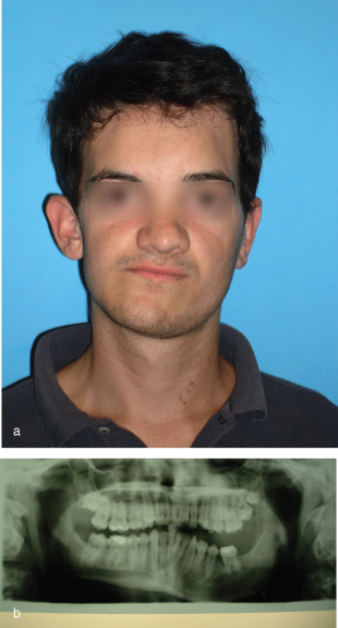
In contrast, hemi-mandibular hyperplasia presents with a three-dimensional enlargement of one side of the mandible during adolescence (Figure 19.2). As a consequence of the vertical component of abnormal growth, there is an increase in height on the affected side of the face, the dentition on that side overerupts to maintain occlusal contact, with a resultant cant in the maxillary occlusal plane and an increase in alveolar height above the inferior dental canal, and the face develops a twisted appearance. If abnormal growth is rapid, a lateral open bite may develop on the affected side as the rate of eruption is outpaced by the rate of vertical skeletal growth. Radiographically, a panoramic tomogram will show that the ascending ramus is elongated vertically with enlargement of the condyle (Figure 19.2b). There is also an elongation and thickening of the condylar neck. The angle of the mandible is rounded, while the lower border is bowed downwards to a lower level compared with the opposite side. There is an increase in the height of the mandibular body, which appears to increase the distance between the molar roots and the mandibular canal. The unaffected side appears to have a normal height. This growth defect is clearly demarcated by the symphysis (Figure 19.2b).
Figure 19.2a,b A case of left-sided hemi-mandibular hyperplasia with the corresponding dental panoramic tomograph.

Hybrid forms of hemi-mandibular elongation and hyperplasia exist where the patient exhibits features of both conditions. The differences between the two conditions are summarised in Table 19.2.
Table 19.2 Comparison of hemi-mandibular elongation and hyperplasia
| Hemi-mandibular elongation | Hemi-mandibular hyperplasia |
| Unilateral horizontal enlargement of mandible | Unilateral three-dimensional enlargement of mandible |
| Transverse displacement of chin point | Vertical displacement of chin and mandible. Chin may be rotated |
Stay updated, free dental videos. Join our Telegram channel

VIDEdental - Online dental courses


